Ebook Hematologic problems in the critically ill: Part 2
Bạn đang xem bản rút gọn của tài liệu. Xem và tải ngay bản đầy đủ của tài liệu tại đây (1.85 MB, 87 trang )
Chapter 6
The Critically Ill Patient
with Abnormal Platelet Count
Luca G. Mascaretti and Paola Pradella
Abnormal platelet counts are a common finding in critically ill
patients. Whereas thrombocytopenia, defined as a platelet count
less than 150*109/L, affects 13–60 % of Intensive Care Unit
(ICU) patients [1] and has been extensively studied, the occurrence of thrombocytosis (platelet counts >400*109/L) is
observed less frequently and has not been studied to the same
extent.
In this chapter, the main causes of thrombocytopenia and
thrombocytosis in critically ill patients will be illustrated, and
their implications on morbidity and mortality will be discussed.
Due to its importance in the ICU setting, a section in this chapter will be dedicated to heparin-induced thrombocytopenia
(HIT).
L.G. Mascaretti, MD ( )
Transfusion Medicine Department, University Hospital Trieste,
Strada di Fiume 447, Trieste 34149, Italy
e-mail:
P. Pradella, MSc
Hemostasis and Blood Coagulation Laboratory,
Transfusion Medicine Department, University Hospital Trieste,
Strada di Fiume 447, Trieste 34149, Italy
e-mail:
G. Berlot, G. Pozzato (eds.), Hematologic Problems in the Critically Ill,
DOI 10.1007/978-88-470-5301-4_6, © Springer-Verlag Italia 2015
59
60
6.1
L.G. Mascaretti and P. Pradella
Thrombocytopenia: A Classification
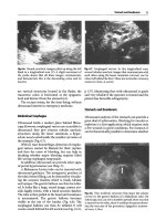
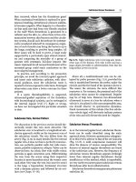
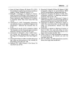
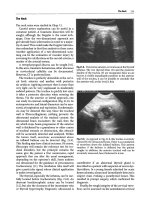
Before addressing the issues related to “true” thrombocytopenia, pseudo (or spurious) thrombocytopenia must be defined. In
some conditions such as liver diseases, neoplasia, autoimmune
disease, or in healthy subjects, antibodies mediated by anticoagulants such as EDTA are responsible for platelet clumping,
which, not being detected by cell counters, will lead to falsely
low platelet counts [2]. Pseudothrombocytopenia is not clinically significant and is diagnosed by microscopic examination
of the blood smear (Fig. 6.1) and by repeating the whole blood
count in tubes with a different anticoagulant (heparin- or citratebased solutions).
“True” thrombocytopenia, to a variable degree, affects all
types of ICU patients in all parts of the world; adult medical ICU
patients are mostly affected, but it is also observed in surgical
and pediatric patients. These observations underlie the comment
made by R.I. Parker in his recent review [1] that thrombocytopenia in ICU patients is “a truly universal occurrence.”
Although a threshold value of 150*109/L is generally
accepted to indicate thrombocytopenia, stable platelet counts
between 150 and 100*109/L are not necessarily considered
pathological. Moreover, it is now recognized that the risk of
clinically spontaneous bleeding is significantly high when platelet counts fall below 20–10*109/L [3].
The two main mechanisms responsible for thrombocytopenia
are reduced production and increased destruction of platelets;
less frequently, a reduced platelet count may also be due to
sequestration and hemodilution [1, 2].
Table 6.1 summarizes the main classification criteria for
thrombocytopenia, the most frequent pathological mechanisms
and the associated clinical conditions. The table does not include
causes of thrombocytopenia in pregnancy and postpartum, since
these conditions go beyond the scope of this chapter.
6
The Critically Ill Patient with Abnormal Platelet Count
61
Thrombocytopenia
Microscopic examination of
blood smear
Spurious or pseudothrombocytopenia, not
clinically significant
YES
PLT
clumping?
NO
Consider hereditary
thrombocytopenia
YES
Giant PLTs,
family history?
NO
YES
Consider TTP
Schistocytes?
NO
Consider bone
marrow disorders
YES
Blasts?
Consider: ITP, DITP, HIT,
DIC, viral infections
NO
YES
Consider infection
Lympho-cytosis,
atypical
lymphocytes?
NO
Isolated
Thrombocytopenia
Fig. 6.1 Diagnostic algorithm based on blood smear (Adapted from Stasi [3])
It should always be remembered that in a significant number
of cases, thrombocytopenia is due to multiple factors, such as
for example in sepsis.
The diagnostic workup for thrombocytopenia must include,
in addition to laboratory tests discussed in this chapter, a family
Enhanced
destruction
Nonimmune: platelet aggregation
Immune: platelet specific
auto-antibodies
Immune: immune complexes
Nonimmune: microangiopathic
Infiltration of bone marrow due to
neoplastic diseases
Infiltration of bone marrow due to
storage disorders
Drug-related marrow suppression
Marrow failure due to radiation therapy
Nonimmune: mechanical
Secondary bone-marrow failure
Main
classification
criteria
Pathological mechanism
Decreased
Primary bone-marrow failure
production
Table 6.1 Causes of thrombocytopenia
[10]
[11]
[12]
[13]
Gaucher’s disease
Chemotherapy, other drugs
Internal radiation, external radiation
Intravascular devices such as central venous
catheters, intraaortic balloon pump
Thrombotic thrombocytopenic purpura (TTP),
disseminated intravascular coagulation
(DIC), subacute bacterial endocarditis (SBE),
vasculitis
[11]
[16]
[17]
Drugs
Immune thrombocytopenic purpura (ITP)
Autoimmune disorders
[14, 15]
[ 8]
[9]
References
[4–7]
Examples of clinical conditions
Myelodisplastic disorders, Fanconi’s anemia,
congenital amegakaryocytic
thrombocytopenia
Sepsis, severe idiopathic aplastic anemia, severe
malnutrition
Acute leukemia, widespread marrow metastases
62
L.G. Mascaretti and P. Pradella
Hemodilution
Sequestration
[11]
Drug related immune thrombocytopenia:
antiepileptics, gold compunds, vancomycin,
thiazides, quinine/quinidine
Heparin-Induced Thrombocytopenia (HIT)
Sepsis
Portal hypertension leads to the redistribution of
platelets from the circulating pool to the
splenic pool
Transfusion of platelet-poor blood products,
infusion of colloids and crystalloids
Secondary to fluid infusion in case of
massive hemorrhage
[22]
[20]
[8]
[21]
[19]
Posttransfusion purpura
Immune:
platelet-specific allo-antibodies
Immune: Drugs
Immune: Heparin-induced
Immune: sepsis
Congestive splenomegaly
[18]
Hypersplenism, hemophagocytic
lymphohistiocytosis
Immune: cell-mediated
6
The Critically Ill Patient with Abnormal Platelet Count
63
64
L.G. Mascaretti and P. Pradella
history for thrombocytopenia, the evaluation of its “dynamics,”
meaning if it is a new finding, if it is chronic or whether it has a
relapsing presentation. Information on bleeding episodes is also
very important, as is the history of concomitant diseases such as
infections, tumors, or autoimmune diseases. Finally, it is of
paramount importance to collect the history related to recent
medication (heparin, antibiotics) and blood transfusion since
especially for hospitalized patients, drug-induced thrombocytopenia (DITP) is among the most common causes of low platelet
counts. Since the aim of this chapter is to discuss thrombocytopenia in critically ill patients, it goes without saying that it is
challenging to understand this condition in these patients also
because a complete history may be difficult to obtain.
Whereas by definition, the Whole Blood Count is the basic
laboratory test for diagnosing thrombocytopenia, the microscopic examination of the blood smear gives additional, important information on the pathogenetic mechanism involved [3].
Figure 6.1 illustrates an algorithm that guides the hematologist
in the diagnosis of isolated thrombocytopenia. Other tests
employed in the diagnosis of the causes of thrombocytopenia
are liver and renal function tests, coagulation tests including
d-dimers, lactate dehydrogenase, and bone marrow aspirate and
biopsy.
Platelet antibody assays and other tests such as reticulated
platelets have a limited specificity and therefore their use is
debatable [16].
Before describing the clinical conditions associated with
thrombocytopenia, the importance of the rate of decline in platelet counts must be pointed out. When a constant, slow reduction
in platelet number is observed with minimum (nadir) counts
falling below 20*109/L, a DITP due to marrow inhibition is the
probable cause. On the other hand, when there is a fast rate of
decline (24–48 h) in platelet numbers, an immune mechanism is
suspected. A variable rate in platelet reduction is suggestive of
consumptive coagulopathy [1].
6
The Critically Ill Patient with Abnormal Platelet Count
6.1.1
65
Thrombocytopenia Due to Reduced
Production
Thrombocytopenia caused by bone marrow suppression may be
due to acquired or congenital conditions. In the latter category
are comprised Fanconi’s anemia, congenital amegakaryocytic
thrombocytopenia, thrombocytopenia, and absent radii syndrome; a comprehensive review of these clinical conditions has
been recently published by Parikh and Bessler [4]. The inherited
bone marrow failure syndromes are genetic disorders affecting
blood cell lineages. They are characterized by a wide spectrum
of symptoms ranging from aplastic anemia to symptoms related
to the suppression of one or two cell lines. Congenital amegakaryocytic thrombocytopenia is an inherited bone marrow failure syndrome usually diagnosed at birth, and characterized by
insufficient production of megakaryocytes due to a defect in the
thrombopoietin receptor [5].
Acquired bone marrow failure is often due to myelodysplastic syndromes, a heterogeneous group of clonal bone marrow
disorders characterized by ineffective hematopoiesis, morphological and functional abnormalities of hematopoietic cells, and
increased risk of malignant transformation. The prevalence of
thrombocytopenia in these diseases varies from 40 to 65 % [6],
and together with platelet dysfunction, is responsible for the
increased hemorrhagic risk in these patients.
Sepsis is a condition affecting a significant number of
patients admitted to hospitals; a recent review reports that in the
USA, 2 % of patients corresponding to 750,000 per year are
septic, half of which are admitted to ICUs [8]. Clinical signs of
sepsis are diverse and depend on the microorganism, site of
original infection, and health condition of the patient.
Thrombocytopenia in sepsis is a common finding and severe
forms of sepsis are associated with coagulation disorders that
can lead to disseminated intravascular coagulation (DIC).
66
L.G. Mascaretti and P. Pradella
Thrombocytopenia can also be caused by drugs that suppress
the bone marrow, and in particular megakaryocyte proliferation
and maturation. Whereas antimetabolytes, cytotoxic drugs, and
alkylating agents exert a toxic effect on all bone marrow cell
lines, some antibiotics such as linezolid, may cause a selective
suppression of platelet cell lines [11].
Other causes of thrombocytopenia due to decreased production (Table 6.1) are storage disorders [10], infiltration of bone
marrow due to neoplastic diseases [9] and radiation therapy [12].
Thrombocytopenia due to reduced production is not a frequent cause of admission to the ICUs, since it is more often
preexistent.
6.1.2
Thrombocytopenia Due to Enhanced
Destruction or Consumption
6.1.2.1
Thrombocytopenia Due to Enhanced
Destruction: Nonimmune Mechanisms
Medical devices such as mechanical heart valves, left-ventricular
assistance devices, and aortic balloon pumps may be responsible
for the destruction of platelets. In a study on 1,302 patients who
underwent percutaneous coronary intervention (PCI) with baseline normal platelet counts (≥150*109/L), 3.1 % developed postPCI thrombocytopenia. Multivariate analysis showed that the use
of intra-aortic balloon pump was an independent predictor of
thrombocytopenia, with an odds ratio of 2.8, confidence intervals
1.1–6.8, p = 0.024. Post-PCI thrombocytopenia was significantly
associated with major adverse cardiovascular events at 6 months
(hazard ratio 2.7, CI 1.3–5.5, p = 0.0069) [13].
Microangiopathic processes such as thrombotic thrombocytopenic purpura (TTP), hemolytic uremic syndrome (HUS),
and disseminated intravascular coagulation (DIC) may be
6
The Critically Ill Patient with Abnormal Platelet Count
67
responsible for thrombocytopenia due to enhanced platelet
destruction.
TTP is characterized by microvascular platelet clumping,
which leads to thrombocytopenia and microangiopathic hemolytic anemia. Common findings are “broken” erythrocytes or
schistocytes (see algorithm reported in Fig. 6.1), neurological
disorders, renal failure, and fever [14]. The disease is due to a
congenital or acquired deficiency in ADAMTS13, a metalloprotease which cleaves von Willebrand factor. ADAMTS13 deficiency is responsible for microvascular thrombosis and
thrombocytopenia. Plasma exchange is the optimal therapy, and
its effectiveness is probably due to the removal of antiADAMTS13 autoantibodies and large von Willebrand factor
multimers.
HUS is similar to TTP in that microvascular thrombosis,
thrombocytopenia, microangiopathic hemolytic anemia, renal
insufficiency, and altered mental status are common features.
However, ADAMTS13 is normal and the disease is generally
due to endothelial cell damage caused by a toxin produced by
pathogenic strains of Escherichia or Shigella. In HUS, thrombocytopenia is usually not severe but dialysis may be required to
treat renal insufficiency [23].
DIC does not occur as an isolated event but is practically
always associated with an underlying condition such as tissue
damage (trauma, burns, hemolytic transfusion reaction, acute
transplant rejection), neoplasia, systemic infection, obstetric
conditions (abruption placentae, placenta previa, amniotic fluid
embolism), and other clinical conditions such as shock, cardiac
arrest, and aortic aneurysm. DIC is the result of an overstimulation
of the coagulation system and its clinical presentation varies
from severe hemorrhage to thrombosis (or both simultaneously).
Thrombocytopenia, abnormal prothrombin time and activated
partial thromboplastin time (PT and aPTT), decreased fibrinogen
and elevated fibrinogen degradation products are common laboratory features of DIC. DIC-associated mortality is mostly due to
68
L.G. Mascaretti and P. Pradella
the original disease, which is complicated by hemorrhage or
thrombosis. Multiorgan dysfunction syndrome is a frequent consequence of DIC and is due to hemorrhagic or thrombotic events
in liver, heart, kidneys, central nervous system, and lungs [15].
The main therapeutic goal in DIC is that of treating the
underlying condition. As far as transfusion of blood products is
concerned, there has been a lot of debate on its benefit and
potential harm; generally, platelet counts should be kept more
than 20*109/L in presence of mild bleeding and more than
50*109/L when there is active bleeding. Plasma or cryoprecipitate should be considered when bleeding is associated with low
fibrinogen levels. The aim of fibrinogen replacement is to maintain levels more than 100 mg/dl to prevent or treat bleeding [24].
6.1.2.2
Thrombocytopenia Due to Enhanced
Destruction: Immune Mechanisms
(Except HIT)
In addition to Heparin-Induced Thrombocytopenia (HIT) which
will be discussed in the following section, primary Immune
Thrombocytopenia (ITP), post-transfusion purpura (PTP), and
drugs may lead to immune platelet destruction.
ITP is an acquired disorder mediated by immunological
mechanism, characterized by low platelet counts in the absence
of any possible known cause of thrombocytopenia. It affects
children and adults (with a slight prevalence in women) and
symptoms range from massive bleeding (gastrointestinal, skin–
mucosal, and intracranial) to minimal bruising or only alterations
in whole blood count. Evaluation of the blood smear is
important in the diagnosis of ITP (Fig. 6.1) and antiplatelet
antibody assays are not routinely performed due to the low
specificity of this test. Adult ITP is treated with corticosteroids
or IVIg and platelet transfusions are recommended only for
emergency cases in presence of active bleeding [16].
6
The Critically Ill Patient with Abnormal Platelet Count
69
PTP is a rare complication of transfusion occurring 7–10
days after a red blood cell or platelet transfusion and is characterized by a dramatic fall in platelet count reaching a nadir less
than 10*109/L. Thrombocytopenia is caused by platelet alloantibodies in the recipient which at first destroy the transfused
platelets, but successively also react with self-platelets. PTP is
managed by administering IVIg or if available, compatible
platelets (usually HPA-1a negative) [19].
Drug-induced thrombocytopenia (DITP) may either be
caused by drugs suppressing bone marrow (see previous
section) or by drugs eliciting diverse types of antibodies.
Table 6.2 summarizes the main types of antibodies implicated in
DITP [11]. DITP may be hard to diagnose in critically ill
patients, since thrombocytopenia may become evident several
days after the beginning of therapy, and has to be distinguished
from other causes of thrombocytopenia.
Other causes of thrombocytopenia include platelet sequestration and hemodilution. Thrombocytopenia is a common feature
of liver cirrhosis and is attributable to portal hypertension with
sequestration of platelets in the enlarged spleen [21].
In massive transfusion, defined as the transfusion of one
blood volume in 24 h, coagulation abnormalities are almost
always present and are in part due to hemodilutional thrombocytopenia. However, coagulopathy associated with massive
transfusion has many additional components, among which are
coagulation factor dilution, hypothermia, type of solutions used
for volume replacement, and DIC [22].
6.1.2.3
Clinical Significance of Thrombocytopenia
in Critically Ill Patients
Hui and coworkers published a review in 2011 [25] aimed at
better understanding the clinical role of thrombocytopenia in
critically ill patients. It analyzed 24 studies for a total of 6,894
Fab-binding monoclonal Ab
Fiban-dependent Ab
Hapten-induced Ab
Type of antibody (Ab)
Drug-dependent Ab
Mechanism and features
“Classic” mechanism: severe thrombocytopenia occurs
5–10 days after beginning of new drug, which binds
to platelet glycoproteins (IIb/IIIa, or IbIX). Abs bind
to glycoproteins and sensitized platelets are
eliminated by the reticuloendothelial system,
resulting in severe thrombocytopenia. Abs react with
glycoproteins only when the drug is present.
Drug acts like a hapten, i.e., small molecules that elicit
an immune response when linked to larger carrier
molecule. Abs will bind to the platelet membrane
glycoproteins only when the hapten (drug) is
covalently linked to them.
Drugs bind to glycoprotein IIb/IIIa and may form a
neo-epitope: autoantibodies are either preexistent
and therefore naturally occurring, or may be
stimulated by exposure. Thrombocytopenia occurs
after a few hours from beginning of therapy.
Abciximab is a Fab fragment monoclonal antibody
which reacts with glycoprotein IIIa, preventing
fibrinogen binding. Thrombocytopenia associated
with this drug is due to naturally occurring
antibodies which recognize the murine portions of
the monoclonal Ab.
Abciximab
Tirofiban and eptifibatide
Penicillin
Main drugs
Quinine
Table 6.2 Antibodies and mechanisms implicated immune drug-induced thrombocytopenia
70
L.G. Mascaretti and P. Pradella
Some drugs may elicit autoantibodies which do not
require the presence of the drug to bind to platelet
antigens. Thrombocytopenia can therefore persist
after the drug is no longer administered.
Heparin-induced Thrombocytopenia (see following
chapter)
Some platelet Abs in addition to binding to circulating
platelets may also react with megakaryocytes, thus
inhibiting platelet production.
Based on data from Arnold et al. [11]
Anti-megakaryocyte Abs
Immune complexes
Drug-induced autoantibody
formation
Eptifibatide, quinine
Heparin
Gold, l-dopa, procainamide,
sulphonamides,
alemtuzumab
6
The Critically Ill Patient with Abnormal Platelet Count
71
72
L.G. Mascaretti and P. Pradella
patients; whereas 8.3–67.4 % of patients had low platelet counts
at admission, the proportion of patients which developed thrombocytopenia during their stay in the ICU ranged from 13 to
44 %. Major risk factors for the development of thrombocytopenia were high illness severity, organ dysfunction, sepsis, and
renal failure. The review was unable to show convincing evidence for an association between thrombocytopenia and bleeding, but multivariate analysis conducted by six studies indicated
that thrombocytopenia was an independent predictor of mortality. This finding is confirmed by Stansbury and coworkers [26]
in their study on the prognostic significance of platelet counts in
the first 24 h after severe injury.
6.2
Heparin-Induced Thrombocytopenia (HIT)
Critically ill patients are often suspected of having HIT, because
both thrombocytopenia and heparin treatment are common in
the ICU setting. Nevertheless, a recent study demonstrated that
the diagnosis of HIT was confirmed in only 0.5 % of these
patients [27].
Heparin-Induced Thrombocytopenia (HIT) is a particular type
of drug-induced thrombocytopenia that is associated with a prothrombotic condition, despite a low circulating platelet count.
Although this disorder may occur with any molecular-weight
heparin, the incidence of HIT is higher with unfractionated
heparin compared to low-molecular-weight heparin [28, 29].
Other risk factors are host-related, with the female sex more
affected than the male [30] and the surgical population more
affected than the medical [31].
Two types of HIT are described with different clinical
features. Type 1 HIT is likely induced by a nonimmune mechanism, with circulating platelet clumping in the presence of
heparin and their sequestration in the spleen. The consequent
thrombocytopenia develops usually in 2–3 days after starting
6
The Critically Ill Patient with Abnormal Platelet Count
73
heparin, is mild and resolves spontaneously with no thrombotic
or hemorrhagic complications. Unlike the former, Type 2 HIT is
an immunomediated disorder, in which the anticoagulant binds
to Platelet Factor 4 (PF4), a protein released from activated
platelets, and triggers the development of specific antibodies
[32]. The macromolecular complex constituted by the antibody
and heparin-PF4 binds a specific receptor on the platelet surface
leading to further platelet activation [33] and to thrombin generation [34]. Activated platelets are cleared from circulation
with consequent thrombocytopenia and a paradoxical enhanced
risk for arterial and especially venous thrombosis.
Different laboratory methods are available to identify the
presence of HIT antibodies:
• Functional assays with the HIT patient serum activating normal platelets in the presence of heparin
• Antigen assays to detect the binding of HIT antibodies to
their target heparin/PF4
Functional assays are more specific for clinically relevant
antibodies, but require specialized personnel, so antigen assays
are the most widely used [35].
A typical feature of Type 2 HIT is the reduction of more than
50 % in the platelet count, leading to a moderate thrombocytopenia with a median platelet nadir of 50–60*109/L; unlike other
drug-mediated thrombocytopenias, a platelet number less than
20*109/L is very uncommon. In naïve patients, the typical onset
of thrombocytopenia is 5–14 days after the beginning of heparin
exposure; in patients treated in the past 3 months, it may occur
early within 24 h (early onset). Seldom platelet counts begin to
fall after more than 15 days from the beginning of heparin treatment, sometimes after heparin discontinuation with a delay
onset [36].
When HIT is strongly suspected, any heparin treatment (even
exposure to heparin flushes or lines washing procedure) must be
discontinued and replaced with another anticoagulant, for
example, direct thrombin or activated FX inhibitors [37]. In a
74
L.G. Mascaretti and P. Pradella
few days, platelet count returns to normal values or to
pretreatment values.
Actual guidelines suggest a clinical evaluation with a scoring
system to test the likelihood of the disorder [38, 39]. The most
widely used is the 4 T’s score, based on clinical traits of HIT such
as the degree of thrombocytopenia, the timing of the onset, the
presence of a new or enlarged thrombosis, and an eventual different cause of platelet count decrease, as is shown in Table 6.3 [38].
With a low score (≤3), HIT can be excluded without any
laboratory assay and the heparin treatment may be continued; if
the score is moderate or high (4–6), all heparin exposure should
be discontinued to avoid HIT complications and an alternative
anticoagulant should be chosen [40]. Recently, two new methods have been proposed for assessing the clinical probability of
HIT in the early management of patients suspected of having
HIT [41, 42] but they need further validation. Whichever
method is used, a careful evaluation is necessary in HIT exclusion or confirmation in order to prevent bleeding risks in thrombocytopenic patients.
6.3
Thrombocytosis in Critically Ill Patients
Elevated platelet counts (>400*109/L) are not a common finding
among critically ill patients and contrary to thrombocytopenia,
thrombocytosis in hospitalized patients has not been investigated at great length. From the etiological point of view,
thrombocytosis may be classified as primary or secondary.
Whereas the former group includes myeloproliferative or
myelodysplastic syndromes, the latter may be either secondary or paraneoplastic. In the ICU patient, the main underlying
clinical conditions responsible for thrombocytosis are infection, trauma, splenectomy, hemolysis, bleeding, and drugs such
as antifungals, amoxicillin/clavunate, enoxaparin [43]. Two
Points = 1
50 % fall
or
Platelet nadir 10–19*109/L
Points = 0
<30 % fall
or
Platelet nadir
<10*109/L
Timing of platelet count Clear onset between day 5
Consistent with immunization but not Platelet count fall
fall
and 10
clear (e.g., missing platelet counts)
≤4 days (without
recent heparin
or
or
exposure)
Platelet fall ≤1 day (if heparin Onset of thrombocytopenia after
exposure within past 30
day 10
days)
or
Fall ≤1day if heparin exposure 30–100
days ago
Thrombosis (or other
New thrombosis (confirmed); Progressive or recurrent thrombosis;
None
sequelaes, e.g., skin Skin necrosis;
Erythematous skin lesions;
lesions)
Acute systemic reaction
Suspected thrombosis not yet proven
postintravenous
unfractionated heparin
bolus
Other causes of
No other cause for platelet
Possible other cause is evident
Definite other cause
thrombocytopenia
count fall is evident
is present
Thrombocytopenia
Points = 2
>50 % platelet fall
and
Platelet nadir ≥20*109/L
Table 6.3 The 4 T’s score (adapted from [38])
6
The Critically Ill Patient with Abnormal Platelet Count
75
76
L.G. Mascaretti and P. Pradella
other main conditions leading to thrombocytosis are familial
(hereditary), due to a mutation responsible for an increase in
the production of thrombopoietin [44], and essential thrombocythemia, a condition which may eventually lead to myelofibrosis or leukemia.
Differential diagnosis between primary and secondary thrombocytosis is not always straightforward in the ICU setting.
Generally speaking, if thrombocytosis occurs during the stay in
an ICU, it is most probably of secondary nature. However, if the
patient is admitted urgently and no previous whole blood count
is available, hematological consultation and further testing
(e.g., JAK2) might be useful for the characterization of thrombocytosis [43]. For patients affected by secondary thrombocytosis, risk of thrombotic or hemorrhagic complications is <2 %,
irrespective of platelet count.
As far as therapy is concerned, there is no threshold above
which platelet removal by apheresis or antiaggregation therapy
should be initiated. Risk of thrombosis in these patients must
consider associated clinical conditions such as sepsis, trauma,
and rheumatic disease, which themselves predispose to venous
clot formation. Platelet apheresis is able to reduce platelet
counts significantly and is used primarily in patients with
myeloproliferative diseases in which either a thrombotic or
bleeding event has occurred. Aspirin is administered in thrombocytosis (both primary and secondary) patients who have had
a thrombotic event.
References
1. Parker RI (2012) Etiology and significance of thrombocytopenia in
critically ill patients. Crit Care Clin 28:399–411
2. Rice TW, Wheeler AP (2009) Coagulopathy in critically ill patients.
Part 1: Platelet disorders. Chest 136:1622–1630
3. Stasi R (2012) How to approach thrombocytopenia. Hematology 2012:
191–197
6
The Critically Ill Patient with Abnormal Platelet Count
77
4. Parik S, Bessler M (2012) Recent insights into inherited bone marrow
failure syndromes. Curr Opin Pediatr 24:23–32
5. Ballmaier M, Germeshausen M (2009) Advances in the understanding of
congenital amegakaryocytic thrombocytopenia. Br J Hematol 146:3–16
6. Kantarjian H, Giles F, List A et al (2007) The incidence and impact of
thrombocytopenia in myelodysplastic syndromes. Cancer 109(9):
1705–1714
7. Cazzola M, Malcovati L, Invernizzi R (2011) Myelodysplastic/myeloproliferative neoplasms. Hematology 2011:264–272
8. Finfer SR, Vincent JL (2013) Severe sepsis and septic shock. NEJM
369(9):840–851
9. Kwaan HC (2007) Thrombosis and bleeding complications in malignant hematologic disorders. Hematology 2007:151–157
10. Grabowski GA (2012) Gaucher disease and other storage disorders.
Hematology 2012:13–18
11. Arnold DM, Nazi I, Warkentin TE et al (2013) Approach to the diagnosis and management of drug-induced immune thrombocytopenia.
Transfus Med Rev 27(3):137–145
12. Mac Manus M, Lamborn K, Khan W (1997) Radiotherapy-associated
neutropenia and thrombocytopenia: analysis of risk factors and development of a predictive model. Blood 89(7):2303–2310
13. Shenoy C, Orshaw P, Devarakonda S et al (2009) Occurrence, predictors,
and outcomes of post-percutaneous coronary intervention thrombocytopenia in an unselected population. J Interven Cardiol 22:156–162
14. Fontana S, Kremer Hovinga JA, Lammle B et al (2006) Treatment of
thrombotic thrombocytopenic purpura. Vox Sanguinis 90:245–254
15. Kitchens CS (2009) Thrombocytopenia and thrombosis in disseminated intravascular coagulation (DIC). Hematology 2009:240–246
16. Provan D, Stasi R, Newland AC et al (2010) International consensus
report on the investigation and management of primary immune thrombocytopenia. Blood 115(2):168–186
17. McKenzie CGH, Guo L, Freedman J et al (2013) Cellular immune dysfunction in immune thrombocytopenia (ITP). Br J Haemat 163:10–23
18. Usmani NG, Woda A, Newburger PE (2013) Advances in understanding the pathogenesis of HLH. Br J Haemat 161:609–622
19. Rozman P (2002) Platelet antigens. The role of human platelet alloantigens (HPA) in blood transfusion and transplantation. Transplant Immunol
10:165–181
20. Warkentin TE, Sheppard JA, Heels-Ansdell D et al (2013) Heparin-induced
thrombocytopenia in medical surgical critical illness. Chest 144(3):
848–858
21. Peck-Radosavljevic M (2001) Hypersplenism. Eur J Gastroenterol
Hepatol 13:317–323
78
L.G. Mascaretti and P. Pradella
22. Hardy JF, de Moorloose P, Samama M (2004) Massive transfusion and
coagulopathy: pathophysiology and implications for clinical management. Can J Anesth 51(4):293–310
23. Thiele T, Selleng K, Selleng S et al (2013) Thrombocytopenia in the
Intensive Care Unit – diagnostic approach and management. Semin
Hematol 50(3):239–250
24. Dunn AL (2009) Disseminated intravascular coagulopathy. In: Hillyer
CD, Shaz BH, Zimring JC, Abshire TC (eds) Transfusion Medicine and
hemostasis: clinical and laboratory aspects. Elsevier, Burlington/London
25. Hui P, Cook DJ, Lim W et al (2011) The frequency and clinical significance of thrombocytopenia complicating critical illness. Chest 139(2):
271–278
26. Stansbury LG, Hess AS, Thompson K et al (2013) The clinical significance of platelet counts in the first 24 hours after severe injury.
Transfusion 53(4):783–789
27. Trehel-Tursis V, Louvain-Quintard V, Zarrouki Y et al (2012) Clinical
and biologic features of patients suspected or confirmed to have heparininduced thrombocytopenia in a cardiothoracic surgical ICU. Chest
142(4):837–844
28. Warkentin TE, Levine MN, Hirsch J et al (1995) Heparin-induced
thrombocytopenia in patients treated with low-molecular-weight heparin or unfractionated heparin. N Engl J Med 332:1330–1335
29. Martel N, Lee J, Wells PS (2005) Risk for heparin-induced thrombocytopenia with unfractionated and low molecular-weight heparin thromboprophylaxis: a meta-analysis. Blood 106:2710–2715
30. Warkentin TE, Sheppard JAI, Sigoin CS et al (2006) Gender imbalance
and risk factor interactions in heparin-induced thrombocytopenia. Blood
108:2937–2941
31. Chong BH (2003) Heparin induced thrombocytopenia. J Thromb Haemost
1:1471–1478
32. Amiral J, Bridey F, Dreyfus M et al (1992) Platelet factor 4 complexed
to heparin is the target for antibodies generated in heparin-induced
thrombocytopenia. Thromb Haemost 68:95–96
33. Newman PM, Chong BH (2000) Heparin-induced thrombocytopenia:
new evidence for the dynamic binding of purified anti-PF4–heparin
antibodies to platelets and the resultant platelet activation. Blood 96:
182–187
34. Tardy-Poncet B, Piot M, Chapelle C et al (2009) Thrombin generation and
heparin-induced thrombocytopenia. J Thromb Haemost 7:1474–1481
35. Greinacher A, Warkentin TE (2006) Recognition, treatment, and prevention of heparin-induced thrombocytopenia: review and update.
Thromb Res 118:165–176
6
The Critically Ill Patient with Abnormal Platelet Count
79
36. Warkentin TE (2002) Platelet count monitoring and laboratory testing for
heparin-induced thrombocytopenia. Recommendations of the College of
American Pathologists. Arch Pathol Lab Med 126:1415–1423
37. Backhoul T, Greinacher A (2012) Recent advances in the diagnosis and
treatment of heparin-induced thrombocytopenia. Ther Adv Hematol
314:237–251
38. Linkins LA, Dans AL, Moores LK et al (2012) Treatment and prevention of heparin-induced thrombocytopenia: antithrombotic therapy and
prevention of thrombosis, 9th ed: America College of Chest Physicians
Evidence-based Clinical Practice Guidelines. Chest 141:e495S–e530S
39. Watson H, Davidson S, Keeling D (2012) Guidelines on the diagnosis
and management of heparin induced thrombocytopenia: second edition. Br J Haematol. doi:10.1111/bjh.12059
40. Lo GK, Juhl D, Warkentin TE et al (2006) Evaluation of pretest clinical
score (4 T’s) for the diagnosis of heparin-induced thrombocytopenia in
two clinical settings. JTH 4:759–765
41. Cuker A, Arepally G, Crowther MA et al (2010) The HIT Expert
Probability (HEP) Score: a novel pre-test probability model for
heparin-induced thrombocytopenia based on broad expert opinion.
J Thromb Haemost 8:2642–2650
42. Messmore HL, Fabbrini N, Bird NL et al (2011) Simple scoring system
for early management of heparin-induced thrombocytopenia. Clin Appl
Thromb Hemost 17:197–201
43. Powner DJ, Hoots WK (2008) Thrombocytosis in the NICU. Neurocrit
Care 8:471–475
44. Vannucchi A, Barbui T (2007) Thrombocytosis and thrombosis.
Hematology 2007:363–370
Chapter 7
Adverse Transfusion Reactions
in Critically Ill Patients
Federica Tomasella and Luca G. Mascaretti
As transfusion entered routine clinical practice in the
mid-twentieth century, it was apparent that the benefits were
counterbalanced by unwanted reactions both of infectious and
noninfectious nature [1, 2]. Whereas the former received wide
attention also by the general population [3], the latter mainly
remained of restricted interest to transfusion scientists (and
naturally to the patients). It is a well-known fact that in the past
25 years, blood testing and donor selection have had a notable
impact on reducing infectious complications [4, 5] and today,
noninfectious adverse reactions to transfusion (NIART) are
prevalent. If we look at the UK’s Serious Hazards of Blood
Transfusion hemovigilance data for 2012 [6], of the 538 cases
analyzed only 3 were transfusion-transmitted infections; 372
acute transfusion reactions, 42 hemolytic transfusion reactions,
11 transfusion-related acute lung injuries, and 82 transfusionassociated circulatory overload. Hemovigilance data for our
region, Friuli Venezia Giulia (North East Italy), are presented in
Table 7.1 [7].
F. Tomasella, MD ( ) • L.G. Mascaretti, MD
Transfusion Medicine Department, University Hospital Trieste,
Strada di Fiume 447, Trieste 34149, Italy
e-mail:
G. Berlot, G. Pozzato (eds.), Hematologic Problems in the Critically Ill,
DOI 10.1007/978-88-470-5301-4_7, © Springer-Verlag Italia 2015
81
Adverse transfusion reactions
Febrile nonhemolytic transfusion reactions
(FNHTR)
Allergic transfusion reactions (ATR)
Circulatory overload
Hypotension
Severe dyspnea
Delayed hemolytic transfusion reactions (DHTR)
Anaphylaxis
Transfusion-associated graft versus host disease
(TA-GVHD)
Transfusion errors
Transfusion-related acute lung injury (TRALI)
Septic complications
Others
Total adverse reactions
Total transfused units
Frequency of adverse reaction per unit (%)
2010
52
39
8
3
4
0
2
0
0
0
1
15
124
71,147
0.17
2007–2009
195
140
20
14
4
6
6
1
7
2
0
77
472
219,129
0.22
Table 7.1 Adverse transfusion reactions in Friuli Venezia Giulia 2007–2012
5
0
0
20
146
72,728
0.20
42
1
3
3
1
1
0
2011
70
1
0
0
12
120
70,488
0.17
49
3
3
1
0
4
0
2012
47
13
2
1
124
862
433,492
0.20
270
32
23
12
7
13
1
Total
2007–2012
364
82
F. Tomasella and L.G. Mascaretti
7 Adverse Transfusion Reactions in Critically Ill Patients
83
The critically ill patient can be affected by both infectious
and noninfectious adverse reactions after a transfusion therapy
and the importance of diagnosis is remarkable for the severity of
clinical conditions usually treated in an intensive care unit.
Transfusion reactions, in fact, can be masked by the severity of
the main illness and the lack of active collaboration of the
patient [8].
The aim of this chapter is to give an overview of the most
common adverse transfusion reactions.
7.1
Infectious Adverse Reactions to Transfusion
(IARTs)
IARTs can be caused by viruses, bacteria, and protozoa.
Potentially, an undefined number of infective agents are liable
to transmit a disease after a transfusion, but we shall consider
the most frequent and pathogenic. In this field, it is important to
know that not all infectious reactions have the same incidence in
different countries, and for this reason the policy of detecting
tests varies from USA [9] and Europe, and at the same time
among European countries (EU). In this paper, we will focus on
Italian policy, which is harmonized with EU regulations.
7.1.1
Viruses
The transmission of viruses after a transfusion therapy is usually
due to the presence of the infective agent in the circulation of the
donor.
In the past 30 years, the risk of transmitting a virus infection
with transfusion has greatly decreased because of the development of microbiological research and new detection techniques
(serological and nucleic acid testing (NAT)). At the same time,
F. Tomasella and L.G. Mascaretti
84
more restrictive donor selection criteria and pathogen reduction
or inactivation technologies are usually employed to further
reduce the risk of infection [10]. Residual risk is due to asymptomatic donors who donate in the “window period.”
Table 7.2 summarizes information related to the principal
virus infections potentially transmitted by transfusion.
7.1.1.1
Management
It is useful for ICU specialists to know the main transfusionrelated viral infections. In fact, differently from the main immunological adverse reactions, the symptoms of IARTs can appear
some days after transfusion and can be confused with the main
disease. Particularly, it is necessary to pay attention to patients
with a compromised immunological system who need immediate therapy to stop virus replication.
7.1.2
Bacteria
Bacteria infections following transfusion (Table 7.3) are often
derived from microbial flora present on donor skin which
contaminate blood products. They can also be due to systemic
bacterial infections, though this is a rare event. From 2008, the
Italian National Blood Center recommends using the first 40 ml of
collected blood for testing, diverting it in tubes during withdrawal.
Regarding the kind of blood components, platelet concentrates are more frequently involved in IARTs, because their
storage is at room temperature (22 ± 2 °C). However, medical
and nursing staff must inspect the blood component before
administration to check for integrity of bags, hemolysis, change
in color, gas formation, and clots. Any of these findings must be
communicated to the transfusion center to which the product
must be returned.