Nghiên cứu thực trạng, một số yếu tố liên quan đến nhiễm giun lươn strongyloides spp và kết quả điều trị bằng ivermectin tại huyện đức hoà, tỉnh long an (2017 – 2018) tt tếng
Bạn đang xem bản rút gọn của tài liệu. Xem và tải ngay bản đầy đủ của tài liệu tại đây (643.38 KB, 24 trang )
1
INTRODUCTION
Strongyloides spp is a chronic causative infectious agent. This agent is
considered to be one of the neglected tropicals infectious pathogens, but it is an
important public health problem due to the characteristic of the autoimmune
cycle, leading to hyper infection and death in some patients.
People are acquired Strongyloides spp agent through contact with
contaminated soil sources directly such as agricultural cultivation, recreation
activities, etc. The adult worms usually located in the intestine wall, causing
abdominal pain, prolonged diarrhea, or colitis, .... In addition to disease at the
gastrointestinal tract, the stage when infective larvae of Strongyloides spp enter
the human body, can move to many organs, causing very diverse clinical diseases.
The problem of accurate diagnosis of the case therefore also faces many
difficulties.
Treatment strongyloidiasis cases is more difficult than other intestinal
helminths, especially with hyper infection. The treatment of cases has still been
inconsistent with the timing of treatment and drug selection.
Although strongyloidiasis was discovered in Southern Vietnam at first time,
but there has not been many studies on this pathogen recently. According to the
study results in Cu Chi district, HCM city previously, the prevalence of
Strongyloides spp infection in the community was quite high. While Duc Hoa
district of Long An province was located bordering with Cu Chi district, HCM
city, there still has not been any research on these pathogens.
Stemming from this reality, we conducted the thesis: Studying the actual
situation, various factors related to human Strongyloides spp infection and
ivermectin effectiveness in Duc Hoa district, Long An province (2017 –
2018).”with the objective:
1. Determine the actual situation and factors related to human Strongyloides
spp infection in Duc Hoa district, Long An province in 2017-2018.
2. Determine component species of Strongyloides in human
strongyloidiasis disease by morphological characterization and
molecular biology.
3. Describe the clinical symptoms, para-clinical and evaluate results of
treatment for strongyloidiasis by ivermectin single dose.
2
NOVELTY, SCIENTIFIC AND PRACTICAL SIGNIFICANCE OF THE
THESIS
The thesis provides details of valuable scientific data on the status of
Strongyloides spp infection and related factors in many research sites of Duc Hoa
district, Long An province.
Provide details of the presence of symptoms, signs and value of para-clinical
test in cases of gastrointestinal strongyloidiasis.
Determine the clinical efficacy of ivermectin and the effectiveness of larval
clearance, which is the basis for developing an appropriate intervention program.
For the first time, a new species of Strongyloides spp was identified from
patient in Duc Hoa district that derived from animal origin, by using molecular
biology techniques.
THESIS STRUCTURE
The thesis consists of 136 pages divided into the following sections:
Introduction (02 pages), Chapter 1: Literature review (31 pages), Chapter 2: Study
subjects and methods (29 pages), Chapter 3: Study results (34 pages), Chapter 4:
Discussions (36 pages), Conclusions (02 pages), and Recommendations (1 page).
There are 36 tables, 13 figures, and 115 references (12 pages, including 28
Vietnamese documents and 87 English documents).
Chapter 1
LITERATURE REVIEW
1.1. History of discovering strongyloidiasis
In July 1876, Louis Normand found the parasite in the stool samples of French
diarrhea patients with a history of coming to the Southern Vietnam. He named
this agent Anguillula stercoralis and the corresponding disease has been called
Cochin -China diarrhea. Coming to 1915, the council named scientific unification
named this pathogen Strongyloides stercoralis.
1.2. Pathogen
There are about 104 species of Strongyloides spp, including 52 common
species, some of which cause disease in domestic pets and other animals. The
main pathogen cause human disease is S. stercoralis, and less is S. fuelleborni.
Some other species such as S. procyonic (host is raccoons), S. myopotami and S
ratti (hosts are rats and rodents) are considered to be parasitic diseases transmitted
from animals to humans.
1.2.1 Morphology
The development stages of Strongyloides spp including: parasitic female
living worm, free-living female worm, free-living male worm, the first stage
larvae (rhabditiform), the second stage larvae (filariform) and eggs. Parasitic
male worm has still been not found.
1.2.2. Biological development cycle of Strongyloides spp
3
Strongyloides spp have two stages of the pathogenesis cycle: the parasitic
cycle and the free living cycle. Free living cycle often happen in the tropics due
to adaptive with conditions of external environment. In addition, strongyloidiasis
also has autoinfection cycle. The autoinfection cycle occurs when all or some of
rhabditiform larvae reside in the intestinal wall, rapidly molt to the infected stage,
establishing a parasitic development stage inside the host and this phenomenon
may remain maintenance of host life. This phenomenon also often occurs in
patients who have immunosuppression status. This autoinfection process leads to
two severe status of strongyloidiasis: hyperinfection syndrome and disseminated
strongyloidiasis.
1.3 Epidemiological characteristics
The infection prevalence is usually less than 1% in temperate zone, but may
be above 25% in many parts of the tropics.
1.3.1. The situation of Strongyloides spp infection in the world
Strongyloidiasis is an infectious disease in many countries, especially in West
Africa, the Caribbean, Southeast Asia, tropical regions of Brazil, Cambodia and
Spain. Southeast Asia area has the highest prevalence of the disease.
1.3.2. The situation of Strongyloides spp infection in Vietnam
According to a Galliard survey in 1940, in the northern Vietnam, the
prevalence of Strongyloides spp infection was from 0.2 to 2.5% of the population.
Recent studies using ELISA technique have shown that the infection rate is higher
than 7.6-10.9%.
Strongyloides spp are widely distributed in the southern provinces of Vietnam,
provinces such as Long An, Binh Duong, Tay Ninh, Dong Nai ... and Ho Chi
Minh City (Cu Chi, Thu Duc and Hoc Mon districts) and some provinces in the
Central region and Highlands.
1.4. Strongyloidiasis pathology
The strongyloidiasis has an incubation period approximately 1 month. The
majority of cases in endemic areas often have no symptoms or vague symptoms.
Strongyloidiasis is divided into two forms:
1.4.1 Chronic, uncomplicated Strongyloidiasis: happen in normal individuals,
without immunodeficiency, possible symptoms include:
Skin manifestations: The cutaneous larvae migrans, skin bruises, nonspecific
urticaria, ...
Gastrointestinal manifestations: Abdominal pain, diarrhea, weight loss, anal
itching.
Other manifestations: Patient coughing, pneumonia, signs of cachexia, ...
1.4.2 Severe disease, complications
This form of the disease is common in immunocompromised individuals who
use corticosteroids, immunosuppressing or accompanied by other chronic
diseases such as chronic obstructive pulmonary disease, chronic kidney failure,
malignancy, diabetes, alcoholism, malnutrition.
4
1.4.3. Hyperinfection syndrome and disseminated strongyloidiasis
Hyperinfection syndrome is manifestted by a phenomenon in which the rapid
increasing the number of pathogens leads to an excessive burden without the
spread of external larvae. Disseminated strongyloidiasis is the ultimate
consequence of hyperinfection syndrome. Affected organs include the lungs,
liver, heart, kidneys, endocrine organs and central nervous system.
Strongyloidiasis is severe and lead to death.
1.5 Paraclinical diagnosis
Diagnostic indicators can be based on nonspecific tests: increased number of
eosinophils, increased CRP, increased serum IgE, etc.
1.5.1 Direct test
Strongyloides spp larvae are usually found in faeces or in gastric, duodenum,
sputum, biopsy tissue at a gastric ulcer or other tissue, etc. Direct tests include as
follows: direct stool test, Baermann concentration technique, Formalin - ether,
Harada - Mori stool culture or agar plate culture.
1.5.2 Indirect diagnosis
Currently, serological testing methods are popular and widely used due to their
high sensitivity and general applicability. The most widely used technique is the
ELISA test to detect immunoglobulin G (IgG) antibodies.
1.5.3 Molecular biology diagnosis
The PCR test detects parasitic DNA in feces, especially Strongyloides spp,
which has the advantage of high sensitivity and specificity. PCR techniques have
been used include as follows: real-time PCR, nested PCR, multiplex real-time
PCR and multiplex PCR.
1.6 Treatment
Treatment human strongyloidiasis was based on using current drugs:
Ivermectin 150-200 µg/kg/ day single dose, albendazole 10-15 mg/kg/day and
thiabendazole 50 mg/kg/day. The dose of each drug is determined but no
consistent guide about the number of treatment days.
CHAPTER 2
STUDY SUBJECTS AND METHODS
2.1 Study subjects, sites and duration
2.1.1Study subjects
People residing in Duc Hoa district, Long An province meet the selection
criteria for the sample.
Patients infected with gastrointestinal strongyloidiasis.
Strongyloides larvae were collected from a patient's stool sample test.
Ivermectin pills single dose.
2.1.2. Study sites (location)
5
In the field works: 4 communes Duc Lap Thuong, My Hanh Nam, Hiep Hoa,
An Ninh Tay and 1 Duc Hoa town of Duc Hoa district, Long An province. In each
commune or town, the location is people's residence and health station.
- Laboratory of medical parasitology, Pham Ngoc Thach university of
medicine, Laboratory of Hematology and Immunization, Hospital for Tropical
Diseases in Ho Chi Minh city.
- Genome sequencing at First BASE Laboratories-Axil Scientific, Malaysia.
2.1.3. Study duration
The study was conducted from July 2017 to November 2018.
2.2 Study methods
2.2.1. Study design
Cross-sectional descriptive study: describe infection prevalence and analyzing
related factors.
Laboratory experiments.
Cross-sectional descriptive study describe case series and non-controlled
treatment interventions.
2.2.2 Research methods
2.2.2.1 Objective 1: Determine the actual situation and factors related to human
Strongyloides spp infection in Duc Hoa district, Long An province in 2017-2018.
Research content
Determining the prevalence of intestinal strongyloidiasis in each commune /
town of Duc Hoa district, Long An province.
Description and analysis: A number of demographic factors related to the
prevalence of Strongyloides spp infection in people such as sex, age group,
economic status and education level. Some of the related behaviors such as
agriculture job, toilet use and contact soil directly in daily life.
Techniques used in the study
Interview technique of data collection
Technique for diagnosis of Strongyloides spp infection
Modified Harada Mori culture (Sasa, 1986)
Stool direct smear technique
2.2.2.2. Objective 2: Determine component species of Strongyloides in human
strongyloidiasis disease by morphological characterization and molecular
biology.
Research content
Describe the detection ability of direct stool test and modified Harada Mori
culture technique in the first times.
Describe morphological characteristics of development stages of
Strongyloides spp that collect from human feces.
Determine component species of Strongyloides in human strongyloidiasis
disease in Duc Hoa district by a multiplex real-time PCR and gene sequencing.
Techniques used in the study
6
Diagnostic technique: size measurement of morphological structures of
Strongyloides spp larvae stage 1,2 or adults.
Multiplex real-time PCR technique, 2 steps nested - PCR and gene sequencing.
2.2.2.3 Objective 3: Describe the clinical symptoms, paraclinical and evaluate
results of treatment for Strongyloides spp by ivermectin single dose
Research content
Describe the presence rate of clinical symptoms in patients who were
diagnosed gastrointestinal strongyloidiasis before and after treatment: intermittent
loosing diarrhea, abdominal pain, urticaria, weight loss, headache, cutaneous
larvae migrans.
Describe and analyze the para-clinical parameters of patients who were
diagnosed gastrointestinal strongyloidiasis: the number, ratio of eosinophils and
serum ELISA diagnosis.
Determining the clinical and para-clinical therapeutic effect of single dose
ivermectin and the presence rate of side effects.
Techniques used in the study
Blood test technique to determine eosinophils.
ELISA test for detect specific antibody (IgG) against Strongyloides spp.
Stool test technique to evaluate treatment results: Apply a combination of 2
direct smear technique and stool culture.
2.2.3 Data processing
Analyze the relation among the variables using 2 test, Fisher exact test, t test,
OR with p <0.05 by SPSS 22.0 software in windows.
Genetic sequences were obtained after sequencing, will be processed by
Bioedit v.2.6 (Tom Hall, 2017) and MEGA 6 (Temura, 2013) sofware.
2.3 Ethics in research
Strictly compliance with regulations in biomedical study.
CHAPTER 3
STUDY RESULTS
3.1 Determine the actual situation and factors related to human Strongyloides
spp infection in Duc Hoa district, Long An province in 2017-2018.
3.1.1 Characteristics of study subjects
The total number of samples collected was 1,190 samples, distributed in 5
research sites including 4 communes and 1 town. The study site was Duc Lap
Thuong commune had the largest sample size corresponding to the largest
population (314), followed by Duc Hoa town (233).
The percentage of women participating in the study was 54.1%, more than
men. People between the ages of 15 and 60 participated in the majority (66.8%).
The number of the study participants with education level below high school was
69.1%. People with poor and nearby poverty status accounted for 14.3%. Farmer
accounted for 24.7% and the proportion of unhygienic toilet using was 11.5%.
7
3.1.2 Actual situation of Strongyloides spp in Duc Hoa district
3.1.2.1 The prevalence of Strongyloides spp infection
Table 3.1 Prevalence of Strongyloides spp infection in study site (n = 1,190)
No Commune/town No. of Tests No. (+)Percentage (%)
1 My Hanh Nam
216
16
7.4
2 Hiep Hoa
224
10
4.5
3 An Ninh Tay
203
9
4.4
4 Đuc Lap Thuong
314
39
12.4
5 Duc Hoa town
233
5
2.1
Total
1,190
79
6.64
The overall prevalence of Strongyloides spp infection in Duc Hoa district is
6.64%
3.1.3 Related factors with Strongyloides spp infection
Table 3.2 Relation between Strongyloides spp infection and sex
Strongyloides inf.
Inf. (+)
Non – inf.
Total
Sex
Male
Female
Total
60
486
546
19
625
644
79
1,111
1,190
p < 0.001; OR = 4.06; CI 95%: 2.39 – 6.89
There was a relation between Strongyloides spp infection and sex (p <0.001).
Male was at 4.06 times higher risk of infection than women.
Table 3.3 Relation between Strongyloides spp infection and age
Strongyloides inf.
Inf. (+)
Non – inf.
Total
Age group
< 15
0
240
240
15 – 60
55
740
795
Over 60
24
131
155
Total
79
1,111
1,190
p < 0.01 (Fisher test). OR = 2.46; CI 95%: 1.47 – 4.12
People over 60 years of age were 2.46 times higher risk about Strongyloides spp
infected than the other groups.
Table 3.4 Relation between Strongyloides spp infection and education
Strongyloides inf.
Inf. (+)
Non – inf.
Total
Education
Below High school
64
758
822
High school and more
15
353
368
Total
79
1,111
1,190
p < 0.05; OR = 1.98 ; CI 95%: 1.12 – 3.54
8
People with education level below high school was 1.98 times risk to be infected
Strongyloides spp.
Table 3.5 Relation between Strongyloides spp infection and economic status
Strongyloides inf.
Inf. (+)
Non – inf.
Total
Economic status
Poor and nearby poverty
45
125
170
Average
18
504
522
Well and above
16
482
498
Total
79
1,111
1,190
p < 0.001; OR = 10.84; CI 95%: 5.93 – 19.83
People who had poor and nearby poverty status, was 10.84 times more likely
to be infected Strongyloides spp. than those in the average economy group or
above.
Table 3.6 Relation between Strongyloides spp infection and farmer
Strongyloides inf.
Inf. (+)
Non – inf.
Total
Job
Farmer
45
249
294
Other
34
862
896
Total
79
1,111
1,190
p < 0.001; OR = 4.58; CI 95%: 2.87 – 7.31
Farmer was 5.58 times risk to be infected Strongyloides spp than other job.
Table 3.7 Relation between Strongyloides spp infection and using toilet
status
Strongyloides inf.
Inf. (+)
Non – inf.
Total
Using toilets
Unhygienic
41
96
137
Hygienic
38
1.015
1.053
Total
79
1,111
1,190
p < 0.001; OR = 11.40; CI 95%: 6.99 – 18.59
People who used unhygienic toilets was 11.4 times risk to be infected
Strongyloides spp. than group used hygienic toilets.
Table 3.8 Relation between Strongyloides spp infection and the habit
contact with the soil directly
Strongyloides inf. Inf. (+)
Non – inf.
Total
Habit contact with the soil
Yes
70
513
583
No
9
598
607
Total
79
1,111
1,190
p < 0.001; OR = 9.07; CI 95%: 4.48 – 18.33
People who contact direct with soil in daily activities was more 9.07 times risk
to be infected Strongyloides spp than other.
9
Table 3.9 Multivariate analysis of factors related to Strongyloides spp
infection
Variable
Relation
P
OR
value correction
Sex (male)
Yes
3.26
< 0.01
Age group (> 60)
Yes
< 0.01
2.89
Educational level (below high school)
No
1.03
> 0.05
Economic status
Yes
< 0.01
2.08
Farmer
Yes
< 0.05
2.07
Using toilets (Unhygienic)
Yes
3.30
< 0.01
Living habits (contact with soil)
Yes
< 0.05
2.69
Strongyloides spp infection in Duc Hoa district was associated with: male,
over 60 of age, poor and nearby poverty economic status, farmer, using
unhygienic toilets and contacted soil habits in daily life.
3.2 Determine component species of Strongyloides in human strongyloidiasis
disease
3.2.1 Survey pathogens by morphology
Table 3.10 Analysis stool tests in human strongyloidiasis (n = 79)
Percentage (%)
Name of technique
Number
Direct smear
46
58.2
Modified Harada Mori culture
74
93.7
Coordinate both techniques
79
100
The direct smear test alone was only able to detect 58.2% of total cases,
much lower than the culture technique.
Table 3.11: Morphology index of larvae stage 1 (n = 79)
Structure
Mean ± SD
Min – max
Body length (µm)
279,9 ± 17,5
240.6 – 320.3
Horizontal size (µm)
18.47 ± 0.61
16.5 – 20.0
Length of esophagus (µm)
75.7 ± 5.1
64 – 90.1
Bucal cavity length (µm)
4.4 ± 0.3
3.9 – 5.3
Ratio esophagus length/body length (%)
27.1 ± 2.1
21.0 – 34.0
Pointed tail shape
79/79 (100%)
1st stage larvae: 100% with pointed tail, average length 279m, esophageal
length averaged 27.1% compared to body length.
Table 3.12: Morphology index of larvae stage 2 (n = 79)
Structure
Mean ± SD
Min – max
Body length (µm)
576.4 ± 24.9
510.0 – 632.0
Horizontal size (µm)
16.9 ± 1.1
15.3 – 19.6
Length of esophagus (µm)
244.7 ± 17.9
210.3 – 132.0
Bucal cavity length (µm)
4.5 ± 0.5
4.0 – 6.0
10
Ratio esophagus length/ body length (%)
42.5 ± 3.8
36.0 – 53.0
Horizontal size at endpoint of tail (µm)
2.6 ± 0.2
2.2 – 3.4
Endpoint of tail (blunt pointed/split 2)
11/68 (13.9 %/ 86.1 %)
When cultured at day 3, 2nd larvae stage has slender shape, the endpoint
of tail has blunt pointed or split 2 in shaped
Table 3.13: Morphology index of free-living male (n = 5)
Structure
Mean ± SD
Min – max
Body length (µm)
778.8 ± 27.7 740.8 – 812.6
Horizontal size (µm)
45.1 ± 1.7
43.4 – 47.6
Length of esophagus (µm)
131.3 ± 6.9 120.0 – 136.2
Bucal cavity length (µm)
7.1 ± 0.6
6.6 – 8.1
Ratio esophagus length/ body length (%)
17.0 ± 1.0
16.0 – 18.0
Length of genital spines (µm)
33.4 ± 0.9
32.1 – 34.4
Pointed tail shape
(100%)
Free-living male of Strongyloides spp had 778.8 µm average length,
pointed tail.
Table 3.14: Morphology index of free-living female (n = 3)
Structure
Mean ± SD
Min – max
Body length (µm)
916.7 ± 21.6 892.6 – 934.2
Horizontal size (µm)
46.2 ± 1.7
44.2 – 47.5
Length of esophagus (µm)
130.6 ± 4.6
127.4 – 135.9
Bucal cavity length (µm)
6.8 ± 0.4
6.5 – 7.2
Ratio esophagus length/body length (%)
14.3 ± 1.2
14.0 – 15.0
Distance between vulva with head (% of
49 ± 1.0
48.0 – 50.0
body length)
Free-living female of Strongyloides spp had 916.7µm average length, vulva
was located near the middle of the body, slightly forward from 0 to 1% of the
body length.
3.2.2 Results of real-time PCR in identification of Strongyloides spp
In 79 samples of 2nd stage larvae were collected from 79 patients who infected
with Strongyloides spp in Duc Hoa district. DNA extraction was conducted
according to the manufacturer's procedure, but only 70/79 samples response the
requirements (88.6%). A total of 70 samples were included in the real-time PCR
test.
Perform real-time PCR DNA Strongyloides spp on collected samples to
identify genus of Strongyloides based on 28S rRNA gene sequences U3949. The
identification of species S. stercoralis based on Stro 18S gene sequences
AF279916 and identified species S. ratti based on the sequence Srat 28S gene
location DQ14570.
Table 3.15 Components of Strongyloides spp determined
by real-time PCR (n = 70)
11
Species
No. Percentage (%)
94,2
S. stercoralis
66
S. ratti
2
2,9
Co-infection S. stercoralis, S. ratti
2
2,9
70
100
Total
The ratio of S. stercoralis was 97.1% (68/70) dominantly, of which 2.9%
was co-infected with S. ratti.
3.2.3 Results of Nested - PCR and genetic sequencing
1002 bp
975 bp
500 bp
500 bp
B
A
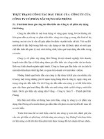
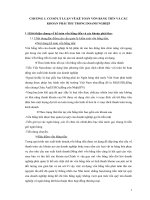
Figure 3.1 Electrophoresis products of PCR I (A) and PCR II on agarose gel
1,5%; M: scale of DNA 100 bp;
C: Negative control (H2O); S: DNA sample of Strongyloides spp
All 14 products of 2-step nested PCR included 4 samples with S. ratti
presence and 10 S. stercoralis random samples (obtained from real-time PCR),
were sequenced genome.
Table 3.16 Analyzing results of sequence of 14 larvae samples in the study
No
1
2
3
4
5
6
1
7
11
15
20
Highest
similarity (%)
99,5
98,6
99,4
99,7
95,6
25
98,5/98
Code
Gene code
Species
AB923888.1
AB923888.1
AB923888.1
AB923888.1
MK369923.1
AB923888.1/
AB453329.1
S. stercoralis
S. stercoralis
S. stercoralis
S. stercoralis
S. stercoralis
S. stercoralis/ S.
ratti
12
7
8
9
10
11
12
13
26
35
42
47
50
54
91,3
100,0
100,0
99,2
100,0
98,0
LL999104.1
S. stercoralis
LL999088.1
S. stercoralis
LL999110.1
S. stercoralis
AB923888.1
S. stercoralis
MK369923.1
S. stercoralis
AB923889.1
S. ratti
AB923888.1/
S. stercoralis/ S.
65
99,3/98,0
AB453329.1
ratti
14
66
98,0
LN609412.1
S. ratti
The species components were similarity very high to the isolates that
published in the gene bank.
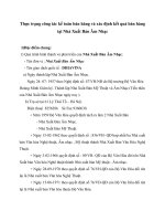
Figure 3.2 Phylogenetic tree was built on group 10 S. stercoralis larvaes
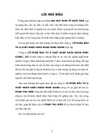
Figure 3.3 Phylogenetic tree was built on group 4 S. stercoralis larvaes
3.3 Describe the clinical symptoms, paraclinical and evaluate results of
treatment for strongyloidiasis by ivermectin single dose
The total number of patients tested positive with Strongyloides spp was 79
cases. Average age: 52.97 ± 27.64 (min - max = 22 - 84)
3.3.1 Clinical and paraclinical symptoms
13
The number of patients infected with Strongyloides spp completely without
clinical symptoms was 10.1%.
Table 3.17 Clinical symptoms in human strongyloidiasis (n = 79)
Symptoms
No. Percentage
Detail
No./Percentage
(%)
(%)
Abdominal
58
73.4
Epigastric
33/79 (41.7%)
pain
A round the 17/79 (21.5%)
navel
Hypogastrium
8/79 (10.1%)
Diarrhea,
33
41.8
Urticaria
45
57.0
The arms
36/79 (45.6%)
Body
9/79 (11.4%)
Headache
49
62.0
Lose-weight
9
11.4
CLM
3
3.8
Gastrointestinal symptoms had a high rate include: abdominal pain accounted
73.4% and diarrhea symptom was 41.8%
Table 3.18 Percentage of patients with hyper-eosinophilia (n = 79)
Value
Number
Percentage
(%)
Normal (< 500)
32
40.5
Eosinophil/µl Increase (≥ 500)
47
59.5
blood
Total
79
100
(E)
Mean = 694.56 ± 461.92. t test= 3.744; p value < 0.01;
Distance of mean = 194.5; CT 95% (91.1 –298.0)
Level
of Normal (<500)
32
40.5
hyperMild increase (500 - 1500)
41
51.9
eosinophilia
High increase (>1500)
6
7.6
Total
79
100
There was 59.5% of patients had hyper- eosinophil in their blood. The mean
of eosinophil was 694.56, significantly different from the normal threshold p
<0.01.
3.3.2 Effectiveness of treatment: Clinical – paraclinical
Table 3.19 ELISA test results in gastrointestinal strongyloidiasis (n = 79)
Value
Number
Percentage (%)
ELISA test
Positive
76
96.2
Negative
3
3.8
total
79
100
Mean of positive value = 32.37 ± 23.26 NTU.Test t = 15.2; p < 0.01;
Distance of mean: 22.4; CI 95% (18.6 – 24.2)
14
There was only 96.2% of strongyloidiasis patient had positive results that found
antibodies againts to Strongyloides spp.
Table 3.20 Responds of clinical symptoms in patients after 6 weeks of
treatment (n=57)
Sign/symptom
Before
Post – treatment
treat.
Cured Reductio No reduction
(%)
n (%)
(%)
Three
Abdomina
48
24 /48
12/48
12/48 (25)
classical
l pain
(50)
(25)
symptoms of Diarrhea
26
10/26
8/26
8/26 (30.8)
strongyloidias
(38.4)
(30.8)
is
Urticaria
39
4 /39
20/39
15/39 (38.5)
(10.3)
(51.2)
Headache
42
10/42
2/42
30/42 (71.4)
(23.8)
(4.8)
Lose-weight
8
2/8
0/8 (0)
6/8 (75.0)
(25.0)
Cutaneous larva migrans
2
2/2
0 (0)
0 (0)
(100)
The symptoms improved with more cured level than reduction level,
conversely, urticaria was reducced more than cured level.
Table 3.21 Ratio of larvae clearance post-treatment (n = 79)
Progress of test results post- treatment
Before treat.
2 weeks
4 weeks
6 weeks
Number of sample
79
75
61
57
Number of infected
79
2
3
3
cases (%)
(2.7%)
(4.9%)
(5.3%)
Number of larvae
73/75
58/61
54/57
cleared cases (%)
(97.3%) (95.1%)
(94.7%)
The prevalence of larvae clearance in faeces was 94.7% at 6 weeks post-treatment.
Table 3.22 Ivermectine effectiveness in the treatment (n = 57)
Detail
Number Percentage (%)
Cured
Stool test (-) and clinical
18
31.6
symptoms (cured)
Reduction
No reduction
Stool test (-) and clinical
symptoms (Reduction)
Stool test (+)
Stool test (-) and clinical
symptoms (No reduction)
Total
32
56.1
3
4
5.3
7.0
57
100
15
Effectiveness of Ivermectine reached 87.7% at level from reduced to cured,
while the efficacy of larvae cleaning reached 94.7%.
3.3.3 Adverse effects of ivermectin
Table 3.23 Ratio of side effects of ivermectin (n = 79)
Side effects
Number
Percentage (%)
Dizziness, increased headache
1
1.3
Nausea
1
1.3
Diarrhea, loose stools (increased)
4
5.1
Erythema rash on the skin
1
1.3
Increased itching
2
2.5
The adverse symptoms: diarrhea, loose stools (increased) accounted for
5.1%, nausea, dizziness accounted for a lower percentage (1.3%) and also
recovered.
CHAPTER 4
DICUSSIONS
4.1 Determine the actual situation and factors related to human Strongyloides
spp infection in Duc Hoa district, Long An province in 2017-2018.
4.1.1 Actual situation of Strongyloides spp in Duc Hoa district
4.1.1.1 The prevalence of Strongyloides spp infection
Summing up the data at 5 study sites, determine the general prevalence of
Strongyloides spp infection in Duc Hoa district is 6.64%, which was classify as
an endemic of the disease (table 3.1).
In 2 study at Phu My Hung and Phu Hoa Dong communes of Cu Chi district,
Ho Chi Minh city, located adjacent to the East with Duc Hoa district, by the same
technique with this study, the authors determined the prevalence of infection turn
were 12.6% (n = 294) and 9.2% (n = 766), higher than our study.
Myo Pa Pa (2018) studied in Myanmar to determine the overall infection rate
of 5.7%, nearly equal to prevalence of this study. This similarity was explained
by the same culture techniques to applicable in diagnosis, although the author
Myo Pa Pa applied the technique of agar culture while we used the technique of
culture with filter paper.
The infection rate found in Duc Hoa district in this study was still lower than
P. Laoraksawong (2017) in Thailand, Virak Khieu (2014) in Cambodia,
Senephansiri P. (2017) in Laos with the infection rate were 23%, 21% and 17.1%
respectively. The high prevalence of this infection may lead to the conclusion that
those in Southeast Asia were the endemic areas of Strongyloidiasis.
Table 3.1 shows that Duc Lap Thuong commune has the highest prevalence of
Strongyloides spp infection at 12.4%, the lowest is in Duc Hoa town (2.1%). An
Ninh Tay and Hiep Hoa communes have approximately equal rate: 4.4% and
4.5%. This result shows that, even within a district, each other study site had
16
different results, possibly because the relevant factors have an impact and needed
to analyze clearly in the subsequent results in the study.
4.1.1.2 Related factors with Strongyloides spp infection
The study data was collected according to the design at each site, aggregated
for the main target of Duc Hoa district. Therefore, to eliminate the general bias
factors, after univariate analysis of each factor, the multivariate analysis model
was included in the analysis of the relation between Strongyloides spp infection
and related factors in Duc Hoa district.
There was relation between Strongyloides spp infection and male (p <0.001)
in table 3.2. Table 3.9 of additional multivariate analysis showed that sex was
associated with Strongyloides spp infection (p <0.01) and had adjusted OR index.
Thus, in the community of Duc Hoa district, sex was a related factor and male
was 3.26 times at risk to be infected than women. This result was similar to study
result of Laoraksawong P. et al (2018) in Thailand (n= 526 ) with 4 times higher
risk in male.
Compared to the study in Cu Chi in 2004, men were 2.96 times at risk than
women. In Cambodia, in two studies at different districts, Virak Khieu et al (2014)
also identified that men was 1.7 times at risk more than women. Thus, from the
data of this study, in collaboration with many of other studies found an association
between male sex and the prevalence of Strongyloides spp infection, may lead to
sex is a related factor with Strongyloides spp infection in community.
This study surveyed the relation between Strongyloides spp infection and
groups of age (below 15, 15 - 60 and > 60). The results of multivariate analysis
also noted a significant relation (p <0.01), the risk in people over 60 years was
2.89 times higher than others. This result was different from the three studies in
Cu Chi in 2001, 2004 and 2017. In that studies, the authors did not identify the
related to age although the studies only interested in two age groups in and out of
labor.
There was relation between Strongyloides spp infection and p <0.05 in table
3.4, those with education level below high school was 1.98 times at risk compared
to the group had higher educational level. However, when included in the
multivariate analysis model, Table 3.9 showed that education levels above and
below high school are not related to the Strongyloides spp infection situation in
Duc Hoa district, the OR index was adjusted equal to 1.03. Thus, the relation
found in univariate analysis is not strong enough, or due to other factors affecting
and causing interference. In 2018, Myo Pa Pa studied in Myanmar, Suntaviritun
P. et al studied in Thailand, determined that there was no relation in education
level and Strongyloides spp infection. This study gave similar results, although
the mentioned authors used secondary school level to divide group in that studies.
In tables 3.5 and 3.9, there was relation between economic status and
Strongyloides spp infection (p <0.01). Infection was more present in the poor and
nearby-povety group with 2.08 times at risk than other. Although Duc Hoa district
17
has been developing economic strongly in recent years, but the index identifying
poor, neaby-povety and average households applied in the study, was generally
prescribed for the rural level nationwide. This index may not really suitable for
fast changing of economic conditions. But the study results found consistent with
the result from many studies in the world showed that the Strongyloides spp
infection was associated with poverty.
There was a relation between Strongyloides spp infection and farmer. The
results in tables 3.6 and 3.9 showed that farmer was really related to this infection
(p <0.05) and the adjusted OR index was 2.08 times. Thus, when working in farm,
the probability of larvae from contaminated soil to invasive the body and causing
disease would be higher. This result was similar to the study of Senephansiri P. in
Laos (2017), two studies of Virak Khieu et al (2014) at 2 different locations in
Cambodia, identified that farmer was at higher risk for Strongyloides spp infection
than other jobs.
Table 3.7 and Table 3.9 showed that there was a significant different between
the using unhygienic toilet with other group in Strongyloides spp infection (p
<0.01). People who used unhygienic toilets was 3.3 times at risk higher than using
hygienic toilets. So, in Duc Hoa district, using toilets was the related factor.
Investigation of the relation between the direct contact with soil in daily
activities and the status of Strongyloides infection, identified the relevance and
OR index as 2.69. This result is similar to the authors V.T.L Binh studied in 2
communes of Cao Dien, Phu Tho and Duong Thanh in Thai Nguyen province in
2014, Senephansiri P in Laos and Myo Pa Pa in Myanmar. Therefore, the direct
contact with soil in daily activities is an important risk factor for the infection of
Strongyloides spp in Duc Hoa district.
4.2 Determine component species of Strongyloides in human strongyloidiasis
disease
4.2.1 Survey pathogens by morphology
Table 3.10 shows that in 79 patients with a gastrointestinal strongyloidiasis,
the first times direct smear test is only able to detect 58.2%. This prove that the
detection ability of direct smear technique in strongyloidiasis diagnosis is quite
low. Therefore, this is not recommended as the main technique to be used to study
to screen Strongyloides spp infection for community.
Modified culture technique (Sasa 1986) in the study detected 93.7% cases at
the first test. This result was higher than 78.4% in a study at Cu Chi district (2004),
47.8% of Rayzan H. Z et al (2012) in Egypt. There were still 5 cases (6.3%) in the
first culture had negative result test while the direct smear technique had positive
result. For that reason, the combination of the two techniques has resulted in better
detection and proves that no technique was absolute perfect.
4.2.1.1 1st stage larvae (rhabditiform)
Table 3.11 shows that the larvae has average length of 279.9 µm, an average
width of 18.47 µm. Thus, compared to Grove DI (1989), Prayong R. et al. (2013),
18
the body length of larvae in this study tend to be longer because that authors
recorded that the length is from 200 - 250 µm, while the horizontal size of larvae
is similar with this study. The reason of difference is explained by the larvae that
cause disease in the community are often chronic, the density of larvae is low, the
symptoms cause not massively equivalent to the longer time of larvae in human
colon, will grow longer leading to body length is longer.
The average length of the esophagus is 75.7 µm, with the bulge forms, and the
average ratio compare to the body length is 27.1%, completely consistent with the
structure of stage 1 larvae. 100% larvae have pointed tail, indicating that all
measured larvae ear stage 1 larvae.
The average length of 1st stage larvae bucal cavity is 4.4 µm, the min - max is
3.9 µm - 5.3 µm. This is an important structure to distinguish with 1st stage
hookworm larvae that have long length bucal cavity, suitable for the authors
Grove D. I (1989), T. T. Hong (2017) and Prayong R. (2013). From the above
results, they are confirmed that all surveyed larvae were 1st stage larvae of the
Strongyloides spp are confirmed that all surveyed larvae are 1st larvae of the
Strongyloides spp.
4.2.1.2 2nd stage larvae
2nd stage larve of Strongyloides spp has 576.4 µm body length in average, the
average horizontal size is 16.9 µm. The results is consistent with Grove D.I
(1989), Prayong R. (2013), which reported from 450 - 600 µm, and horizontally
slender than 1st stage larvae. Average length of esophagus is 244.7 µm, also
tubular, and has an average ratio with body length as 42.5%. This is entirely
consistent with the structure of 2nd stage larvae with tubular esophagus and over
one-third of the body length from literature. 100% of the larvae has not pointed
tail, of which 86.1% has split tail in 2, indicating that all larvae molted in past.
Thus, these 2nd stage larvae had blunt or split 2 in shape(100%) , an average
endpoint width is 2.6 µm, there is a necessary indicator to show that their tail was
not as sharp as the tail of hookworm larvae. This indicator accurately identify all
surveyed larvae collected from the culture sample are belonged to Strongyloides
spieces.
4.2.1.3 Free-living adult worms males and females
Table 3.13 shows that the average length of the male is 778.8 µm, the average
horizontal size is 45.1 µm. Although this result was higher than reporting of
Prayong R. (about 0.7mm), completely consistent with Grove D.I. (1989) is from
700 to 900 µm. Different from the 2nd stage larvae, the esophagus of adult worms
grow to be horizontal and shorter in length. The average length of the esophagus
is 131.3 µm, accounted for the average ratio 17% compared with the body length,
the intercourse spines are 33.4 µm average of length, determining the sex of the
worm as an adult male.
Table 3.14 shows that the average length of female worms is 916.7 µm, the
average horizontal size is 46.2 µm. This result is within the threshold but at a low
19
level compared to the reported of Grove D.I. (1989). The esophagus of female
worms has 130.6 µm of an average length, similar to those of male worm, but the
average ratio compared to the average body length is only as 14.3% because the
female's body length is longer. Two uterine branches contain eggs lying
symmetrically through the vulva.
According to Grove D.I. (1989), the distinction of Strongyloides spieces
including S. stercoralis, S. ratti, S. fuellebornii, etc. and others, can only be based
on the shape of the oral structure, which is difficult to observe. For the above
reasons, in terms of morphology corresponding to the design in this study,
accurate samples were identified as 1st, 2nd stage larvae, male and female adult
worm of Strongyloides spp in limited.
4.2.2 Results of real-time PCR in identification of Strongyloides spp
Table 3.15 statics 70 samples, records that S. stercoralis account for 97.1%,
of which 2.9% is co-infected with S. ratti. Results were also found in components
species with 2.9% of S. ratti infection alone.
The dominant S. stercoralis (97.1%) were consistent with the N.V.DE (2017)
and D.T. Hong (2018) identified 100% as S. stercoralis, although the authors did
not use the same real-time PCR technique. Domestic and world literature reported
as a majority of S. stercoralis, this study found result in Duc Hoa district, the
majority of Strongyloides spp component is S. stercoralis.
In addition to traditional species, in the first times, the study has found the
traces of the S. ratti that cause disease in humans by molecular biology.
Morphology diagnose of this species is extremely difficult because the
morphology structure of Strongloides spieces larvae others are almost similar.
4.2.3 Results of Nested - PCR and genetic sequencing
For the aims of reasserting the species, we divided the sequencing samples into
2 groups based on the species identified by real-time PCR: group 1 include 10
random samples with S. stercoralis results and group 2 include 4 samples had S.
ratti positive result by real-time PCR technical with the specific gene segment
28S.
Figure 3.1 shows the electrophoresis product image of nested PCR technique.
In both step 1 (Fig. A) and step 2 (Fig. B), specific target gene segments for
Strongyloides spp are appeared clearly. The result shows that the target gene
segments have been successfully multiplied.
Table 3.16 shows the sequencing results about identification of the species
component, have high similarity to the DNA gene codes for 18S rRNA of human
pathogen Strongyloides spp in gene bank. The sequence of DNA genes coding for
18S rRNA of the Strongyloides spp upon accession was registered on the world
gene bank AB453329.1, AB923889.1, LN609412.1, AB923888.1, LL999065.1,
LM528082.1, LL999063.1, MK369923.1 and LL999126.1 were used for
comparison in this study. The high level of similarity between the gene sequence
20
of samples in the study and the sequence in the gene bank is high (91.3% - 100%)
for S. stercoralis and over 98% for S. ratti.
The similarity about S. stercoralis result with gene bank in this study is similar
to the rate of 100% by N.V. De (2017) and D.T. Hong (2018). However, those
authors are experimented in groups 2 and 7 samples, so the rate of variation will
be lower than this study is inevitable.
The phylogenetic tree at figure 3.2 shows that group of S. stercoralis samples
has a high level of species similarity over 91% with the gene code registered on
genbank.
The phylogenetic tree at figure 3.3 shows the S. ratti species in the study
completely close to the species originating from the rat Rattus novegicus. The
study results are consistent with Polanco Campo L F. (2018) in Brazil, this author
also found the presence of S. ratti inherently from rats as hosts.
In two co-infection samples S. stercoralis and S. ratti (samples 25 and 65), the
results comparing the highest similarity belong to S. stercoralis species (table
3.16). Thus, the gene segment was replicated and sequenced in the result of S.
stercoralis. This result could be explained by the highest number of S. stercoralis
pathogens in the sample, or the S. ratti gene segment not being replicated through
reaction. In this situation, although the sequencing results for these two coinfection samples did not confirm the species with certainty, although contributed
to the success of the multiplex real-time PCR technique that was experimentally
applied to the co-infection samples in the study, especially with the traditional S.
stercoralis species.
Thus, with the result of identifying a new species - S. ratti, was detected by
real-time PCR with a gene segment (28S), the genetic sequence determined the
98% similarity of S. ratti at another gene segment (18S) in the gene bank. This
study provided solid evidence to confirm the presence of S. ratti causing
infectious disease on the molecular level. In the future, there are necessary to have
newer studies that will be conducted in different local areas in Vietnam, in order
to compare this study results. At higher level, it is necessary to use other specific
genetic markers apply in new studies to describe the phylogenetic tree of
Strongyloides spp more specifically.
4.3. Describe the clinical symptoms, paraclinical and evaluate results of
treatment for strongyloideiasis by ivermectin single dose
4.3.1 Clinical and paraclinical symptoms
There were 8 (10.1%) cases without any symptoms. Usually, patients will go
to hospital if they have significant symptoms, 89.9% of patients appear symptoms
in this study also noted that the symptoms were mild. People still live and work
normally. This is the characteristic of the population of infected Strongyloidiasis
in the community.
Table 3.17 reports that the classic symptoms of triad infection are abdominal
pain (73.4%), urticaria (57%) and intermittent episodes diarrhea (41.8%). This
21
result shows that gastrointestinal signs are the main manifestation of
Strongyloidiasis in the community, in which abdominal pain accounts for the
highest proportion and is higher than rerults of H.H. Quang et al. (71.4%) but
lower than 81.8% of T. T. K. Dung (2009).
Besides, the symptoms of urticaria and intermittent episodes diarrhea were
57% and 41.8% respectively, indicating that the pathogen existing in the
gastrointestinal tract of the patient group has resulted high symptom rate.
In addition, when compared to the pathogenic development cycle of
Strongyloides spp, there are periods when larvae move through the tissue, they
are not present in the gastrointestinal tract continuously, which can affect the
presence rate of these classic symptoms.
In this study, the location of abdominal pain were divided into 3 areas,
epigastric pain accounted for the highest rate of 41.7%, around the navel 21.5%
and the hypogastric 10.1%. Thus, epigastric is the main position in the symptoms
of abdominal pain, similar to Forrer A. et al (2017) recorded epigastric pain as
51.7%.
Weight loss symptoms accounted for 11.4%, showing the systemic effects of
infection on patients. Headache symptoms accounted for 62%. The data of weight
loss and headache in the study has partly shown the impact of the disease not only
localized in the gastrointestinal tract but also other systemic harm.
The cutaneous larva migrans accounted 3 cases (3.8%) at the lower limb area,
suitable for Strongyloides spp larvae which have the ability to penetrate through
the skin similar to hookworm.
Table 3.18 shows a significant increase in the number of eosophil (average of
694.56 /mm3) compared to the average of less than 500. The table shows that
59.5% of people infected with strongyloidiasis have hyper-eosophil. The
different has significant p<0.01. Thus, among infected patients with
gastrointestinal strongyloidiasis, there were phenomenon eosophilia, increasing
the absolute number in the blood, similar to T.T.K. Dung (2009) recorded the
average value of BCAT as 640.27/mm3.
When dividing the level of BCAT increase into 3 levels: normal, mild and high
increasing, the results showed that the mild increase level from 500 to 1500
accounted for the highest proportion (51.9%), the high level of increasing
accounted for the lower rate (7.6%).
Table 3.19 shows the percentage of positive ELISA test results in patients with
gastrointestinal strongyloidiasis reach 96.2%. This is higher than the sensitivity
of the test kit is 89.47% by manufacturer's report. Compared to the normal
threshold value <10 NTU, the difference had strong significant (p <0.01), shows
that the amount of antibodies in strongyloidiasis patient is pretty high.
4.3.3 Effectiveness of ivermectin single dose
4.3.3.1 Clinical response
22
During the follow-up of treatment, only 57 cases participated in the evaluation
stool test. Therefore, Table 3.20 is built on the basis of 57 cases and results in
abdominal pain, diarrhea, and cutaneous larva migrans respond at higher level of
cured than the reduction. Whereas the symptoms of urticaria was a higher
reduction rate than cured (51.2% compared to 10.3%). This reason can be
explained by the symptoms of urticaria in the classical symptoms of
Stronglyloidiasis but the nature is a systemic reaction, involving many different
organ systems of the body and the recovery of symptoms. need more time after
treatment. Table 3.20 also shows a high proportion of headaches and weight loss
(71.4% and 75%, in contrast to the remaining symptom groups.
4.3.3.2 Paraclinical response
Table 3.21 shows that the percentage of patients who follow the test after
treatment decreases gradually over time. At 6 weeks, the proportion of patients
still participating in the study is 57 cases. At 2 weeks after treatment by a single
dose of ivermectin, the larval clearance rate was 97.3%, equivalent to the number
of infected is 2. However, at 4 weeks after treatment, the one more test case has
positive. The number of positive cases remains the same until 6 weeks. Thus, at
the time of 6 weeks after treatment, although no more cases appeared, but on the
total number of samples tested, Ivermectin's larval clean rate was 94.7%.
The results of larval clearance of 94.7% with the single dose of ivermectin in
this study were similar to the results of 95.2% in the research of Barda B. et al
(2017) and higher than Adenusi (84.1%), shows that the drug has a good effect.
4.3.3.3 Effectiveness of treatment: Clinical – paraclinical
The effectiveness of the drug also needs to be considered from the perspective
of coordination between clinical and paraclinical improvement. Table 3.21 was
built from combination of clinical and paraclinical responde include cure,
reduction and non- reduction shows: there are 18 cases meeting the standard of
cured accounting for 31.6%, lower than the reduction rate of 56.1 %. 12.3% did
not responde, of which laboratory failure accounted for 5.3% and clinical failure
of 7%. However, if calculated based on the combined effect of treatment from a
complete reduction to a complete cure, Table 3.21 shows effectiveness of
ivermectin reaching 87.7%. This result is not much higher than H.H. Quang et al
(84.6%) but also showed that ivermectin has a good effect in treating
Strongyloidiasis.
4.3.4 Adverse effects of ivermectin
Side effects of the single dose ivermectin were recorded with 4/79 (5.1%)
diarrhea, an increased itching 2/79 (2.5%); symptoms of dizziness, nausea,
erythema rash on the skin have the same proportion of 1.3% (1/79). The study did
not report any other severe effects. Thus, these adverse effects are mild and selfrelieving are not as significant, similar to Barda B. noted in a comparative study
between ivermectin and moxidectin in 2017.
23
CONCLUSIONS
Studying the actual situation of Strongyloides spp infection on 1,190 people
in Duc Hoa District, Long An Province in 2017-2018, we obtained the following
results:
1. Actual situations and factors related to human Strongyloides spp infection
1.1 The prevalence of Strongyloides spp infection
The prevalence of Strongyloides spp infection in Duc Hoa district was 6.64%,
classified as endemic area of the disease
The highest prevalence in Duc Lap Thuong commune was 12.4% and the
lowest in Duc Hoa town was 2.1%. At the My Hanh Nam, Hiep Hoa and An Ninh
Tay commune, the prevalence were 7.4%, 4.5% and 4.4%, respectively.
The prevalence of Strongyloides spp infection in male was 11.0%, higher than
female. The farmer was infected 15.3%, higher than other jobs. The prevalence of
Strongyloides spp infection in group over 60 of age was 15.5%, higher than group
from 15 to 60 (6.9%).
1.2 Related factors
There was a relation between the Strongyloides spp infection and the economic
status, farmer, situation of using toilets and the habit of soil contact in daily life,
in which
Poor – nearby poverty status was at risk 2.08 times higher than the others
groups.
Farmer was 2.07 times at risk comparing with other jobs.
People who had using unhygienic toilets was at risk 3.3 times higher than
using hygienic toilets.
The habit contact with the soil directly was at risk higher than remaining
groups 2.69 times.
2. Determine component of Strongyloides species
Detected and determined two species of Strongyloides by using molecular
biology techniques: S. stercoralis and S. ratti. The main agent was S. stercoralis
accounted for 97.1%, co-infection was 2.9% and single infection of S. ratti was
2.9%.
The morphological diagnosis was limited to identify Strongyloides spp. In
morphology characterization of Strongyloides spp: the length of rhabditiform was
279. 9 ± 17.5 µm, filariform 576.4 ± 24.9 µm, free - living adult worm male and
female were 778. 8 ± 27.7 µm and 916.7 ± 216 µm.
3. Clinical manifestations and effectiveness of treatment by ivermectin single
dose 0.2mg/kg.
3.1 Clinical and paraclinical symptoms
The classical symptoms of strongyloidiasis: abdominal pain, urticaria and
diarrhea accounted for 73.4%, 57% and 41.8%. Other symptoms such as
headache, weight loss, larva migrans cutaneous accounted for a lower proportion.
24
10.1% of strongyloidiasis patients had none clinical symptoms.
In strongyloidiasis patients, the proportion of positive ELISA test was
96.2%.
Eosinophil value did not increase in 40.5% of strongyloidiasis cases. Hyper
- eosinophil was 59.5% with mild increase counted 51.9%.
3.2 Effectiveness of treatment by Ivermectin with single dose 0.2 mg/kg body
weight
After 6 weeks, the effectiveness of treatment from reduced to completely cure
was 87.7%, of which:
- The proportion of eliminated larvae in stool test was 94.7%.
- Clinical signs had completely cured higher than reduce include as follows:
abdominal pain 50%, diarrhea 38.4% and larva migrans cutaneous 100%.
Headache and weight loss were completely cured 23.8% and 25%. Reducing of
urticaria accounted 51.2%, higher than completely cure.
3.3 Adverse effects of ivermectin single dose
Diarrhea was the highest rate 5.1%, pruritus was 2.5%. The symptoms of
dizziness, nausea and skin rash had the same proportion 1.3%.
The side effects were mild, disappears spontaneously and none necessary to
intervene.
RECOMMENDATIONS
From the study results obtained, we have the following recommendations
1. Increasing the prevention of Strongyloides spp in Duc Hoa district
community, Long An province. Based on the factors related to the disease such
as: economic status, agricultural occupation, toilet use status and living habits was
identified, as a basis data to apply to build an effective prevention program.
2. Application of the real-time PCR procedure that identified in the study to
diagnose strongyloidiasis. Continuing to develop research and application of realtime PCR technique with other types of specimens.
3. Application of good treatment effect of ivermectin to human
strongyloidiasis to case treatment in hospital and mass treatment for community.
4. Using the source of larvae, serum, and fecal samples of this study, as a basis
for developing new studies on Strongyloides spp pathogens in the Southern
provinces.