Tissues-based chemical profiling and semi-quantitative analysis of bioactive components in the root of Salvia miltiorrhiza Bunge by using laser microdissection system combined with UPLC-q-TO
Bạn đang xem bản rút gọn của tài liệu. Xem và tải ngay bản đầy đủ của tài liệu tại đây (2.4 MB, 13 trang )
Xie et al. Chemistry Central Journal (2016) 10:42
DOI 10.1186/s13065-016-0187-7
RESEARCH ARTICLE
Open Access
Tissues‑based chemical profiling
and semi‑quantitative analysis of bioactive
components in the root of Salvia miltiorrhiza
Bunge by using laser microdissection system
combined with UPLC‑q‑TOF‑MS
Wenjian Xie1, Hongjie Zhang1, Jianguo Zeng2, Hubiao Chen1, Zhongzhen Zhao1* and Zhitao Liang1*
Abstract
Background: The dry root of Salvia miltiorrhiza Bunge (Danshen in Chinese) is an used-widely traditional Chinese
herbal medicine with and promising efficacy. This herbal plant has been extensively cultivated in China. Currently,
people usually rely on its morphological features to evalaute its pharmaceutical quality. In this study, laser micro-dissection system (LMD) was applied to isolate single fresh tissue of root of S. miltiorrhiza. Under fluorescent microscopic
model, five tissues namely cork, cortex, phloem, xylem ray and vessel were well recognized and isolated accurately by
LMD, respectively and then the distribution pattern of the major bioactive compounds in various tissues was investigated by ultra-performance liquid chromatography-quadrupole/time of flight-mass spectrometry, which aims to
validate the traditional experience on evaluating pharmaceutical quality of Danshen by morphological features.
Results: Total 62 chemical peak signals were captured and 58 compounds including 33 tanshinones, 23 salvianolic acids
and 2 others were identified or tentatively characterized in micro-dissection tissues. Further semi-quantitative analysis
indicated that the bioactive components such as tanshinones and salvianolic acids were mainly enriched in cork tissue.
Conclusion: In the present study, analysis of metabolic profile in different tissues of roots of S. miltiorrhiza is reported
for the first time. The distribution pattern of major bioactive components could enable medicinal vendors and consumers to relatively determine the pharmaceutical quality of Danshen by morphological features.
Keywords: Tanshinones, Salvianolic acids, Salvia miltiorrhiza Bunge, Tissues-based analysis, Pharmaceutical quality
evaluation
*Correspondence: ;
1
School of Chinese Medicine, Hong Kong Baptist University, Kowloon,
Hong Kong, Special Administrative Region, People’s Republic of China
Full list of author information is available at the end of the article
© 2016 The Author(s). This article is distributed under the terms of the Creative Commons Attribution 4.0 International License
( which permits unrestricted use, distribution, and reproduction in any medium,
provided you give appropriate credit to the original author(s) and the source, provide a link to the Creative Commons license,
and indicate if changes were made. The Creative Commons Public Domain Dedication waiver ( />publicdomain/zero/1.0/) applies to the data made available in this article, unless otherwise stated.
Xie et al. Chemistry Central Journal (2016) 10:42
Background
The dry root of Salvia miltiorrhiza Bunge, namely Danshen in Chinese, which is an important traditional Chinese herbal medicine. Over two thousand years ago,
Danshen has been categorized as a superior grade herbal
medicine by The Divine Husbandman’s Classic of Materia Medica (Shen Nong Ben Cao Jing), which means that
it can be beneficial to human’s health and it is safe, even
it is taken for a long time [1]. Today, it has been used as
a principal drug in many proprietary Chinese medicines
for treating coronary heart disease, cerebrovascular disease, irregular menstruation and hepatosplenomegaly [2].
Around 20 kinds of proprietary Chinese medicines such
as compound Danshen capsules, compound Danshen
tablets, Danshen injection and compound Danshen dripping pills (CDDP) have been developed and some of its
relative products have also been used as over the counter medicine (OTC) in Japan [3, 4]. Moreover, CDDP has
been approved to carry out phase III clinical trial for preventing and treating stable angina and diabetic retinopathy by U.S. FDA [5].
Due to the increasing demands of this plant resources
and extensive application in clinic, S. miltiorrhiza has
been widely cultivated in Sichuan, Shangxi, Shanxi,
Henan, Hebei, Shandong, Anhui, Hubei, Jiangsu and
Zhejiang provinces of China and the supply of Danshen
has been dominated by cultivated resource. According to
the traditional experiences on morphological evaluation
and classification of Danshen, it is divided into different
grades by their size of main root and the color of outer
bark for better transaction in the commercial markets
[3]. As we know, however, the pharmaceutical quality of
herbal medicines may be easily affected by some factors
such as producing areas, harvest season and even cultivation technologies. Up to now, no objective evidences have
been found to prove that the bigger size of main root and
deeper brown–red of appearance of this medicinal plant
could indicate the better pharmaceutical quality. It is no
doubt that it is still unclear whether such simple quality
classification criteria can really reflect its pharmaceutical quality or not. In addition, for quality evaluation of
Danshen, although modern chromatographic methods
involving HPLC fingerprint and determination of main
components by HPLC have been established [4, 6], it is
hard for medicinal vendors and consumers to equip with
modern instruments to evaluate the quality of Danshen.
On the other hand, it is well known that evaluating the
quality of various grades of Chinese herbal medicines by
morphological features is a convenient, quick and practical method compared with other methods that mostly
rely on modern instruments.
Several pharmacological studies have demonstrated
that bioactive effects of Danshen are mainly attributed
Page 2 of 13
to its secondary metabolites including diterpene quinones and salvianolic acids such as tanshinone I (Tan I),
dihydrotanshinone I (DHTan I), tanshinone IIA (Tan IIA),
cryphtotanshinone (CTan) and salvianolic acid B (SaB)
[7–9]. Mapping the distribution of these bioactive components and carrying out semi-quantitative analysis in
various herbal tissues can help to evaluate pharmaceutical quality of herbal medicine. Laser micro-dissected
system (LMD) plus with ultra-performance liquid
chromatography-quadrupole-time of flight-mass spectrometry (UPLC-Q-TOF-MS) has been demonstrated
as a powerful tool to establish an objective relationship
between major bioactive second metabolites and morphological features of herbal medicine [10–13]. Here,
this strategy was firstly applied to validate the traditional
experience and judge them as true or false views, with
regard to pharmaceutical quality, which is important
for the quality evaluation and classification of different
grades of Danshen.
Experiment section
Plant materials
The plant materials (Table 1) were collected from eight
cultivation bases and one natural habitat in China. All
of them were authenticated as S. miltiorrhiza Bunge by
Dr. Zhitao Liang from school of Chinese Medicine, Hong
Kong Baptist University and the specimens were deposited in the Bank of China (Hong Kong) Chinese Medicines Centre of Hong Kong Baptist University.
Chemicals and reagents
Chemical markers including Tan I, DHTan I, Tan II, CTan
and Sa B were purchased from Chengdu Must Bio-Technology Co., Ltd. (Chengdu, People’s Republic of China)
(Fig. 1). The purity of each standard was over 98 %. Both
acetonitrile and methanol (HPLC grade) were purchased
from E. Merck (Darmstadt, Germany) and formic acid
(HPLC grade) was ordered from Tedia, USA. Water for
analyzing was prepared by a Mili-Q water purification
system (Millipore, Bedford, MA, USA).
Materials and instruments
Cryotome (Thermo Shandon As620 Cryotome, Cheshire,
UK), Cryogen (Thermo Shandon, Cheshire, UK), Nonfluorescent polyethylene terephthalate (PET) microscope
steel frame slide (76 × 26 mm, 1.4 μm, Leica Microsystems, Bensheim, Germany), Leica Laser microdissection 7000 system, 500 μL micro-centrifuge tube (Leica),
Centrifuge (Centrifuge 5417R, Eppendorf, Hamburg,
Germany), Ultrasonic instrument (CREST 1875HTAG
Ultrasonic Processor, CREST, Trenton, NJ), HPLC grade
vial (1.5 mL, Grace, Hong Kong), Glass-lined pipe with
plastic ring (400 μL, Grace, Hong Kong), Electronic
Xie et al. Chemistry Central Journal (2016) 10:42
Page 3 of 13
Table 1 Sample information of S. miltiorrhiza in the present study
Sample no.
Colour of outer barka
Sizeb (cm)
Sources
Collection date
S1
Brownish–red
0.8
Cultivation, Zhongjiang County, Sichuan province
2014.11.19
S2
Dark brownish–red
1.3
Cultivation, Shangluo City, Shanxi province
2014.11.19
S3
Dark brownish–red
0.75
Cultivation, Fangcheng County, Henan province
2014.11.19
S4
Brownish–red
1.4
Wild, Henan province
2014.11.19
S5
Brownish–red
0.7
Cultivation, Linqu County, Shandong province
2014.11.19
S6
Brownish–red
1.0
Cultivation, Beijing
2014.11.19
S7
Brownish–red
0.5
Cultivation, Beijing
2014.11.19
S8
Brownish–red
0.65
Cultivation, Beijing
2014.11.19
S9
Brownish–red
1.0
Cultivation, Nanjing, Jiangsu province
2015.05.31
a
Colour of outer bark refer to Fig. 6
b
Size calculated by diameter of main root of S. miltiorrhiza
O
O
O
O
O
O
R
O
O
Tanshinone I (Tan I)
O
Dihydrotanshinone I (DHTan I) Tanshinone II A (Tan II A )
C18H12O3
MW: 276.2861
C18H14O3
MW: 278.3020
C19H18O3
MW: 294.3444
HO
OH
OH
O
OH
O
O
S
S
R
OH
O
HO
O
OH
R
C19H20O3
MW: 296.3603
E
OH
O
OH
O
Salvianolic acid B (Sa B)
O
O
R
C36H30O16
MW: 718.1534
Fig. 1 Chemical structures of 5 chemical markers
balance (Mettler Toledo MT5 style), Agilent 6540 ultradefinition accurate mass quadrupole time-of-flight
spectrometer equipped with a mass hunter workstation
software (Agilent version B.06.00 series, Agilent Technologies, USA), Acquity UPLC BEH C18 column (2.1
mm × 100 mm, 1.7 μm) coupled with a C18 pre-column
(2.1 mm × 5 mm, 1.7 μm, Waters, USA).
Samples preparation
The protocol of samples preparation for analysis was usually divided into three stages. Firstly, each prepared fresh
root was fixed by cryogen and frozen on a −35 °C cryo
bar, before being cut into 30 μm cross-section of tissue
and attached on a non-fluorescent polyethylene terephthalate. At the next stage, each prepared cross-section of
Xie et al. Chemistry Central Journal (2016) 10:42
Page 4 of 13
tissue was exposed to a Leica LMD-BGR fluorescence filter system at 6.3 magnification for microscopic authentication (field of 6, color saturation of 1.20, exposure time of
777 μs, gain of 2.5, and IFW1 light intensity of green and
diaphragm of 5), after then 5 different target tissue, around
1 × 106 μm2 per each (Table 2), were individually isolated
by Laser Micro-dissection system (7000 V 7.5.0.5112 edition) with an optimal parameters (DPSS laser bean wavelength of 349 nm, power of 53 μJ, aperture of 46, speed of
2, specimen balance of 41, head current of 100 %, plus frequency of 4046 Hz), before collecting it by a cap of 500 μL
micro-centrifuge tube. Finally, each prepared sample was
sent to centrifuge 5 min (12,000 rpm, 20 °C) in order to
ensure it fell into the bottom from the cap, and then added
100 μL methanol into each micro centrifuge tube for ultrasonic extraction 60 min and then centrifuged again 10 min
(12,000 rpm, 20 °C). 90 μL supernatant was transferred
into a glass-lined pipe with a plastic ring accommodated
by a HPLC grade vial and stored at a 4 °C refrigerator.
According to the results of preliminary experiment, the
optimal running parameters of UPLC were set as follows: the mobile phase consisted of water with 0.1 %
formic acid (A) and acetonitrile with 0.1 % formic
acid (B) with an procedure of linear gradient elution:
0-8 min (40 % B), 8–20 min (40–75 % B), 20–22 min
(75–100 % B), 23–25 min (100 % B), the injection volume was 3 μL and the flow rate was set at 0.35 mL/
min. Salvianolic acids were more sensitive in negative
ion scanning mode while tanshinones were more sensitive in positive ion scanning mode, so the mass spectra were acquired in both positive and negative modes
by scanning from 100 to 1700 in mass to charge ratio
(m/z), the scanning of MS was performed under the
following operation parameters: dry gas temperature
of 325 °C, dry gas (N2) flow rate of 8 L/min, nebulizer
pressure of 45 psi, V-cap of 4500, nozzle voltage 500 V,
and fragmentor 150 V.
Standard solution preparation
Results and discussion
Each standard including Tan I, DHTan I, Tan IIA, CTan
and Sa B was accurately weighed and dissolved individually in methanol to produce mixed stock solution with
concentrations at 0.96 mg/mL of Sa B, 0.992 mg/mL of
DHTan I, 0.954 mg/mL of Tan I, 0.991 mg/mL of CTan,
1.028 mg/mL of Tan IIA. The series concentrations of
mixed working solution were prepared by diluting the
mixed stock solution with methanol. In addition, due to
the high sensitive requirement in UPLC-QTOF-MS, here
a blank control containing solvent was set to exclude the
negative impact on analyzing process.
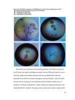
Microscopic characteristics and separation of tissues
Method of UPLC‑QTOF‑MS
Here, sample seven was used as a representative to present
the microscopic characteristics of whole cross-section of
root observed under bright filed and fluorescence mode
(Fig. 2). Under the bright filed, the anatomical features of
Table 2 Total micro-dissected area in different tissues
Sample no.
Special tissue/total micro-dissected area (μm2)
Cork
Cortex
Phloem
Xylem ray
Vessel
S1
1,006,611
1,003,330
1,063,204
1,022,559
1,020,931
S2
1,000,990
1,000,072
1,000,320
1,000,791
1,000,276
S3
1,000,160
1,003,816
1,000,051
1,000,830
1,000,686
S4
1,000,011
1,000,962
1,000,249
1,000,343
1,000,589
S5
1,000,583
1,000,699
1,000,983
1,000,300
1,000,349
S6
1,000,736
1,001,599
1,000,860
1,001,172
1,000,058
S7
1,003,180
1,000,194
1,000,901
1,000,148
1,001,122
S8
1,000,609
1,000,606
1,000,728
1,000,407
1,000,354
S9
1,000,402
1,000,365
1,000,629
1,000,310
1,000,291
Fig. 2 Cross-sections of the root of S. miltiorrhiza (S7) a observed
under the bright filed mode b observed under the fluorescent mode
Xie et al. Chemistry Central Journal (2016) 10:42
root were found to be mainly composed of cork, cortex,
phloem, cambium, xylem ray and vessel (from external to
internal part). Cork was brownish–red and consisted of
several layers of narrow cells and cortex showed brownish–yellow color and lied with several layers of fat cells.
The boundary between phloem and cambium was unclear.
Wide xylem ray were found at the middle between each
two grouped or single vessels. When observed by fluorescence mode, cork also showed similar color as observed in
bright filed. Cortex exhibited brownish–yellow. Phloem
and xylem showed the similar fluorescence while vessels
showed yellowish–white. According to structural characteristics of tissues under fluorescence mode, fives tissues
namely cork, cortex, phloem, xylem ray and vessels were
isolated for analyzing, respectively.
Page 5 of 13
in other tissues from all of samples. In the BPC chromatograms of various tissues, the chemical profiles of 9 samples were dissimilar (Table 4). Peaks 1 and 2, peaks 6 and
7, peak 8 only could be detected in cortex of S5, in cork
of S2, in cork of S1, respectively while peak 38 (DHTan
I) could be detected in all micro-dissected tissues except
for xylem ray of S5. SaB, DHTan I, Tan I, CTan and Tan
IIA were found as common peaks in cork from all of samples and some of them also could be detected in other tissues. The results demonstrated that SaB, DHTan I, Tan I,
CTan and Tan IIA were the main components in the tissues of root of S. miltiorrhiza. Thus, further quantitative
analysis of them in various tissues was also carried out by
UPLC-QTOF-MS.
Identification of chemicals in various tissues
Quantitative analysis of tanshinones and salvianolic acids
in various tissues
Mapping chemical profiles in micro-dissection tissues
was performed by UPLC-QTOF-MS and the representative base peak chromatograms (BPC) showing all the
detected peaks of cork and cortex tissues from S1, S2
and S5 were showed in Fig. 3. The BPC chromatograms
of others were showed in the Additional file 1. Total 62
chromatographic peaks were detected (Table 3). Peaks
of tanshinones could be recognized by their generated
molecular ions of [M+Na]+ and [M+H]+ while peaks
of salvianolic acids were easily generated their molecular
ions of [M−H]−. Peaks 4, 38, 48, 49 and 59 were identified as SaB, DHTan I, Tan I, CTan and Tan IIA by their
accurate mass and corresponding mass ions as well
as comparison of chemical markers, respectively. The
molecular ions of SaB (717.1406 [M−H]− m/z), DHTan
I (301.0834 [M+Na]+ and 279.1015 [M+H]+ m/z), Tan
I (299.0684 [M+Na]+ and 277.0867 [M+H]+ m/z),
CTan (319.1307 [M+Na]+ and 297.1488 [M+H]+ m/z)
and Tan IIA (317.1158 [M+Na]+ and 295.1333 [M+H]+
m/z) were detected in marker and sample solutions. The
molecular ions of others were identified or tentatively
characterized by their accurate mass data in comparison
with literature reports [14–22]. From the Fig. 4, the number of chemicals in cork was more abundant than those
Linear regression analysis in statistics including calibration curve and correlation coefficients of determination
(R2), limits of detection (LOD, S/N > 3) and limits of
quantification (LOQ, S/N > 10) were investigated under
the above conditions for the quantitative analysis. The
peak areas as the dependent variable (y axis) and the concentration as the independent variable (x axis, ng/mL)
was used to generate the calibration curves of each reference, All of the R2 value were over 0.9996 (n = 9). The
LOD is 44.31, 3.88, 7.73, 3.87 and 8.03 ng/mL to Sa B,
DHTan I, Tan I, CTan and Tan IIA and the LOQ is 75.00,
12.90, 12.42, 12.89 and 26.74 ng/mL to Sa B, DHTan I,
Tan I, CTan and Tan IIA, respectively (Table 5).
The results (Fig. 5) demonstrated that the amounts
of major tanshinones and Sa B in various tissues were
different and it could be seen that the contents of
Sa B (Fig. 5a) and major tanshinones (Fig. 5b, calculated by DHTan I, Tan I, CTan and Tan IIA) in cork
were much higher than those in other herbal tissues as
well. In addition, Sa B could also be quantified in cortex of samples 5 and 8. It suggested that the growing
area and/or harvest season could influence tissue-specific chemical profiles, especially affect the amounts
of major tanshinones. In detail, the total contents of
Xie et al. Chemistry Central Journal (2016) 10:42
Page 6 of 13
Fig. 3 The represent BPC chromatograms from cork tissue of S1 and S2 detected under positive mode (a), cork tissue of S1 and cortex tissue of S5
(b) as well as cork tissue of S2 and S5 (c) detected under negative mode.1SP solvent peak
Xie et al. Chemistry Central Journal (2016) 10:42
Page 7 of 13
Table 3 Characteristics of bioactive components in various tissues
Peak no.a Rt (min)
Polarity
1
2.63
313.0718 [M−H]−
3.59
−
−
−
−
2
3
4
5
6
7
8
9
10
11
12
13
14
3.96
4.43
4.56
4.98
6.04
7.18
7.40
7.63
7.77
535.1818 [M−H]
359.0732 [M−H]
717.1406 [M−H]
137.0242 [M−H]
−
193.0479 [M−H]
+
+
335.0894 [M+Na] , 313.1071 [M+H]
−
297.1118 [M−H]
+
+
319.0944 [M+Na] , 297.1124 [M+H]
−
117.0193 [M−H]
−
357.0588 [M−H]
Formula
Identification
C17H14O6
Salvianolic acid Fb
C26H32O12
(+)1-hydroxypinoresinol-1-O-β-D-glucosideb
C18H16O8
Rosmarinic acidb
C36H30O16
Salvianolic acid Bc
C7H6O3
Protocatechualdehydeb
C10H10O4
Ferulic acidb
C18H16O5
Tanshindiol Cb
C19H22O3
Arucadiolb
C18H16O4
Danshenxinkunb
C4H6O4
Succinic acidb
C18H14O8
Prolithospermic acidb
7.84
335.1252 [M + Na] , 313.1432 [M+H]
C19H20O4
Miltionone IIb
8.99
+
+
317.0786 [M+Na] , 295.0969 [M+H]
C18H14O4
Trijuganone Ab
9.12
+
+
C18H16O4
Tanshinone VIb
C19H18O4
Isotanshinoneb
+
+
319.0944 [M+Na] , 297.1124 [M+H]
−
15
9.27
383.9794 [M−H]
16
9.98
333.1097 [M+Na]+, 311.1279 [M+H]+
Unknown
10.07
+
+
303.0996 [M+Na] , 281.1162 [M+H]
C18H16O3
Methylene dihydrotanshinoneb
18
10.21
+
+
335.1252 [M+Na] , 313.1434 [M+H]
C19H20O4
Miltionone Ib
19
10.35
491.1039 [M−H]−
C26H20O10
Salvianolic acid Cb
20
10.41
333.1098 [M+Na]+, 311.1282 [M+H]+
C19H18O4
Tanshinone IIbB
21
10.64
333.1100 [M+Na]+, 311.1282 [M+H]+
C19H18O4
3α-hydroxytanshinone IIA/3β-hydroxytanshinone IIbA
22
10.87
333.1099 [M+Na]+, 311.1283 [M+H]+
C19H18O4
3α-hydroxytanshinone IIA/3β-hydroxytanshinone IIbA
23
10.98
327.0872 [M−H]−
C18H16O6
Methylsalvianolate Fb
24
11.38
363.1202 [M+Na]+, 341.1380 [M+H]+
C20H20O5
Cryptomethyltanshinoateb
25
11.57
295.0958 [M−H]−
C18H16O4
Tanshinol Bb
26
11.75
325.1079 [M−H]−
C14H14O9
Monocaffeoyltartaric acidb
12.01
−
17
27
28
12.02
C20H30O
Ferruginolb
+
C18H22O3
Epicryptoacetalide/Cryptoacetalideb
C18H22O3
Epicryptoacetalide/Cryptoacetalideb
C19H22O4
Tanshinone Vb
C18H14O3
Methylenetanshinquinoneb
285.1853 [M−H]
+
309.1125 [M+Na] , 287.1642 [M+H]
−
29
12.18
487.3401 [M−H]
30
12.24
309.1125 [M+Na]+, 287.2002 [M+H]+
31
32
12.35
12.38
−
313.1438 [M−H]
+
−
12.46
485.3274 [M−H]
34
12.57
537.1038 [M−H]−
12.82
−
36
12.88
+
301.0838 [M+Na] , 279.1016 [M+H]
33
35
Unknown
Unknown
C18H14O4
3-hydroxymethylenetanshinoneb
C19H22O3
Miltiodiolb
C18H14O3
Dihydrotanshinone Ic
C18H14O3
1,2-dihydrotanshinone Ib
321.1646 [M+Na] , 299.1642 [M+H]
−
37
12.96
555.3268 [M−H]
38
13.00
301.0834 [M+Na]+, 279.1015 [M+H]+
39
40
41
42
43
44
45
46
47
48
49
13.11
Lithospermic acidb
+
293.0819 [M−H]
+
C27H22O12
+
Unknown
+
301.0834 [M+Na] , 279.1015 [M+H]
C20H26O4
Salviolb
13.41
+
+
319.1306 [M+Na] , 297.1491 [M+H]
C19H20O3
Isocryptotanshinoneb
13.84
+
+
303.0998 [M+Na] , 281.1173 [M+H]
C18H16O3
Danshenxinkun Bb
14.25
+
+
C20H18O5
Methyl tanshinoateb
C18H14O8
Prolithospermic acidb
C19H24O3
Miltipoloneb
C18H18O2
Methylenemiltironeb
13.16
14.32
14.62
14.75
−
329.1750 [M−H]
361.1045 [M+Na] , 339.1230 [M+H]
−
357.0616 [M−H]
+
+
333.1089 [M+Na] , 301.1800 [M+H]
−
265.1470 [M−H]
C17H16O6
5,3′-dihydroxy-7,4′-dimethoxyflavanoneb
15.97
+
+
299.0684 [M+Na] , 277.0867 [M+H]
C18H12O3
Tanshinone Ic
16.00
+
+
C19H20O3
Cryptotanshinonec
15.45
−
315.0846 [M−H]
319.1307 [M+Na] , 297.1488 [M+H]
Xie et al. Chemistry Central Journal (2016) 10:42
Page 8 of 13
Table 3 continued
Peak no.a Rt (min)
50
51
52
53
54
55
56
57
58
59
60
61
62
16.15
16.35
16.64
Polarity
Formula
Identification
−
C20H28O3
1-phenanthrenecarboxylic acidb
−
C20H26O2
5-dehydrosugiolb
C20H28O2
Sugiolb
C18H12O3
Isotanshinone Ib
315.1949 [M−H]
297.1830 [M−H]
−
299.2018 [M−H]
+
+
16.79
299.0684 [M+Na] , 277.0867 [M+H]
17.25
+
301.0834 [M+Na] , 279.1015 [M + H]
C18H14O3
Dihydroisotanshinone Ib
17.75
+
+
315.1001 [M+Na] , 293.1179 [M+H]
C19H16O3
1,2 -didehydrotanshinone IIbA
18.18
+
+
289.1204 [M+Na] , 267.1386 [M+H]
C17H14O3
Dihydrotanshinlactoneb
18.87
+
+
C19H20O2
Δ1 -dehydromiltironeb
+
303.1306 [M+Na] , 281.1539 [M+H]
C21H26O3
2-(7-Dihydroxyl)-benzofuranyl-,ferulic acidb
19.30
+
+
317.1158 [M+Na] , 295.1333 [M+H]
C19H18O3
Tanshinone II cA
20.01
+
+
317.1151 [M+Na] , 295.1332 [M+H]
C19H18O3
Isotanshinone II bA
20.39
+
+
C19H22O2
Miltironeb
C44H60O6
3,4-Dihydroxy-(1α,3α,4α,5β)-1-carboxy-4-hydroxy1,3,5-cyclohexanetriyl ester-benzenepropanoicb
19.13
21.31
−
325.1824 [M−H]
305.1515 [M+Na] , 283.1700 [M+H]
−
683.4317 [M−H]
Rt retention time
a
The peak numbers referred to Fig. 3
b
Identified by previously reported from Salvia species
c
Identified by chemical markers
Fig. 4 The profile of chemicals in various tissues from S1 to S9
4, 14, 16, 20, 23, 30, 32, 36, 38, 39,
1–5, 9–11, 14, 15, 17, 22–24, 28, 32,
41–46, 48, 49, 52, 54, 55, 59, 61, 62
36, 38, 39, 41, 48, 49, 53, 58
4, 23, 30, 32, 38, 39, 41, 42, 46, 48,
49, 54, 59, 61
4, 12, 14, 16, 18, 20, 23, 24, 30, 32,
36, 38, 41–43, 45, 46, 48–50, 52,
54, 59, 61
4, 12, 14, 16, 18, 20, 23, 24, 30, 32,
36, 38, 41–43, 45, 48–50, 52, 54,
59, 61
4, 12, 17, 22–33, 35, 37–42, 48, 49,
59
S5
S6
S7
S8
S9
The peak numbers referred to Table 3 and Fig. 3
4, 5, 9, 14, 20, 30, 32, 36, 38, 39,
41–43, 45, 48, 49, 53–55, 59, 61
S4
a
4, 9, 12–14, 16–18, 20–22, 24, 28, 30, 9, 17, 22, 32, 38, 39, 41 48, 49, 53, 54 9, 32, 38, 39, 41, 48, 49, 53, 54
32, 36, 38, 39, 41–43, 45, 48, 49,
51–55, 59, 61, 62
S3
23, 30, 32, 36, 38, 39, 41
4, 23, 24, 30, 32, 38, 39, 41, 49
9, 16, 23, 24, 30, 32, 37–39, 41, 42,
45, 48, 49, 53
9, 23, 24, 30, 32, 36, 38, 39, 41, 49
23, 30, 32, 36, 38, 39, 41
23, 24, 30, 32, 36, 38, 39, 41, 46, 49
9, 23, 24, 30, 32, 38, 39, 41, 46, 49,
63
23, 24, 30, 32, 38, 39, 41, 46, 48
9, 17, 22, 24, 28, 32, 38, 39, 41, 46,
48, 49, 53
9, 14, 20, 32, 38, 39, 41 48, 49, 53, 54 9, 17, 32, 38, 39, 41, 48, 49, 53, 54
9, 32, 36, 38, 39, 41, 48, 49, 53, 54
4, 6, 7, 9, 17, 20, 24, 28, 31, 32, 36, 38, 9, 32, 36, 38, 39, 41, 48 49, 53, 54
39, 41–43, 45, 46, 48, 49, 52–56,
59, 61
9, 17, 24, 32, 36, 38, 39, 41, 42, 46,
48, 49, 53, 54, 59
S2
9, 17, 32, 36, 38, 39, 41, 46, 48, 49,
53
Phloem
3, 4, 8, 9, 12–14, 16–22, 24–39,
41–57, 59–61
Cortex
S1
Cork
Sample no. Herbal tissues/peak No.a
Table 4 The distribution of bioactive components in various tissues from different samples
23, 30, 32, 38, 39
23, 30, 32, 38, 39
9, 23, 24, 30, 32, 38, 39, 41, 49
9, 23, 24, 30, 32, 38, 39, 41, 46
16, 24, 39
9, 32, 38, 39, 41, 48, 49, 53, 54
9, 32, 38, 39, 41, 48, 49, 53, 54
9, 17, 23, 28, 32, 36, 38, 39, 41, 48,
49, 53, 54
9, 17, 20, 32, 36, 38, 39, 41–43, 48,
49, 53, 54, 59, 61
Xylem ray
23, 30, 32, 36, 38, 39, 41
4, 23, 30, 32, 38, 39, 49
23, 30, 32, 38, 39, 41, 49
23, 30, 32, 36, 38, 39, 41, 46
9, 10, 32, 38, 39, 41, 48, 49, 53, 54, 59
9, 32, 38, 39, 41, 48, 49, 53,
9, 17, 24, 28, 32, 38, 39, 41, 48, 49,
53, 54
9, 17, 24, 28, 32, 36, 38, 39, 41, 48,
49, 53, 54
10, 14, 32, 36, 38, 39, 41, 46, 48, 49,
53, 54, 59
Vessel
Xie et al. Chemistry Central Journal (2016) 10:42
Page 9 of 13
Xie et al. Chemistry Central Journal (2016) 10:42
Page 10 of 13
Table 5 Methodological validation data of chemical markers
Chemical markers
Calibration curve
R2
LOD (ng/mL)
LOQ (ng/mL)
Sa B
Y = 34.82X−5199.5
0.9997
44.31
75.00
DHTan I
Y = 903.46X+2021.7
0.9996
3.88
12.90
Tan I
Y = 245.31X+1718.4
0.9997
3.73
12.42
CTan
Y = 1410.80X+1063.5
1.0000
3.87
12.89
Tan II A
Y = 1531.80X+12447
0.9998
8.03
26.74
major tanshinones in cork from different samples were
distinct. The amounts of major tanshinones in S1 were
highest and those in S9 were much lower than other
samples. For Tan IIA, the same phenomenon was also
found. Even from the same growing area, it was also
different. S6, S7 and S8 were from Beijing growing
area, the size of S8 was smaller than S6 but it contained
higher amounts of major tanshinones, reaching around
sixfold to S6 while the size of S8 and S7 was similar but
it contained higher contents than those of S6 (Fig. 6;
Table 1). This may be connected to the cultivation
technologies. Distinctly, even though S1 was not the
biggest size of main root in research samples, the total
contents of major tanshinones were the highest among
all of samples. Modern studies on quality evaluation
have demonstrated that the roots of S. miltiorrhiza
from Zhong Jiang county located in Sichuan province
of China have the best pharmaceutical quality and
this production district has been regarded as one of
geo-authentic habitats of S. miltiorrhiza [3]. Principal
component analysis was used to compare amounts of
major tanshinones in different tissues from all herbal
samples in order to further verify experimental results.
The loading plot (Fig. 7) showed that the cork and
other tissues were obviously separated by the two most
important principal components. Moreover, only cork
showed brown–red or dark brown–red whether it was
observed in bright filed or in fluorescence mode and
the total contents of major tanshinones in cork were
much higher than those of other tissues among all of
samples. Thus, tanshinones may be responsible for the
unequal fluorescence characteristics between cork and
other tissues.
Conclusions
In conclusion, different tissues from the same sample
and different samples have various chemical profiles. The
total contents of salvianolic acid B and major tanshinones
varied in samples from the same or different growing
areas and different harvest seasons.
As mentioned before, traditional experience on quality
evaluation of Danshen considers that the main root with
bigger size and deeper brown–red has better pharmaceutical quality [23]. Now, the present study has revealed
that its major active components such as tanshinones
and salvia acids are mainly accumulated in cork tissue
and higher amounts of tanshinones in cork would exhibit
deeper brown–red. Thus, Danshen with thinner main
root, more lateral roots and deeper brown–red of outer
bark would contain higher tanshinone components. The
results support one of the criteria of traditional pharmaceutical quality evaluation of Danshen that samples
with deeper brown red of outer bark have better quality.
However, it is contradicted with another criterion which
samples with bigger size of main root have better quality.
It is to say that bigger main root of this herbal medicine
cannot ensure better pharmaceutical quality. Also, the
factors of influencing the pharmaceutical quality involve
production district, harvest season and cultivation technologies. For the quality evaluation by morphological
features with size of main root and color of outer bark
should be restricted to the samples from the same growing area with the same harvest season and cultivation
technique. Therefore, comprehensive quality evaluation
system of Danshen including morphological features as
well as qualitative and quantitative analysis of chemicals
should be established.
Xie et al. Chemistry Central Journal (2016) 10:42
Fig. 5 Methodological validation data of chemical markers
Page 11 of 13
Xie et al. Chemistry Central Journal (2016) 10:42
Page 12 of 13
Fig. 6 The appearance of 9 research samples (S1–S9, from left to right)
Provincial Key Laboratory of Crop Germplasm Innovation and Utilization
and National Chinese Medicinal Herbs Hunan Technology Center, Hunan
Agricultural University, Changsha, China.
Acknowledgements
We acknowledge Mr. Alan Ho from the School of Chinese Medicine, Hong
Kong Baptist University for his technical supports. This work is supported by
the National Natural Science Foundation of the People’s Republic of China
(Project No. 81303219) and Innovation and Technology Fund (ITS/185/13FX).
Competing interests
The authors declare that they have no competing interests.
Received: 11 April 2016 Accepted: 20 June 2016
Fig. 7 A loading plot obtained from principal component analysis of
the contents of major tanshinones contained in different tissues from
all of samples
Additional file
Additional file 1. Supplementary data involving the BPC chromatograms
of various micro-dissected tissues from samples 1–9 were provided.
Authors’ contributions
WX has carried out the experimental study and drafted the manuscript. ZL
and ZZ initiated and have been significantly involved by contributing their
intellectual content for the research work, analyzing the results and correcting
the manuscript accordingly. HZ, JZ and HC have made their intellectual contributions in revising the manuscripts with their knowledgeable suggestions. All
authors read and approved the final manuscript.
Author details
1
School of Chinese Medicine, Hong Kong Baptist University, Kowloon, Hong
Kong, Special Administrative Region, People’s Republic of China. 2 Hunan
References
1. Yan SX, Ji SF (1991) The divine husbandman’s classic of materia medica.
Shan Xi Science Press, Tai Yuan, p 29
2. State Pharmacopoeia Committee, Pharmacopoeia of the People’s Republic of China, China Medical Science and Technology Press, Beijing, China,
2010, pp 71
3. Zhao ZZ, Xiao PG (2007) Encyclopedia on contemporary medicinal
plants. World Publishing Corporation, Shang Hai, p 358
4. Yuan D, Pan YN, Fu WW, Toshiaki M, Yoshihiro K (2005) Quantitative
analysis of the marker compounds in Salvia miltiorrihiza root and its
phytomedicinal preparations. Chem Pharm Bull 53:508–514
5. Clinical Trials.gov, This site provides a registry and results database of
publicly and privately supported clinical studies of human participants
conducted around the world. />Compound+Danshen+dripping+pills&Search=Search
6. Liu AH, Lin YH, Yang M, Guo H, Guan SH, Sun JH, Guo DA (2007) Development of the fingerprints for the quality of the roots of Salvia miltiorrhiza
and its related preparations by HPLC-DAD and LC-MSn. J Chromatogr B
846:32–41
7. Chen CP, Yokozawa T, Chung HY (1999) Inhibitor effect of caffeic acid
analogs isolated from Salviae miltiorrhizae radix against 1,1-diphenyl2-picrylhydrazyl radical. Exp Toxicol Pathol 51:59–63
8. Liu GT, Zhang TM, Wang BE, Wang YW (1992) Protective action of seven
natural phenolic compounds against peroxidative damage to biomembranes. Biochem Pharmacol 43:147–152
Xie et al. Chemistry Central Journal (2016) 10:42
9. Yagi A, Fujimoto K, Tanonaka K, Hirai K, Takeo S (1989) Possible active
components of tan-shen (Salvia miltiorrhiza) for protection of the myocardium against ischemia-induced derangements. Planta Med 55:51–54
10. Yi L, Liang ZT, Peng Y, Yao X, Chen HB, Zhao ZZ (2012) Tissue-specific
metabolite profiling of alkaloids in Sinomenii Caulis using laser microdissection and liquid chromatography-quadrupole/time of flight-mass
spectrometry. J Chromatogr A 1248:93–103
11. Liang ZT, Oh KY, Wang YQ, Yi T, Chen HB, Zhao ZZ (2014) Cell type-specific
qualitative and quantitative analysis of saikosaponins in three Bupleurum
species using laser microdissection and liquid chromatography-quadrupole/time of flight-mass spectrometry. J Pharm Biomed Anal 97:157–165
12. Liang ZT, Sham TT, Yang GY, Yi L, Chen HB, Zhao ZZ (2013) Profiling of
secondary metabolites in tissues from Rheum palmatum L. using laser
microdissection and liquid chromatography mass spectrometry. Anal
Bioanal Chem 405:4199–4212
13. Chen YJ, Liang ZT, Zhu Y, Xie GY, Tian M, Zhao ZZ, Qin MJ (2014) Tissuespecific metabolites profiling and quantitative analyses of flavonoids in
the rhizome of Belamcanda chinensis by combining laser-microdissection with UHPLC-Q/TOF-MS and UHPLC-QqQ-MS. Talanta 130:585–597
14. Yang M, Liu AH, Guan SH, Sun JH, Xu M, Guo DA (2006) Characterization
of tanshinones in the roots of Salvia miltiorrhiza (Dan-shen) by highperformance liquid chromatography with electrospray ionization tandem
mass spectrometry. Rapid Commun Mass Spectrom 20:1266–1280
Page 13 of 13
15. Liu AH, Guo H, Ye M, Lin YH, Sun JH, Xu M, Guo DA (2007) Detection,
characterization and identification of phenolic acids in Danshen using
high-performance liquid chromatography with diode array detection and electrospray ionization mass spectrometry. J Chromatogr A
1161:170–182
16. Yasumasa I, Izumi M, Yutaka T (1989) Abietane type diterpenoids from
Salvia miltiorrhiza. Phytochemistry 28:3139–3141
17. Asari F, Kusumi T, Zheng GZ, Cen YZ, Kakisawa H (1990) Cryptoacetalide
and epicryptoacetalide, novel spirolactone diterpenoids from Salvia
miltiorrhiza. Chem Lett 19:1885–1888
18. Ayhan U, Gulacti T, Nur T (1995) Diterpenoids from Salvia heldrichiana.
Phytochemistry 40:1473–1475
19. Hui Y, Ip SP, Sun HD, Che CT (2003) Constituents of Salvia trijuga. Pharm.
Biol. 41:375–378
20. Qian TX, Yan ZH, Li LN (1993) Mono-feruloyl-R, R-(+)-tartaric acid from
Salvia chinensis. J Chinese Pharm Sci 2:148–150
21. Yang Y, Zhu B, Sun LN, Wu ZJ, Chen WS (2013) Chemical constituents of
Salvia przewalskii Maxim. Asian J Chem 25:1747–1748
22. Lu YR, Foo LY (2002) Polyphenolics of Salvia-a review. Phytochemistry
59:117–140
23. Xu GJ (1996) Pharmacognosy of Chinese herbal medicine. Chinese Medical Science Press, Beijing, p 393