Banana bunchy top viral coat protein (CP) gene expression studies at molecular level in Hill Banana cv. Sirumalai (AAB)
Bạn đang xem bản rút gọn của tài liệu. Xem và tải ngay bản đầy đủ của tài liệu tại đây (414.61 KB, 14 trang )
Int.J.Curr.Microbiol.App.Sci (2017) 6(6): 398-411
International Journal of Current Microbiology and Applied Sciences
ISSN: 2319-7706 Volume 6 Number 6 (2017) pp. 398-411
Journal homepage:
Original Research Article
/>
Banana Bunchy Top Viral Coat Protein (CP) Gene Expression Studies at
Molecular Level in Hill Banana cv. Sirumalai (AAB)
Chandrasekar Arumugam*, Kalaiponmani Kalaimughilan and Angappan Kathithachalam
Department of Plant Biotechnology, Centre for Plant Molecular Biology and Biotechnology,
Tamilnadu Agricultural University, Coimbatore -641003, India
*Corresponding author
ABSTRACT
Keywords
Banana, BBTV,
CP, PCR,
Cloning,
Southern blot,
Expression vector.
Article Info
Accepted:
04 May 2017
Available Online:
10 June 2017
Bunchy top disease caused by the banana bunchy top virus (BBTV) is a serious
disease in hill banana. Detection of the BBTV infection in the planting material could
help in the effective management of the disease. Primers were designed for the PCR
amplification of BBTV coat protein (CP). In the present study, to confirm the virus
infection and to quantify the viral load in the plants, samples from infected and
healthy banana were subjected to PCR and southern blot hybridization. There was no
distinguishable difference in viral load with respect to age of the plants was observed.
The BBTV capsid protein (CP) gene located on component 3 was cloned in an
expression vector pET28a (+). Expression of the CP in E. coli BL21 cells was induced
by adding isopropyl-3-D-1-thiogalactoside (IPTG) to a final concentration of 1 mm.
The expressed CP which migrated as a protein of approximately 19.5 kDa in Sodium
Dodecyl Sulphate (SDS)-polyacrylamide gel electrophoresis (PAGE) was identified
by its molecular weight. Hence it is concluded that the CP gene of BBTV was cloned
and expressed in E. coli can be used for antibody rising or adding an affinity
purification tag will enable us to produce pure monoclonal antibody in future.
Introduction
various yield limiting factors such as pests,
and diseases. Diseases like black sigatoka
(Mycosphaerella fijiensis), wilt (Fusarium
oxysporum), viruses like banana bunchy top
virus (BBTV), banana streak virus (BSV),
banana bract mosaic virus (BBMV) and
nematodes cause significant yield losses
(Tripathi et al., 2004). Long generation time,
various levels of ploidy, lack of genetic
variability and sterility of most edible
cultivars have hampered the development of
disease-resistant Musa by conventional
breeding.
Banana (Musa spp.) is one of the most
important fruit crop in the world (Dale et al.,
1987). It is the major staple food crop for
approximately 400 million people. In India,
banana is cultivated in Karnataka, Tamil
Nadu, Gujarat, Maharastra, Andhra Pradesh,
Assam and Kerala. The main varieties of
banana are Dwarf Cavendish, Bhusaval Keli,
Basrai, Poovan, Harichhal, Nendran, Safed
velchi, Robusta and Grand naine. Among the
states Gujarat has the highest productivity of
177.5 metric tonnes against India's average of
35.50 metric tonnes. Banana is affected by
398
Int.J.Curr.Microbiol.App.Sci (2017) 6(6): 398-411
Several approaches have been attempted to
manage bunchy top disease in banana and
none of the strategies was able to give 100%
protection. The strategies include planting of
disease free plantlets from disease free stock
clumps, regular inspection for bunchy top
disease, removal and destruction of diseased
plants. Research progress in understanding
the mechanism of resistance to this disease
has been very slow in comparison with many
other serious viral diseases because of the
problems in purifying the virus.
protein of approximately 20 kDa (Thomas
and Dietzen, 1991). Several approaches have
been attempted to manage bunchy top disease
in banana and none of the strategies was able
to give 100% protection (Karan et al., 1994;
Horser et al., 2001). The strategies include
planting of disease free plantlets from disease
free stock clumps, regular inspection for
bunchy top disease, removal and destruction
of diseased plants (Amain et al., 2008).
Research progress in understanding the
mechanism of resistance to this disease has
been very slow in comparison with many
other serious viral diseases because of the
problems in purifying the virus (Wu and Su,
1990). This is because of the presence of large
amounts of latex and phenolic compounds in
banana tissue which interfere with virus
extraction and purification (Dale et al., 1986).
Banana bunchy top disease, caused by BBTV
is the devastating viral disease of banana.
BBTV was first recorded during 1889 in Fiji.
BBTV is transmitted in persistent, circulative
manner by the black banana aphid
(Pentalonia nigronervosa) (Singh et al., 1996;
Valerie et al., 2001). The infected plants with
advanced symptoms have a rosette
appearance with narrow, upright and
progressively shorter leaves, giving rise to the
common name "bunchy top ". The leaf edges
often roll upwards and show a marginal
yellowing. Dark green streaks are often found
on midrib and petiole, extending down into
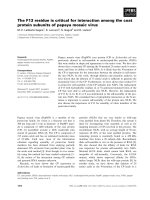
the pseudostem. The most effective diagnostic
symptom is short dark green dots and dashes
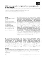
(Fig. 1) along the minor leaf veins. In Tamil
Nadu, it is a very serious disease in lower
Pulney hills where the ecotypes of hill
banana, Virupakshi and Sirumalai (AAB) are
cultivated (Fig. 2).
Successful control of bunchy top disease
depends on the availability of reliable BBTV
detection methods which will help us to select
disease free plantlets. Symptoms of bunchy
top disease are not visible at the time of
planting. Hence, development of a detection
kit for quick detection of BBTV is essential
for diagnosis of the disease at the time of
planting. Enzyme Linked Immunosorbant
Assay (ELISA) is convenient and easy
method but it has a limitation of being less
sensitive when the viral load is low (Landgraf
et al., 1991). In this context, Nucleic acid
based polymerase chain reaction (PCR)
(Edwards and Gibbs, 1994; Grieco and
Gallitelli, 1999; Tettelin et al., 1999; Olmos
et al., 2002; Stellrecht et al., 2004) and
hybridization methods have the advantage of
amplifying the target nucleic acid present
even at very low level and it has become an
attractive technique for the diagnosis of plant
viral diseases (Mullis et al., 1986; Henson and
French, 1993; Xie and Hu, 1994; McManus
and Jones, 1995; Hafner et al., 1997a and b;
Elnifro et al., 2000; Nassuth et al., 2000).
Molecular investigations on BBTV genome
revealed that BBTV has isometric virions
(Harding et al., 1991; Katul et al., 1997; Su et
al., 2000), 18-20 nm in diameter, and a
multicomponent genome consists of at least
six circular single stranded DNA (ssDNA)
component (BBTV DNA-1 to 6) ranging in
size from 1108 bp to 1111 bp (Burns et al.,
1994 and 1995; Karan et al., 1997;
Wanitchakorn et al., 1997; Beetham et al.,
1999). BBTV virions have a single coat
399
Int.J.Curr.Microbiol.App.Sci (2017) 6(6): 398-411
to a new eppendorf tube, and 450 µl of ice
cold IPA (Isopropyl alcohol) was added and
after mixing, the tubes were kept in ice for 20
min. The tubes were centrifuged for 15 min at
12,000 rpm and supernatant was discarded
without disturbing the pellet. The pellet was
washed with 500 µl of 70% ice cold ethanol
and centrifuged for 10 min at 12,000 rpm.
The supernatant was discarded and the pellet
was air dried for 5 min and suspended in 40
µl of 0.1X TE buffer (1mM Tris HCl pH 8.0
and 0.1mM EDTA pH 8.0) and kept at 65ºC
for 3 min (to suspend the pellet well) and
stored at -20ºC. The isolated DNA was
checked for its purity by 0.8% agarose gel
electrophoresis and quantified by UV
Spectrophotometer.
Materials and Methods
Collection of plant sample
Hill banana field trial was conducted at
Horticulture Research Station (HRS), Tamil
Nadu
Agricultural
University,
Thadiyankudisai (Lower Pulney Hills). Thirty
hill banana suckers infected with BBTV were
collected from HRS and 30 tissue culture and
certified hill banana suckers free from BBTV
infection were collected from National
Research Centre for Banana NRCB,
Trichirapalli and used as planting material for
field trial. The leaf samples were collected
from the plants at two different stages viz.,
juvenile stage (3 months after planting) and
vegetative stage (6 months after planting).
Healthy tissue culture derived plants were
grown negative control.
Total genomic
banana leaves
DNA
extraction
Multiple sequence alignment and primer
designing
ClustalW (Thompson et al., 1997) program
was used for multiple sequence alignment of
retrieved sequences of different BBTV
isolates. Conserved blocks in the BBTV
genome sequence were identified after
sequence alignment. Primer 3 software was
used for designing primers (Table 1). Primer
quality parameters like GC percentage,
melting temperature and product size were
taken into consideration and worthiness of the
designed primer was analyzed using Fast PCR
software.
from
Fresh young emerging green leaves with
midribs were collected from the field trial
plants and DNA was isolated and stored at 20°C for further use. Genomic DNA was
isolated using the modified CTAB (Cetyl
Trimethyl Ammonium Bromide) protocol
(Sambrook et al., 1989). Prior to extraction,
100 to 300 mg of midrib of young hill banana
leaves were cut into bits and transferred to a
zip lock bag (7 x 9 cm). Extraction CTAB
buffer 1 ml (0.2 M EDTA, 1.4 M NaCl, 1 M,
CTAB 2 %) was added immediately. The
samples were kept at room temperature and
squeezed by rolling a glass rod over the
sample to extract the cell contents. About 500
µl of the cell extract was transferred into an
eppendorf tube, and then 33 µl of 20 % SDS
was added into the tube and mixed well. The
tube was kept at 65º C (heating blocks) for
10–12 min and then the tube was centrifuged
for 10 minutes at 12,000 rpm and 450 µl of
the supernatant was transferred immediately
PCR screening of BBTV infection in hill
banana
PCR amplification was performed to amplify
the CP gene. DNA samples isolated from the
hill banana leaf samples were used as
template. PCR reaction were performed in a
final volume of 20 µl, 2 µl diluted total
genomic DNA, 2.0 µl of 10X PCR buffer (10
mM Tris-HCl, pH 9.0, 50 mM KCl, 1.5 mM
MgCl2), 0.5 µl of 200 mM dNTPs, 1 µl of 100
ng of respective forward and reverse primers,
400
Int.J.Curr.Microbiol.App.Sci (2017) 6(6): 398-411
0.5 µl of 3U Taq DNA polymerase
(Bangalore Genei Pvt. Ltd., Bangalore, India)
and 14 µl of sterilized double distilled water.
Amplification was performed in a PTC100TM Programmable Thermal Cycler (MJ
Research, Inc., Watertown, USA). The
temperature profile is as follows: Initial
denaturation at 94°C for 5 min, 35 cycles of
denaturation at 94°C for 1 min annealing at
57°C for 1 min, extention at 72°C for 1 min
and final extension for 10 min at 72°C. After
amplification, 8 µl of the product was used for
electrophoretic analysis on 0.8% agarose gels.
Cloning and sequencing of CP gene
Cloning of CP gene
Agarose gel electrophoresis
PCR amplification of CP gene was done using
the designed forward and reverse primer. PCR
amplified product was resolved by agarose
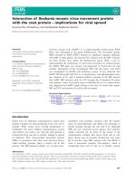
gel electrophoresis. DNA band of expected
513 bp size (Fig. 3) was excised from the gel
and the DNA fragment was eluted using gel
extraction kit by following manufacturer's
instructions (Sigma, USA). The eluted DNA
fragment was ligated into a T-tailed vector
pTZ57R/T by T/A cloning method (catalog #
K1214; MBI Fermentas).
The PCR products were resolved on 0.8%
agarose gel was prepared and to this 2µl
ethidium bromide was added from the stock
(10 mg/ml). After cooling, the mixture was
poured into a preset template using an
appropriate comb. The comb was removed
after solidification and gel with template was
placed in a horizontal electrophoresis unit
containing 1X TBE buffer.
Ligation reaction was set up by mixing 50 ng
of vector DNA, 150 ng of amplified product,
1 U of T4 DNA ligase and 1 µl of 10X buffer
in a 10 µl reaction. The reaction mix was
incubated at 16°C for 16 h and the ligated
product transformed into competent E. coli
(DH5α) cells. The recombinant white colonies
were selected and screened for the
recombinant plasmid by colony PCR analysis.
The PCR product was mixed with 6X loading
dye at 5:1 ratio and loaded. Electrophoresis
was carried out at 60V (Sambrook et al.,
1989). After the gel electrophoresis, the
amplified DNA bands were visualized under
UV transilluminator.
Colony PCR analysis
Colony PCR analysis was done to identify the
recombinant clones by using the CP gene
specific primers. Reactions were performed in
a final volume of 20 µl (2 µl of diluted total
genomic DNA, 2.0 µl of 10X PCR buffer (10
mM Tris-HCl, pH 9.0,50 mM KCl, 1.5 mM
MgCl2), 0.5 µl of 200 mM dNTPs, 1 µl of
100 ng of forward (ATGGCTAGGT
ATCCGAAG) and reverse primer (TCAAA
CATGATATGTAATTC), 0.5 µl of 3U Taq
DNA polymerase (Bangalore Genei Pvt. Ltd.,
Bangalore, India) and 14 µl of sterilized
double distilled water.
PCR Amplification of coat protein (CP)
PCR analysis was performed to amplify the
CP gene in the DNA samples isolated from
the hill banana leaf samples collected from
the field trial (Fig. 2). The reactions were
carried out in a reaction volume of 20µl and
by following the temperature profiles as
described above. After amplification, 10 µl of
the PCR product was electrophoresed in a
0.8% agarose gel and bands were visualized
by ethidium bromide staining.
Amplification was performed in a PTC100TM Programmable Thermal Cycler (MJ
Research, Inc., Watertown, USA).
401
Int.J.Curr.Microbiol.App.Sci (2017) 6(6): 398-411
The temperature profile used in the
amplification was as followes; Initial
denaturation at 94°C for 5 min followed by 35
cycles of 94°C for 1 min, 57°C for 1 min,
72°C for 1 min and final extension for 10 min
at 72°C. After amplification, 8 µl of the
product was used for electrophoretic analysis
on 1% agarose gels.
Southern blot analysis
Restriction digestion of genomic DNA
Restriction digestion was done according to
Sambrook et al., (1989). The banana genomic
DNA was restricted with the restriction
endonucleases, Hind III (2 units of enzyme
per µg of DNA) in recommended buffer, at
37oC, overnight.
Sequencing of the coat protein
The isolated recombinant plasmid was
sequenced
(NCBI
accession
number:
FJ664271.1). The obtained nucleotide
sequences were analyzed through NCBIBLAST search.
Reaction was analyzed by 0.8 % agarose gel
electrophoresis along with undigested banana
DNA as control.
Multiple sequence alignment was performed
using ClustalW program to analyze the
similarity between the cloned insert and the
gene sequence 513 base pair retrieved from
the database.
Equal amount of restricted genomic DNAs
(10 µg/lane) were resolved on a 0.8 % agarose
gel. After electrophoresis, the gel was
immersed in 0.25 N Hydrochloric acid for 15
minutes for partial depurination of DNA.
After depurination, incubating the gel in 0.4
M sodium hydroxide for 20 minutes
denatured DNA. After soaking the gel in 10X
SSC for 30 minutes, the gel was placed
carefully on transfer platform covered with a
Whatman 3 mm filter paper wick with its
margins hanging in 10X SSC poured in a
glass tray, a piece of nylon filter, cut
marginally smaller than the gel, was placed
carefully on the gel.
Southern blotting
NCBI accession number: FJ664271.1
ATGGCTAGGTATCCGAAGAAATCCATC
AAGCAGAGGCGGGTTGGGCGCCGGAA
GTATGGCAGCAAGGCGGTAACGAGCC
ACGACTACTCGTCGTTAGGGTCAATAT
TGGTTCCTGAAAACACCGTCAAGGTAT
TTCGGATTGAGCCTACTGATAAAACAT
TACCCAGATATTTTATCTGGAAAATGT
TTATGCTTCTTGTGTGCAAGGTGAAGC
CCGGAAGAATACTTCATTGGGCTATGA
TCAAGAGTTCTTGGGAAATCAACCAGC
CGACAACCTGTCTGGAAGCCCCAGGTT
TATTTATTAAACCTGAACATAGCCATC
TGGTTAAACTGGTATGTAGTGGGGAAC
TTGAAGCAGGAGTCGCAACAGGGACA
TCAGATGTTGAATGTCTTTTGAGGAAG
ACAACCGTGTTGAGGAAGAATGTAAC
AGAGGTGGATTATTTATATTTGGCATT
CTATTGTAGTTCTGGAGTAAGTATAAA
CTACCAGAACAGAATTACATATCATGT
TTGA
Nylon filter was covered immediately with 5
sheets of whatman 3 mm filter papers and
then with a stack of crude filter papers, cut
exactly to the dimensions of the gel. The stack
of filter papers was covered with a glass plate,
over which a weight of 500 g was placed.
The level of 10X SSC should be below the
surface of the platform. The upward diffusion
of the buffer was allowed for 18 hours. After
transfer, the nylon filter was carefully
removed, cross linked against UV light for 50
seconds, rinsed with 2X SSC and dried.
402
Int.J.Curr.Microbiol.App.Sci (2017) 6(6): 398-411
expression vector pET28a (+) at EcoRI and
HindIII site. The ligated product was
transformed into competent E. coli strain
BL21cells. The recombinant colonies were
selected by colony PCR and restriction
digestion analysis.
Preparation of radiolabeled probes
The DNA was amplified using CP gene PCR
by, following standard procedure (Sambrook
et al., 1989). About 25 ng of DNA fragments
was denatured with 0.5 µg of random
hexanucleotide primers in a boiling water
bath for 5 minutes and chilled immediately on
ice. To the mixture, other reaction compounds
are added to set a 25 µl reaction containing 25
µM each of 0.33 mM dATP, 0.33 mM dTTP,
and 0.33 mM dGTP, 50 µCi of α [32P] dCTP
(BRIT, CCMB campus, Hyderabad) and 10
units of klenow fragments of DNA
polymerase I. The reaction was incubated at
room temperature for three hours.
Induction of coat protein gene in E. coli
The E. coli strain, BL21 harbouring the
plasmid pET 28a (+) CP was streaked
separately on LB agar plate with appropriate
antibiotics and incubated at 37°C for 12-16 h
so as to get isolated individual colonies.
Single colony was inoculated in LB broth
containing appropriate antibiotics and grown
on a rotary shaker (200 rpm) at 37°C for 16 h.
Fifty ml of LB broth was inoculated with 1%
of overnight grown culture and it was
incubated at 37°C and 200 rpm till the OD
reaches 0.3 – 0.5 at 595 nm. One ml of the
aliquot was taken in an eppendorf and
centrifuged at 10,000 rpm for 5 min., and the
pellet was dissolved in 50 µl of the laemmli
buffer (0.06M Tris-HCl, pH6.8, 10%
glycerol, 5% β-Mercaptoethanol and 2%
SDS) and stored at –20°C for later use as
uninduced culture. The remaining culture was
induced by the addition of IPTG (isopropylβ–D–thiogalactopyranoside), to a final
concentration of 1mM. The culture was
allowed to grow at 37°C and the aliquot was
collected at 3 and 6 h., after incubation and
the pellet was dissolved in Laemmli buffer
(Laemmli, 1970) and stored at –20°C.
Southern Hybridization
Southern hybridization was performed as
described by Sambrook et al., (1989) with
required modifications. Nylon filters carrying
electrophoretically resolved DNA from
banana tissue culture plant and infected were
prehybridized in prehybridization solution
(5X SSC, 5X Denhardt’s solution, 1 mg/ml
denatured salmon sperm DNA) at 62ºC for 4
hours. The prehybridized filters were
hybridized in hybridization solution (pre
hybridization solution + radiolabelled probes
at a concentration of 1X 10⁶ cpm/ml.) at 62ºC
for 18 hours. After hybridization, nylon filters
were washed sequentially with 2X SSC
+0.1% SDS for 15 minutes at room
temperature (twice), and 0.1X SSC +0.1%
SDS for 15 minutes at 62ºC. Then filters were
dried at room temperature and exposed for
autoradiography.
SDS-PAGE analysis
The glass plates were cleaned thoroughly with
water followed by alcohol and placed in a
casting unit. The separating gel mixture was
poured in the glass plate assembly and
overlaid with a film of isopropanol to avoid
trapping of any air bubble and uniform gel
surface. After polymerization, the alcohol
layer was removed, rinsed with water and
Expression of recombinant coat protein
gene in E. coli
Cloning in pET vector
To express the coat protein gene in E. coli, the
CP gene of BBTV was subcloned into
403
Int.J.Curr.Microbiol.App.Sci (2017) 6(6): 398-411
blotted with filter paper. The stacking gel
mixture was then poured and the comb was
placed on top of the sandwich. After
polymerization, the comb was carefully
removed and wells were rinsed with electrode
buffer before loading the samples. The protein
samples were mixed with 1× loading dye,
boiled for 2 min and then loaded.
Electrophoresis was carried in a 1× buffer
(Tris 0.025M; Glycine 0.192M and SDS
0.1%) at constant current of 15 mA till the
dye front reached the separating gel. The
current supply was then increased to a
constant supply of 30 mA till the blue dye
reaches the bottom. After the electrophoretic
run, the gel unit was dismantled and gel was
fixed using a fixative (Methanol; acetic acid;
water, 4:1:5 ratio) and stained in a Coomassie
Brilliant Blue solution for 12 hrs. The gel was
destained until the background became
colorless and the gel was documented using
Alphaimager (AlphaInnotech, USA).
enzymes, KpnI and HindIII. The selected
three recombinant clones were sequenced
(Fig. 3). Sequencing has resulted in a length
of 513 bp and the homology search of the
sequence using NCBI blast (Altschul et al.,
1997) showed similarity (99%) to the reported
ten ‘CP’ gene sequences and are 98% similar
to four ‘CP’ gene sequences. The identity
search was made with BLASTX algorithm
using the same nucleotide sequence.
Southern hybridization analysis
To confirm the virus infection and virus load
at two different (juvenile stage and vegetative
stage) stages, the five plant samples each
from infected and healthy banana samples
were subjected to southern blot hybridization
and virus infection was confirmed using CP
gene specific probe of BBTV. DNA of the
banana samples both infected and healthy was
restricted with restriction endonucleases,
HindIII, the products were electrophoratically
resolved, transferred to nylon membrane and
hybridized against radiolabeled probes
prepared using the BBTV CP gene.
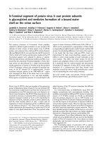
Hybridization of the blot with BBTV CP gene
probe showed that no signal was observed in
case of healthy plant control indicating free
from BBTV infection, whereas the infected
sample showed strong hybridization signal
indicates the presence of BBTV (Fig. 4). Due
to the presence of excessive phenolic
compounds, they formed smeared lanes with
faint bands.
Results and Discussion
Cloning and sequencing of the ‘CP’ Gene
of BBTV
The ‘CP’ gene-specific primers of BBTV
were designed using the conserved region in
the ‘CP’ gene. They are used for the complete
amplification of the ‘CP’ gene in the BBTV
infected hill banana plants. The PCR products
were resolved on 0.8% agarose gel and an
expected size of 513 bp was observed. The
full length ‘CP’ gene (513 bp) was then eluted
from the 0.8% agarose gel and cloned into the
T/A cloning vector, pTZ57R/T and named as
pHBScp. The ligated product was then
transformed into the E. coli strain (DH5α) and
was selected on ampicillin containing
medium. The recombinant colonies were
identified by colony PCR using the same ‘CP’
gene specific primer and one of positive
clones was selected and further confirmed by
releasing the cloned fragment using restriction
Expression of BBTV coat protein in E. coli
The BBTV coat protein expressed (Fig. 5) in
E. coli was subjected to SDS-PAGE analysis.
The expression of BBTV coat protein was
found in very low at 1-6 hour of incubation in
IPTG induction and low level of expression
was found at 7 and 8 hours of incubation in
IPTG induction (Fig. 6).
404
Int.J.Curr.Microbiol.App.Sci (2017) 6(6): 398-411
Figure.1 Dark green patches of BBTV infected leaf sheath
Dark green patches
Figure.2 Field view of healthy and BBTV infected banana
Figure.3 Confirmation of Recombinant clones by Colony PCR analysis, M- 100 bp ladder,
1- Coat protein (CP) gene (513 bp), 2- Negative control (Tissue culture Plantain).
405
Int.J.Curr.Microbiol.App.Sci (2017) 6(6): 398-411
Figure.4 Southern blot analysis at Juvenile stage and Vegetative stage, PC – Positive control
(Cloned Coat Protein Plasmid), NC – Negative control (Tissue culture Certified Plantain), IBBTV infected plantain and H – Healthy plantain
Juvenile Stage
PC NC I1 I2 I3 I4 I5 H1 H2 H3 H4 H5
Vegetative Stage
PC NC I1 I2 I3 I4 I5 H1 H2 H3 H4 H5
Figure.5 Cloning of pET -28a(+) vector with CP gene
Figure.6 BBTV coat protein, M- Protein marker, C –pET 28a(+) vector (control), 3h, 6h
– hours after IPTG induction
406
Int.J.Curr.Microbiol.App.Sci (2017) 6(6): 398-411
Table.1 Primer sequences for amplifying gene-specific sequences.
Primer name
CP gene forward primer
CP gene reverse primer
Sequence
5’-ATGGCTAGGTATCCGAAG-3’
5’-TCAAACATGATATGTAATTC-3’
Banana is one of the most important fruit crop
grown in most of the southern states of India.
Several banana cultivars are under cultivation
in India and hill banana is one of the unique
cultivar grown in the lower Paliney hills in
Tamil Nadu. Hill banana cultivation is
seriously affected by the disease caused by
BBTV. BBTV is transmitted in a persistent
manner by the banana aphid Pentalonia
nigronervosa and causes a yellows disease in
bananas, it was classified as a 'possible
member' of the luteovirus group (Matthews,
1981). Further evidence supporting this
classification is the reported occurrence of
dsRNA in BBTV infected plants but not in
corresponding healthy plants (Dale et al.,
1986). Selection of disease free planting
material is one of the very important
techniques
in
banana
cultivation.
Identification of BBTV infection in the
planting material/suckers is very difficult as
the symptom development is not visible at the
early stage of infection. Several reports are
available for the molecular detection of the
plant virus in different crops using PCR and
ELISA method. Immunological detection
using technique such as ELISA is not
effective as the virus is present in low tire in
the banana plant. There is only one successful
report of ELISA based detection of BBTV.
Most of the recent literature showed that
BBTV detection was effective using PCR
method (Mansoor et al., 2005). PCR based
detection technique, had the advantage of
amplifying the target nucleic acid present
even at very low level and it had become an
attractive technique for the diagnosis of many
plant viral diseases (Henson and French,
1993; Xie and Hu, 1994; Hafner et al., 1997a,
Chandrasekar et al., 2011).
Base pairs
18
20
The CP coded by the DNA-3 component of
the BBTV has been used for virus detection
because of its important role in viral particles
packing and disease establishment and has
been consistently associated with BBTD in
Asian countries group. An attempt was made
to develop BBTV detection protocol utilizing
the CP gene specific primers.
In order to, PCR amplification using CP
specific primer gave amplicon of expected
size (513 bp) with DNA isolated from
infected hill banana plant. A common
problem in the use of PCR for diagnostic
purposes is, that it needs a good quality DNA.
Plant species which contains high phenolic
shows false-negatives i.e., even though the
target viral DNA is present but still fail to
amplify because of the quality of the DNA
taken for PCR analysis. So the amplification
of a PCR by using the internal control primer
will possibly demonstrates the absence of
PCR inhibitors in banana DNA samples and
can provide some level of confidence on a
diagnostic PCR reaction that is negative for
the presence of BBTV. BBTV detection
method using internal control primer and
BBTV Rep gene primer was reported by
Mansoor et al., (2005). Potential problems in
PCR include false negatives due to reaction
failure or false positives due to contamination
(Chandrasekar et al., 2011). False negatives
were often revealed in Southern blot
technique.
Harding et al., (1991) used the Southern blot
technique to detect the presence of BBTV in
banana tissue and in aphids. In the present
study, to confirm the presence of BBTV
(DNA-3) CP gene, Southern hybridization
407
Int.J.Curr.Microbiol.App.Sci (2017) 6(6): 398-411
analysis
was
performed.
Southern
hybridization
analyses
against
the
radiolabeled probes prepared from BBTV CP
gene. The experiment performed with
samples collected at two banana growth
stages viz., juvenile and vegetative stage.
From each stage five samples of infected and
healthy samples were collected. The result
indicates that all found in both stages.
Normally banana plants has more mucilage
and phenolic compound, in Biotic stress
condition the phenolic, abscisic acid,
ethylene, jasmonic acid, and salicylic acid
levels are increased, here the BBTV plays
major role of stress factor, where the phenolic
levels are more elevated. Due to the presence
of excessive phenolic compounds, they
formed smeared lanes with faint bands.
Infected plants had similar level of,
hybridization signal in both stages, in the
healthy plants no hybridization signal was
seen. Harding et al., (1991) have showed the
virus like particles are associated with banana
bunchy top disease contains small singlestranded DNA. They have used southern blot
method to analyze the expression of virus like
particles with extracts from sucrose gradientpurified BBTV, healthy banana plant and
partially purified BBTV. Their studies have
also shown that the expression was seen only
at the infected plants not in the healthy plants.
Coat protein expressed in E. coli showed very
low level of coat protein accumulation as
revealed by SDS-PAGE analysis. The low
level of coat protein accumulation may be due
to the small size of the protein not being
stably present in the E. coli cells. In contrast,
Harding et al., (1991) and Wanitchakorn et
al., (2000) used the SDS-PAGE they have got
expression at a molecular weight of
20100KDa.
banana bunchy top virus (BBTV) infection
study. I confirm that all the research meets the
ethical guidelines, including adherence to the
legal requirements of the study country.
Abbreviations
BBTV- Banana Bunchy Top Virus
CP – coat protein
PCR- Polymerase chain reaction
Acknowledgement
I thank the Chairman of Department of Plant
Biotechnology, Centre on Plant Molecular
Biology and Biotechnology, Tamil Nadu
Agricultural University, Coimbatore for
providing all the facilities for molecular work,
The Head, Horticultural Research Station,
Thadiyankudisai, Tamil Nadu Agricultural
University, Coimbatore for the field facility
and the National Research Centre for Banana
(NRCB) Trichirapalli, for providing the
certified hill banana suckers free from BBTV
infection planting material for field trial.
References
Altschul S.F, Madden T.L, Schaffer A.A,
Zhang J., Zhang Z., Miller W, Lipman
D.J., 1997. Gapped BLAST and PSTBLAST: a new generation of protein
database search programs. Nucleic
Acids Res 25: 3389-3402.
Amain I, Qazi J, Mansoor S, Llyas M,
Briddon R.W., 2008. Molecular
characterization of banana bunchy top
virus (BBTV) from Pakistan. Virus
genes 36:191-198.
Beetham P.R, Harding R.M, Dale J.L., 1999.
Banana Bunchy top virus DNA-2 to 6
are monocistronic. Arch Virol 144: 89105
Burns T.M, Harding R.M, Dale J.L., 1994.
Evidence that banana bunchy top virus
Research involving human participants
and/or animals
It does not involving in the any Human
participants and Animals. It is confirmation of
408
Int.J.Curr.Microbiol.App.Sci (2017) 6(6): 398-411
has a multiple component genome. Arch
Virol 137:371-380.
Burns T.M, Harding R.M, Dale J.L., 1995.
The genome organization of banana
bunchy top virus: analysis of six ssDNA
components. J Gen Virol 76: 14711482.
Chandrasekar A, Kalaiponmani K, Elayabalan
S, Kumar K.K, Angappan K,
Balasubramanian P., 2011. Screening of
banana bunchy top virus through
multiplex
PCR
approach.
Arch
Phytopathol Plant Protect 19:19201925.
Dale J.L., 1987. Banana bunchy top: an
economically important tropical plant
virus disease. Adv Virus Res 33: 301325
Dale J.L, Philllips D.A., Parry J.N., 1986.
Double-stranded RNA in banana plants
with bunchy top disease. J. Gen. Virol.
67:371-375.
Edwardsand M.S, Gibbs R.A., 1994.
Multiplex
PCR:
Advantages
development and applications. PCR
Methods Appl 3:65-75
Elnifro E.M, Ashshi A.M, Cooper R.J,
Klapper P.E., 2000. Multiplex PCR:
Optimization and Application in
Diagnostic Virology. Clin Microbiol
Rev 13:559-570.
Grieco F, Gallitelli D, 1999. Multiplex
reverse transcriptase polymerase chain
reaction applied to virus detection in
globe artichoke. J Phytopathol 147:183185.
Hafner G.J, Harding R.M, Dale J.L., 1997a. A
DNA primer associated with banana
bunchy top virus. J Gen Virol 78:479486.
Hafner G.J, Stafford M.R, Wolter L.C,
Harding R.M, Dale J.L., 1997b. Nicking
and joining activity of banana bunchy
top virus replication protein in vitro. J
Gen Virol 78:1795-1799.
Harding R.M, Burns T.M, Dale J.L., 1991.
Virus-like particles associated with
banana bunchy top disease contain
small single stranded DNA. J Gen Virol
72:225- 230.
Henson J.M, French C.R., 1993. The
polymerase chain reaction and plant
disease
diagnosis.
Annu
Rev
Phytopathol 31:81-109.
Higgins D.G, Sharp P.M., 1988. CLUSTAL.
A package for performing multiple
sequence
alignment
on
a
microcomputer. Gene 73:237-244
Horser C.L, Karan M, Harding R.M, Dale
J.L., 2001. Additional Rep-encoding
DNAs associated with banana bunchy
top virus. Arch Virol 146:71-86
John S.J, Briddon R.M, Mansoor S, Bedford
I.D, Pinner S.M, Saunders K, Stanley
J.Y, Zafar K.A, Malik P., 2001.
Identification of DNA components
required for induction of cotton leaf curl
disease. Virol 285:234-243.
Karan M, Harding R.M, Dale J.L., 1994.
Evidence for two groups of banana
bunchy top virus isolates. J Gen Virol
75:3541-3546.
Karan M, Harding R.M, Dale J.L., 1997.
Association of banana bunchy top virus
DNA components 2 to 6 with bunchy
top disease. Mol Plant Pathol online: 18
Katul L, Maiss E, Morozov S.Y, Vetten H.J.,
1997. Analysis of six DNA components
of the faba bean necrotic yellows virus
genome and their structural affinity to
related plant virus genomes. Virology
233:247-259.
Landgraf A, Reckmann B, Pingoud A., 1991.
Direct analysis of polymerase chain
reaction products using enzyme linked
immunosorbent assay techniques. Annu
Biochem 198:86-91
Laemmli, O. K., 1970. Cleavage of structural
proteins during the assembly of the head
409
Int.J.Curr.Microbiol.App.Sci (2017) 6(6): 398-411
of bacteriophage T4. Nature, 227: 680685.
Mansoor S, Qazi J, Amain I, Khatri I.A, Khan
A.I, Raza S, Zafar Y, Briddon R.W.,
2005. A PCR based method, with
internal control, for the detection of
BBTV in banana. Mol Biotechnol
30:167-169.
Matthews R.E.F., 1981. Classification and
nomenclature of viruses. Intervirol
16:53-60
McManus P.S, Jones A.L., 1995. Detection of
Erwinia amylovora by nested PCR and
PCR-dot-blot
and
reverse
blot
hybridizations. Phytopathol 85:618-623
Menzela W, Jelkmann W, Maiss E., 2002.
Detection of four apple viruses by
multiplex
RT-PCR
assays
with
coamplification of plant mRNA as
internal control. J Virol Methods 99:8192.
Meunier A, Schmit JF, Stas A, Kutluk N,
Bragard C., 2003. Multiplex reverse
transcription PCR for the simultaneous
detection of Beet necrotic yellow vein
virus, Beet soil borne virus, and Beet
virus Q and their vector Polymyxa betae
KESKIN on sugar beet. Appl Environ
Microbiol 69:2356-2360.
Mullis KF, Faloona F, Scharf S, Saiki R,
Horn G, Erlich H., 1986. Specific
enzymatic amplification of DNA in
vitro: the polymerase chain reaction.
Cold Spring Harb Symp Quant Biol
51:263-273.
Nassuth A, Pollari E, Helmeczy K, Stewart S,
Kofalvi S.A., 2000. Improved RNA
extraction and one-tube RT-PCR assay
for simultaneous detection of control
plant RNA plus several viruses in plant
extracts. J Virol Methods 90:37- 49.
Olmos A, Bertolini E, Cambra M., 2002.
Simultaneous
and
cooperational
amplification (Co-PCR): a new concept
for detection of plant viruses. J Virol
Methods 106:51-59.
Pearson W.R, Lipman D.J., 1988. Improved
tools
for
biological
sequence
comparison. Proc Natl Acad Sci
85:2444-2448.
Sambrook J, Fritsch E.F, Maniatis T., 1989. A
Laboratory Manual, 2nd edn. Cold
Spring Harbor, NY: Cold Spring Harbor
Laboratory Molecular Cloning
Singh R.P, Kurz J, Boiteau G., 1996.
Detection of stylet-borne and circulative
potato viruses in aphids by duplex
reverse transcription polymerase chain
reaction. J Virol Methods 59:189-196
Stellrecht K.A, Woron M, Mishrik N.G,
Venezia R.A., 2004. Comparison of
Multiplex PCR Assay with Culture for
Detection of Genital Mycoplasmas. J
Clin Microbiol 42 (4):1528-1533.
Su H.J, Tsao L.Y, Wu M.L, Hung T.H., 2000.
Biological and molecular categorization
of strains of BBTV. J Phytopathol
151:290-296.
Tettelin H, Radune D, Kasif S, Khouri H,
Salzberg S.L., 1999. Optimized
multiplex PCR: efficiently closing a
whole genome shotgun sequencing
project. Genomics 62:500-507.
Thomas J.E, Dietzgen R.G., 1991.
Purification,
characterization
and
serological detection of virus-like
particles associated with banana bunchy
top disease in Australia. J Gen Virol
72:217-224
Thompson J.D, Gibson T.J, Plewniak F,
Jeanmougin F, Higgins D.G., 1997. The
CLUSTALX
windows
interface:
flexible strategies for multiple sequence
alignment aided by quality analysis
tools. Nucleic Acid Res 25:4876-4882
Tien J.E, Xin L.Z., 2005. Cloning and
sequencing of DNA components of
Banana bunchy top virus Hainan isolate.
Chinese J Agri Biotechnol 2:91-97.
Tripathi L, Tripathi J.N, Tushemereirwe
W.K., 2004. Strategies for resistance to
bacterial wilt disease of bananas
410
Int.J.Curr.Microbiol.App.Sci (2017) 6(6): 398-411
through genetic engineering. Afr J
Biotechnol 3 (12):688-692
Valerie K, Thomas J.E, Sharman M,
Mademba-Sy F., 2001. First record of
banana bunchy top disease in New
Caledonia. Australasian Plant Pathol
30:71
Vansluys M.A, Monteiro-Vitorello C.B,
Camargo Menck L.E.A, Da Silva
C.F.M, Ferro A.C.R, Oliveira J.A,
Setubal M.C, Kitajima J.C., 2002.
Comparative genomic analysis of plantassociated
bacteria.
Annu
Rev
Phytopathol 40:169-189.
Wanitchakorn R, Harding R.M, Dale J.L.,
1997. Banana bunchy top virus DNA-3
encodes the viral coat protein. Arch
Virol 142:1673-1680
Wanitchakorn R, Harding R.M, Dale J.L.,
2000. Sequence variability in the coat
protein of two groups of banana bunchy
top isolates. Arch Virol 145:593-602
Wu R.Y, Su H.J., 1990. Purification and
characterization of banana bunchy top
virus. J Phytopathol 128:153-160.
Xie W.S, Hu J.S., 1994. Molecular cloning,
sequence analysis and detection of
banana bunchy top virus in Hawaii. Mol
Plant Pathol 85:339-347.
How to cite this article:
Chandrasekar Arumugam, Kalaiponmani Kalaimughilan and Angappan Kathithachalam. 2017.
Banana Bunchy Top Viral Coat Protein (CP) Gene Expression Studies at Molecular Level in
Hill Banana cv. Sirumalai (AAB). Int.J.Curr.Microbiol.App.Sci. 6(6): 398-411.
doi: />
411