Machine learning and network methods for biology and medicine
Bạn đang xem bản rút gọn của tài liệu. Xem và tải ngay bản đầy đủ của tài liệu tại đây (18.04 MB, 195 trang )
Computational and Mathematical Methods in Medicine
Machine Learning and Network
Methods for Biology and Medicine
Guest Editors: Lei Chen, Tao Huang, Chuan Lu, Lin Lu, and Dandan Li
Machine Learning and Network Methods for
Biology and Medicine
Computational and Mathematical Methods in Medicine
Machine Learning and Network Methods for
Biology and Medicine
Guest Editors: Lei Chen, Tao Huang, Chuan Lu, Lin Lu,
and Dandan Li
Copyright © 2015 Hindawi Publishing Corporation. All rights reserved.
This is a special issue published in “Computational and Mathematical Methods in Medicine.” All articles are open access articles distributed under the Creative Commons Attribution License, which permits unrestricted use, distribution, and reproduction in any medium,
provided the original work is properly cited.
Editorial Board
Emil Alexov, USA
Elena Amato, Italy
Konstantin G. Arbeev, USA
Georgios Archontis, Cyprus
Paolo Bagnaresi, Italy
Enrique Berjano, Spain
Elia Biganzoli, Italy
Konstantin Blyuss, UK
Hans A. Braun, Germany
Thomas S. Buchanan, USA
Zoran Bursac, USA
Thierry Busso, France
Xueyuan Cao, USA
Carlos Castillo-Chavez, USA
Prem Chapagain, USA
Hsiu-Hsi Chen, Taiwan
Ming-Huei Chen, USA
Phoebe Chen, Australia
Wai-Ki Ching, Hong Kong
Nadia A. Chuzhanova, UK
Maria Cordeiro, Portugal
Irena Cosic, Australia
Fabien Crauste, France
William Crum, UK
Getachew Dagne, USA
Qi Dai, China
Chuangyin Dang, Hong Kong
Justin Dauwels, Singapore
Didier Delignières, France
Jun Deng, USA
Thomas Desaive, Belgium
David Diller, USA
Michel Dojat, France
Irini Doytchinova, Bulgaria
Esmaeil Ebrahimie, Australia
Georges El Fakhri, USA
Issam El Naqa, USA
Angelo Facchiano, Italy
Luca Faes, Italy
Giancarlo Ferrigno, Italy
Marc Thilo Figge, Germany
Alfonso T. García-Sosa, Estonia
Amit Gefen, Israel
Humberto González-Díaz, Spain
Igor I. Goryanin, Japan
Marko Gosak, Slovenia
Damien Hall, Australia
Stavros J. Hamodrakas, Greece
Volkhard Helms, Germany
Akimasa Hirata, Japan
Roberto Hornero, Spain
Tingjun Hou, China
Seiya Imoto, Japan
Sebastien Incerti, France
Abdul Salam Jarrah, UAE
Hsueh-Fen Juan, Taiwan
Rafik Karaman, Palestine
Lev Klebanov, Czech Republic
Andrzej Kloczkowski, USA
Xiang-Yin Kong, China
Zuofeng Li, USA
Chung-Min Liao, Taiwan
Quan Long, UK
Ezequiel López-Rubio, Spain
Reinoud Maex, France
Valeri Makarov, Spain
Kostas Marias, Greece
Richard J. Maude, Thailand
Panagiotis Mavroidis, USA
Georgia Melagraki, Greece
Michele Migliore, Italy
John Mitchell, UK
Chee M. Ng, USA
Michele Nichelatti, Italy
Ernst Niebur, USA
Kazuhisa Nishizawa, Japan
Hugo Palmans, UK
Francesco Pappalardo, Italy
Matjaz Perc, Slovenia
Edward J. Perkins, USA
Jesús Picó, Spain
Alberto Policriti, Italy
Giuseppe Pontrelli, Italy
Christopher Pretty, New Zealand
Mihai V. Putz, Romania
Ravi Radhakrishnan, USA
David G. Regan, Australia
José J. Rieta, Spain
Jan Rychtar, USA
Moisés Santillán, Mexico
Vinod Scaria, India
Jörg Schaber, Germany
Xu Shen, China
Simon A. Sherman, USA
Pengcheng Shi, USA
Tieliu Shi, China
Erik A. Siegbahn, Sweden
Sivabal Sivaloganathan, Canada
Dong Song, USA
Xinyuan Song, Hong Kong
Emiliano Spezi, UK
Greg M. Thurber, USA
Tianhai Tian, Australia
Tianhai Tian, Australia
Jerzy Tiuryn, Poland
Nestor V. Torres, Spain
Nelson J. Trujillo-Barreto, UK
Anna Tsantili-Kakoulidou, Greece
Po-Hsiang Tsui, Taiwan
Gabriel Turinici, France
Edelmira Valero, Spain
Raoul van Loon, UK
Luigi Vitagliano, Italy
Liangjiang Wang, USA
Ruiqi Wang, China
Ruisheng Wang, USA
David A. Winkler, Australia
Gabriel Wittum, Germany
Yu Xue, China
Yongqing Yang, China
Chen Yanover, Israel
Xiaojun Yao, China
Kaan Yetilmezsoy, Turkey
Hujun Yin, UK
Hiro Yoshida, USA
Henggui Zhang, UK
Yuhai Zhao, China
Xiaoqi Zheng, China
Yunping Zhu, China
Contents
Machine Learning and Network Methods for Biology and Medicine, Lei Chen, Tao Huang, Chuan Lu,
Lin Lu, and Dandan Li
Volume 2015, Article ID 915124, 2 pages
Detection of Dendritic Spines Using Wavelet-Based Conditional Symmetric Analysis and Regularized
Morphological Shared-Weight Neural Networks, Shuihua Wang, Mengmeng Chen, Yang Li,
Yudong Zhang, Liangxiu Han, Jane Wu, and Sidan Du
Volume 2015, Article ID 454076, 12 pages
An Overview of Biomolecular Event Extraction from Scientific Documents, Jorge A. Vanegas,
Sérgio Matos, Fabio González, and José L. Oliveira
Volume 2015, Article ID 571381, 19 pages
NMFBFS: A NMF-Based Feature Selection Method in Identifying Pivotal Clinical Symptoms of
Hepatocellular Carcinoma, Zhiwei Ji, Guanmin Meng, Deshuang Huang, Xiaoqiang Yue, and Bing Wang
Volume 2015, Article ID 846942, 12 pages
Comparative Transcriptomes and EVO-DEVO Studies Depending on Next Generation Sequencing,
Tiancheng Liu, Lin Yu, Lei Liu, Hong Li, and Yixue Li
Volume 2015, Article ID 896176, 10 pages
ROC-Boosting: A Feature Selection Method for Health Identification Using Tongue Image, Yan Cui,
Shizhong Liao, and Hongwu Wang
Volume 2015, Article ID 362806, 8 pages
A Five-Gene Signature Predicts Prognosis in Patients with Kidney Renal Clear Cell Carcinoma,
Yueping Zhan, Wenna Guo, Ying Zhang, Qiang Wang, Xin-jian Xu, and Liucun Zhu
Volume 2015, Article ID 842784, 7 pages
Survey of Natural Language Processing Techniques in Bioinformatics, Zhiqiang Zeng, Hua Shi, Yun Wu,
and Zhiling Hong
Volume 2015, Article ID 674296, 10 pages
A Systematic Evaluation of Feature Selection and Classification Algorithms Using Simulated and Real
miRNA Sequencing Data, Sheng Yang, Li Guo, Fang Shao, Yang Zhao, and Feng Chen
Volume 2015, Article ID 178572, 11 pages
Identification of Chemical Toxicity Using Ontology Information of Chemicals, Zhanpeng Jiang, Rui Xu,
and Changchun Dong
Volume 2015, Article ID 246374, 5 pages
An Improved PID Algorithm Based on Insulin-on-Board Estimate for Blood Glucose Control with Type
1 Diabetes, Ruiqiang Hu and Chengwei Li
Volume 2015, Article ID 281589, 8 pages
G2LC: Resources Autoscaling for Real Time Bioinformatics Applications in IaaS, Rongdong Hu,
Guangming Liu, Jingfei Jiang, and Lixin Wang
Volume 2015, Article ID 549026, 8 pages
Identifying New Candidate Genes and Chemicals Related to Prostate Cancer Using a Hybrid Network
and Shortest Path Approach, Fei Yuan, You Zhou, Meng Wang, Jing Yang, Kai Wu, Changhong Lu,
Xiangyin Kong, and Yu-Dong Cai
Volume 2015, Article ID 462363, 12 pages
Identifying Novel Candidate Genes Related to Apoptosis from a Protein-Protein Interaction Network,
Baoman Wang, Fei Yuan, Xiangyin Kong, Lan-Dian Hu, and Yu-Dong Cai
Volume 2015, Article ID 715639, 11 pages
Cell Pluripotency Levels Associated with Imprinted Genes in Human, Liyun Yuan, Xiaoyan Tang,
Binyan Zhang, and Guohui Ding
Volume 2015, Article ID 471076, 8 pages
A Model of Regularization Parameter Determination in Low-Dose X-Ray CT Reconstruction Based on
Dictionary Learning, Cheng Zhang, Tao Zhang, Jian Zheng, Ming Li, Yanfei Lu, Jiali You, and Yihui Guan
Volume 2015, Article ID 831790, 12 pages
Multivariate Radiological-Based Models for the Prediction of Future Knee Pain: Data from the OAI,
Jorge I. Galván-Tejada, José M. Celaya-Padilla, Victor Treviño, and José G. Tamez-Peña
Volume 2015, Article ID 794141, 10 pages
Nonsynonymous Single-Nucleotide Variations on Some Posttranslational Modifications of Human
Proteins and the Association with Diseases, Bo Sun, Menghuan Zhang, Peng Cui, Hong Li, Jia Jia, Yixue Li,
and Lu Xie
Volume 2015, Article ID 124630, 12 pages
KIR Genes and Patterns Given by the A Priori Algorithm: Immunity for Haematological Malignancies,
J. Gilberto Rodríguez-Escobedo, Christian A. García-Sepúlveda, and Juan C. Cuevas-Tello
Volume 2015, Article ID 141363, 11 pages
Hindawi Publishing Corporation
Computational and Mathematical Methods in Medicine
Volume 2015, Article ID 915124, 2 pages
/>
Editorial
Machine Learning and Network Methods for
Biology and Medicine
Lei Chen,1 Tao Huang,2,3 Chuan Lu,4 Lin Lu,5 and Dandan Li6
1
College of Information Engineering, Shanghai Maritime University, Shanghai 201306, China
Department of Genetics and Genomics Sciences, Mount Sinai School of Medicine, New York, NY 10029, USA
3
Institute of Health Sciences, Shanghai Institutes for Biological Sciences, Chinese Academy of Sciences, Shanghai 200031, China
4
Department of Computer Science, Aberystwyth University, Aberystwyth, Ceredigion SY23 3DB, UK
5
Department of Radiology, Columbia University Medical Center, New York, NY 10032, USA
6
Gastrointestinal Medical Department, China-Japan Union Hospital of Jilin University, Changchun 130033, China
2
Correspondence should be addressed to Lei Chen; chen
Received 12 October 2015; Accepted 12 October 2015
Copyright © 2015 Lei Chen et al. This is an open access article distributed under the Creative Commons Attribution License, which
permits unrestricted use, distribution, and reproduction in any medium, provided the original work is properly cited.
In recent years, many computational methods have been
proposed to tackle the problems that arise in analyzing
various large-scale high dimensional data in biology and
medicine. Useful techniques have been developed by the use
of conventional statistical modeling and analysis and have
helped to reveal many biological mechanisms. However, with
the rapid development of high throughput technologies, biological and medical data generated nowadays are becoming
increasingly more heterogeneous and complex. It is therefore
necessary to develop more effective and efficient approaches
to analyzing such data, requiring more powerful methods like
advanced machine learning algorithms and network based
methods.
In this special issue, eighteen novel investigations are
presented, including a number of newly proposed techniques
for up-to-date data analysis and application systems for
interesting biological and medical problems.
A computational method was proposed by B. Wang et
al. to identify novel candidate genes related to apoptosis.
This method first applied shortest path algorithm in a large
protein-protein interaction network to search new candidate
genes and then the candidate genes were filtered by a permutation test. Twenty-six genes were obtained and analyzed
regarding their likelihood of being novel apoptosis-related
genes.
F. Yuan et al. proposed a computational method to identify new candidate genes and chemicals based on currently
known genes and chemicals related to prostate cancer
by applying shortest path approach in a hybrid network
which was constructed according to information concerning
chemical-chemical interactions, chemical-protein interactions, and protein-protein interactions.
B. Sun et al. designed an analysis pipeline to study
the relationships between eight types of damaging protein
posttranslational modifications (PTM) and a few human
inherited diseases and cancers. The results suggested that
some human inherited diseases or cancers might be related
to the interactions of damaging PTMs.
Y. Zhan et al. identified a five-gene signature that predicts
prognosis in patients with kidney renal clear cell carcinoma
(KIRC). The RNA expression data from RNA-sequencing and
clinical information of 523 KIRC patients were analyzed. The
AUC (area under ROC curve) of the five-gene signature was
0.783 which showed high sensitivity and specificity.
Z. Ji et al. developed a Nonnegative Matrix Factorization (NMF) based feature selection approach (NMFBFS)
to identify potential clinical symptoms for HCC patient
stratification. The results on 407 HCC patient samples with 57
symptoms showed the effectiveness of the NMFBFS approach
in identifying important clinical features, which will be very
helpful for HCC diagnosis.
2
C. Zhang et al. proposed adaptive weight regularized
ADSIR for low dose CT reconstruction. Three numerical
experiments are carried out for evaluation and comparisons
are made with other algorithms.
J. I. Galv´an-Tejada et al. presented the potential of Xray based multivariate prognostic models to predict the
onset of chronic knee pain. Using X-rays quantitative imageassessments, multivariate models may be used to predict subjects that are at risk of developing knee pain by osteoarthritis.
Y. Cui et al. developed a method called ROC-Boosting
to select significant Haar-like features extracted from tongue
images for health identification. They analyzed the images of
1,322 tongue cases and selected features focused on the root,
top, and side areas of the tongue which can classify the healthy
and ill cases.
S. Wang et al. proposed a novel automatic approach for
dendritic spine identification in neuron image. The method
integrated wavelet based conditional symmetric analysis and
regularized morphological shared-weight neural networks.
Its good performance and the comparison with existing
methods suggest the utility of the method.
S. Yang et al. proposed the use of a combination of edgeR
and DESeq to analyze miRNA sequencing data with a large
sample size.
R. Hu et al. proposed an automated resource provisioning
method, G2LC, for bioinformatics applications in IaaS. It
guaranteed applications performance and improved resource
utilization. Evaluated on real sequence searching data of
BLAST, G2LC saved up to 20.14% of resource.
R. Hu and C. Li proposed an improved PID algorithm
based on insulin-on-board estimate using a combinational
mathematical model of the dynamics of blood glucoseinsulin regulation in the blood system. The simulation results
demonstrated that the improved PID algorithm can perform
well in different carbohydrate ingestion and different insulin
sensitivity situations. Compared with the traditional PID
algorithm, the control performance was improved obviously
and hypoglycemia can be avoided.
J. G. Rodriguez-Escobedo et al. described the use of the “a
priori” algorithm at resolving KIR gene patterns associated
with haematological malignancies, previously unrevealed
through traditional statistical approaches.
Z. Jiang et al. built a new method to predict chemical toxicities based on ontology information of chemicals.
This method was more effective than previous method and
provided new insights to study chemical toxicity and other
attributes of chemicals.
L. Yuan et al. explored the hidden relationship between
miRNAs and imprinted genes in cell pluripotency. They
found that the neighbors of imprinted genes on molecular
network were enriched in modules such as cancer, cell death
and survival, and tumor morphology. The imprinted region
may provide a new look for those who are interested in cell
pluripotency of hiPSCs and hESCs.
T. Liu et al. reviewed the recent discoveries and advance
in the field of evolutional developmental biology in light of
the development in large-scale omics studies.
J. A. Vanegas et al. presented a survey on the state-ofthe-art text mining approaches to extraction of biomolecular
Computational and Mathematical Methods in Medicine
events, which are useful for understanding the underlying
biological mechanisms. The popular natural language processing and machine learning methods and tools have been
analyzed for this task of phases varied from feature extraction,
trigger/edge detection to postprocessing.
Z. Zeng et al. surveyed natural language processing techniques in bioinformatics. First, they searched for knowledge
on biology and retrieved references using text mining methods and reconstructed databases. Then, they analyzed the
applications of text mining and natural language processing
techniques in bioinformatics. Finally, numerous methods and
applications are discussed for future use by text mining and
natural language processing researchers.
In summary, this special issue collects a number of
innovative studies that address various challenging issues
in analyzing data in biology and medicine. We hope that
this publication will become a landmark in the international
development of the relevant literature and also will help
encourage more researchers and practitioners to be engaged
in this ever increasingly important field.
Lei Chen
Tao Huang
Chuan Lu
Lin Lu
Dandan Li
Hindawi Publishing Corporation
Computational and Mathematical Methods in Medicine
Volume 2015, Article ID 454076, 12 pages
/>
Research Article
Detection of Dendritic Spines Using Wavelet-Based
Conditional Symmetric Analysis and Regularized Morphological
Shared-Weight Neural Networks
Shuihua Wang,1,2 Mengmeng Chen,3,4,5 Yang Li,1 Yudong Zhang,2,6
Liangxiu Han,7 Jane Wu,3,4 and Sidan Du1
1
Department of Electronic Engineering, Nanjing University, Nanjing 210024, China
School of Computer Science and Technology, Nanjing Normal University, Nanjing 210023, China
3
State Key Laboratory of Brain and Cognitive Science, Institute of Biophysics, Chinese Academy of Sciences, Beijing 100101, China
4
Department of Neurology, Lurie Cancer Center, Center for Genetic Medicine, Northwestern University School of Medicine,
Chicago, IL 60611, USA
5
University of Chinese Academy of Sciences, Beijing 100101, China
6
Translational Imaging Division, Columbia University, New York, NY 10032, USA
7
School of Computing, Mathematics and Digital Technology, Manchester Metropolitan University, Manchester M1 5GD, UK
2
Correspondence should be addressed to Sidan Du;
Received 17 June 2015; Revised 2 September 2015; Accepted 27 September 2015
Academic Editor: Valeri Makarov
Copyright © 2015 Shuihua Wang et al. This is an open access article distributed under the Creative Commons Attribution License,
which permits unrestricted use, distribution, and reproduction in any medium, provided the original work is properly cited.
Identification and detection of dendritic spines in neuron images are of high interest in diagnosis and treatment of neurological
and psychiatric disorders (e.g., Alzheimer’s disease, Parkinson’s diseases, and autism). In this paper, we have proposed a novel
automatic approach using wavelet-based conditional symmetric analysis and regularized morphological shared-weight neural
networks (RMSNN) for dendritic spine identification involving the following steps: backbone extraction, localization of dendritic
spines, and classification. First, a new algorithm based on wavelet transform and conditional symmetric analysis has been developed
to extract backbone and locate the dendrite boundary. Then, the RMSNN has been proposed to classify the spines into three
predefined categories (mushroom, thin, and stubby). We have compared our proposed approach against the existing methods.
The experimental result demonstrates that the proposed approach can accurately locate the dendrite and accurately classify the
spines into three categories with the accuracy of 99.1% for “mushroom” spines, 97.6% for “stubby” spines, and 98.6% for “thin”
spines.
1. Introduction
Dendritic spines are small “doorknob” shaped extensions
from neuron’s dendrites, which can number thousands to
a single neuron. Spines are typically classified into three
types based on the shape information: mushroom, stubby,
and thin. “Mushroom” spine has a bulbous head with a
thin neck; “stubby” spine only has a bulbous head; “thin”
spine has a long thin neck with a small head. Research has
shown that the changes in shape, length, and size of dendritic
spines are closely linked with neurological and psychiatric
disorders, such as attention-deficit hyperactivity disorder
(ADHD), autism, intellectual disability, Alzheimer’s disease,
and Parkinson’s disease [1–5]. Therefore, the morphology
analysis and identification of structure of dendritic spines are
critical for diagnosis and further treatment of these diseases
[6, 7].
Traditional manual detection approach of dendritic
spines detection is costly and time consuming and prone to
error due to human subjectiveness. With the recent advances
in biomedical imaging, computer-aided semiautomatic or
automatic approaches to detect dendritic spines based on
2
image analysis have shown the efficacy. SynD method proposed by Schmitz et al. [8] is a semiautomatic image analysis
routine to analyze dendrite and synapse characteristics in
immune-fluorescence images. For the fluorescence imaging, the neurite and soma were captured in the separated
imaging channels. In that case, soma and synapse were
detected without intervention from neurite [9–11] based on
the channel information. However, this method cannot be
extended to the images, of which the information is captured in the same channel. Therefore, many other methods
were proposed to solve this problem, for instance, ImageJ
[12], NeuronStudio [13], NeuronJ [14], and NeuronIQ [15].
However, these methods have some limitations. For example, NeuronIQ was designed for the confocal multiphoton
laser scanning. NeuronJ was used to trace the dendrite
growing in the condition of manually marking the dendrite
first. Koh et al. detected spines from stacks of image data
obtained by laser scanning microscopy [16]. The algorithm
first extracted the dendrite backbone defined as the medial
axis and then geometric information was employed to detect
the attached and detached spines according to the shape of
each candidate spine region. Features including spine length,
volume, density, and shape for static and time-lapse images
of hippocampal pyramidal neurons were used as key points
for the detection. The disadvantage of this method is that
it might lose many spines during the detection because of
the thresholding method used in this case. To overcome
this problem, Xu et al. proposed a new detection algorithm
for the attached spines from the dendrites by two grassfire
steps [17]: a global threshold was chosen to segment the
image and then the medial axis transform (MAT) was applied
to find the centerlines of the dendrites. Then some large
spines (noncenterlines) were removed from the centerlines.
After the backbone was extracted, two grassfire procedures
were applied to separate the spine and dendrite. The results
of the proposed method were similar to the results of the
manual method. Cheng et al. proposed a method using an
adaptive threshold based on the local contrast to determine
the foreground, containing the spine and dendrite, and
detect attached and detached spines [18]. Fan et al. used
the curvilinear structure detector to find the medial axis of
the dendrite backbone and spines attached to the backbone
[19]. To locate the boundary of dendrite, an adaptive local
binary fitting (aLBF) energy level set model was proposed
for localization. Zhang et al. extracted the boundaries and
the centerlines of the dendrite by estimating the second-order
directional derivatives for both the dendritic backbones and
spines [20]. Then a classifier based on Linear Discriminate
Analysis (LDA) was built to classify the attached spines
into true and false types. The accuracy of the algorithm
was calculated according to the backbone length, spine
number, spine length, and spine density. Janoos et al. used
the medial geodesic to extract the centerlines of the dendritic
backbone [21]. He et al. proposed a method based on NDE to
classify the dendrite and spines [22]. The principle of their
method was that spine and dendrite had different shrink
rates. Shi et al. proposed a wavelet-based supervised method
for classifying 3D dendritic spines from neuron images
[23].
Computational and Mathematical Methods in Medicine
Existing work is encouraging. However, the problems
remain on how to improve accuracy (e.g., accurate extraction
of backbone, accurate detection of attached and detached
spines). Different from existing approaches, in this paper,
we have proposed new algorithms for efficient detection of
dendritic spines using wavelet-based conditional symmetric
analysis and regularized morphological shared-weight neural
network. Our contributions include the following:
(1) A new extraction model for dendrite backbone and
its boundary localization using wavelet-based conditional symmetric analysis and pixel intensity difference, which can allow accurate extraction of backbone, the first important step for dendritic spines.
(2) A new way for spine detection based on regularized morphological shared-weight neural networks
(RMSNN) to efficiently detect spines and classify
them into right categories, that is, mushroom, thin,
and stubby.
The rest of this paper is organized as follows. Section 2
describes the proposed methods including wavelet-based
conditional symmetry analysis and pixel intensity difference
for the dendrite detection and localization and regularized
shared-weight neural networks for the spine detection. In
Section 3, we have conducted experimental evaluation and
demonstrated the effectiveness of the proposed algorithm.
Section 4 discusses the results. Section 5 concludes the proposed approach and highlights the future work.
2. Methods
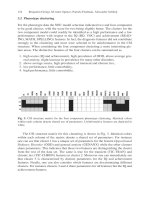
Figure 1 shows the steps of our proposed approach to dendritic spines. In the image acquisition phase, we demonstrated the process for the neuron culture, label, and imaging.
In the second step, we preprocessed the images by reducing
the noise and smoothing the background [24, 25]. Then, we
extracted the dendrite backbone based on the conditional
symmetric analysis and located the dendrite boundary based
on the difference of the pixel intensity. Afterwards, the spines
were detected, classified, and characterized by RMSNN.
2.1. Image Acquisition. The neurons used for imaging in
this paper were cortical neurons, primary cultured from
Embryonic 18th- (E18-) day rat and next cultured until the
22nd day in vitro. Then, the neurons were transfected by
Lipofectamine 2000 and imaged at the 24th day by Leica
SP5 confocal laser scanning microscopy (CLSM) by 63x.
The size of the image is 1024 × 1024, and the resolution
is 0.24 um/pixel at the confocal layer. The images used for
the morphology analysis were obtained by the maximum
intensity projection (MIP) of the original 3D image stack. As
the images were captured as Z-stack series, we projected the
3D image stack onto the 𝑥𝑦, 𝑦𝑧, and 𝑧𝑥 planes, respectively.
Since the slices along the optical direction (𝑧) provided very
limited information and the computation time based on the
3D image stacks is highly increased, it was desired to consider
only the 2D projection onto the 𝑥𝑦 plane. The 2D image
used for analysis was a maximum intensity projection of
Computational and Mathematical Methods in Medicine
3
Image acquisition
phase
Backbone
extraction
Embryonic (E18) rat
Primary cultured
cortical neurons
Dendrite location
phase
Boundary location
Noise reduction,
background
smooth
Transfected (22nd day)
by Lipofectamine
2000
Spine extraction
Spine classification
Imaging (24th day) by
Leica SP5 (CLSM) by
63x
Spine analysis
phase
Spine
characterization
Figure 1: Flowchart of the proposed detection method of the dendritic spines.
the original 3D stack. It was obtained by projecting in the 𝑥𝑦
plane the voxels with maximum intensity values that fall in
the way of parallel rays traced from the viewpoint to the plane
of projection.
We randomly selected 15 different images from Leica SP5
confocal laser scanning microscopy to form the spines library
to test our algorithm. All images contain distinct spines
including mushroom, stubby, and thin types. The typical size
of the image is 1024 × 1024. Most spines in the images are
within a rectangle of 20 × 20 in pixel, but the “thin” spine
is within an about 5 × 20 rectangle in pixel. The spines
have variable gray-level intensities. Spines collected from the
image library were employed to build an image base library.
Spine subimages in the library were taken as samples to
test the classification accuracy of RMSNN. In order to cover
as many cases as possible, the image base library contains
distinct sizes and spines with different orientations.
In order to build the golden-standard spine library, five
experts in the neuroscience field were employed to manually
mark the spines in the collected images and classify the spines
into three predefined categories including “mushroom,”
“stubby,” and “thin” types. For the conflict of the manual
marking, the minority was supposed to be subordinated to
the major. Then according to the marked spines, we computed
the maximum width, length, area, and the center point. The
randomly selected image base library contains about 2700
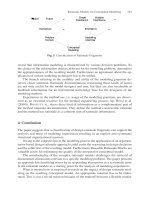
subimage samples, 900 for each type of spines. Figure 2 shows
some image samples in our image base library. As we can see
from the image sample, spines of “mushroom” type contain a
thin neck and head, the stubby type connects directly with the
dendrite without neck, and the thin type is with the smallest
size with only a thin neck and without head.
(a) Mushroom
(b) Stubby
(c) Thin
Figure 2: Samples of the subimages used in the image library.
differential equation (PDE) proposed by Wang et al. [26] to
enhance the image. Figure 3 shows an example of the original
image and the preprocessed result.
2.3. Backbone Extraction Using the Wavelet Transformation Based Conditional Symmetric Analysis. Considering the
attached spines, it is necessary to firstly locate the dendrites in
order to segment the spines from the dendrite. The backbone
extraction and boundary localization are critical for dendritic
spine classification and analysis, which include the following
steps.
Step 1. Remove the noise and small isolated point-set.
Step 2. Locate the backbone of the dendrite.
Step 3. Locate the boundary of the dendrite.
2.2. Image Preprocessing. Considering the limitation of imaging technique, we have employed the 2D median filter to
deal with the noise introduced by the imaging mechanism of
the photomultiplier tubes (PMT) and then used the partial
The backbone is defined as the thinning of the dendrite.
Due to the variance of width of dendrite, attached and
detached spines, it is a challenging task to locate the boundary
4
Computational and Mathematical Methods in Medicine
(a) Original image
(b) Preprocessed image
Figure 3: An example of preprocessed image.
of the dendrite directly from the preprocessed images. Therefore, we have developed a new extraction model utilizing
wavelet transform based conditional symmetric analysis. The
essence of this model is to conduct a local conditional
symmetry analysis of the contour of the region of interest
(ROI) and then compute the center points to produce the
backbone of the dendrite.
Due to the complexity of the dendrites and dendrite
spines’ distribution, we have employed morphological operation to remove the small isolated point-set for the dendrite
in the binary image obtained by local Otsu [27–29] via (1),
which could decrease the disconnection rate of the dendrite
detection:
stands for the partial derivative of 𝑦, respectively. 𝜃(𝑥, 𝑦) is a
low pass filter.
For 𝜑𝑥 (𝑥, 𝑦) and 𝜑𝑦 (𝑥, 𝑦), the scale wavelet transform
(WT) could be written as the following equations:
𝑊𝑥,𝑠 𝑓 (𝑥, 𝑦) = (𝑓 ∗ 𝜑𝑥,𝑠 ) (𝑥, 𝑦) = 𝑠
𝜕
(𝑓 ∗ 𝜃𝑠 ) (𝑥, 𝑦) ,
𝜕𝑥
𝑊𝑦,𝑠 𝑓 (𝑥, 𝑦) = (𝑓 ∗ 𝜑𝑦,𝑠 ) (𝑥, 𝑦)
=𝑠
(3)
𝜕
(𝑓 ∗ 𝜃𝑠 ) (𝑥, 𝑦) .
𝜕𝑦
𝑃
{1,
={
0,
{
more than 𝑛 positive pixels in its 3-by-3 window,
(1)
otherwise,
in which 𝑛 is the threshold of the number of positive pixels.
The value of 𝑛 could be determined by trial and error method
and means that the pixel belongs to the major line if there
are more than 𝑛 positive pixels in its 3 × 3 neighborhood
window. Otherwise, the value of the pixel is forced to be
0, treated as the small isolated point-set. The determination
of the centerline of the dendrite is based on the conditional
symmetric analysis.
The symmetric analysis was accomplished via the wavelet
transform. We have applied the wavelet transform to detect a
pair of contour curves:
𝜑𝑥 (𝑥, 𝑦) =
𝜕
𝑥
,
𝜃 (𝑥, 𝑦) = 𝜙 (√𝑥2 + 𝑦2 )
𝜕𝑥
2
√ 𝑥 + 𝑦2
𝑦
𝜕
𝜑𝑦 (𝑥, 𝑦) =
,
𝜃 (𝑥, 𝑦) = 𝜙 (√𝑥2 + 𝑦2 )
𝜕𝑦
√ 𝑥 2 + 𝑦2
(2)
in which 𝑥 and 𝑦 stand for the coordinate of the contour
curve. 𝜑𝑥 (𝑥, 𝑦) means the partial derivative of 𝑥 and 𝜑𝑦 (𝑥, 𝑦)
Here, 𝜃𝑠 = (1/𝑠2 )𝜃(𝑥/𝑠, 𝑦/𝑠). We can get the modulus of the
gradient vector as
𝑊𝑥,𝑠 𝑓 (𝑥, 𝑦)
),
∇𝑊𝑠 𝑓 (𝑥, 𝑦) = (
𝑊𝑦,𝑠 𝑓 (𝑥, 𝑦)
(4)
2
2
∇𝑊𝑠 𝑓 (𝑥, 𝑦) = √ 𝑊𝑥,𝑠 𝑓 (𝑥, 𝑦) + 𝑊𝑦,𝑠 𝑓 (𝑥, 𝑦) ,
𝐴 𝑠 𝑓 (𝑥, 𝑦) = arctan (
𝑊𝑦,𝑠 𝑓 (𝑥, 𝑦)
𝑊𝑥,𝑠 𝑓 (𝑥, 𝑦)
),
(5)
(6)
where ∇ is the gradient vector and the gradient direction is
given as (6). The contour points (𝑥, 𝑦) are the local maxima of
|∇𝑊𝑠 𝑓(𝑥, 𝑦)| in the direction of 𝐴 𝑠 𝑓(𝑥, 𝑦) at scale 𝑠. However,
the local maxima modulus is not the exact edge point.
We selected (7) as the basis function. We set 𝜑− (𝑥) =
+
−𝜑 (−𝑥) and had 𝜑(𝑥) = 𝜑+ (𝑥) + 𝜑− (𝑥) as the wavelet
function, which had the following properties: gray invariant,
slope invariant, width invariant, and symmetric [29, 30]. The
advantage is to make the extraction of a pair of contours with
accurate protrusions. Consider
Computational and Mathematical Methods in Medicine
5
𝜑+
(1 − 8𝑥2 + 2√1 − 16𝑥2 ) (1 + √1 − 𝑥2 )
1 √
1
2
{
{
−
(4𝑥 ln
( 1 − 16𝑥2 − 3√9 − 16𝑥2 + 8√1 − 𝑥2 )) , 𝑥 ∈ (0, )
{
{
2
2
{
√
𝜋
2𝑥
4
9𝑥 − 8𝑥 + 3 9 − 16𝑥
{
{
{
{
{
{
8𝑥 (1 + √1 − 𝑥2 )
{
1 √
1 3
{
{ 2 (4𝑥 ln
−
𝑥 ∈ [ , ) (7)
(3 9 − 16𝑥2 − 8√1 − 𝑥2 )) ,
= {𝜋
√9 − 16𝑥2
2𝑥
4 4
9
+
3
{
{
{
{
2
√
{
3
{
{ 2 (4𝑥 ln 1 + 1 − 𝑥 − 4 √1 − 𝑥2 ) ,
𝑥 ∈ [ , 1)
{
{
{
𝜋
𝑥
𝑥
4
{
{
{
𝑥 ∈ [1, ∞) .
{0,
The distance between two symmetric points is equal to
the scale of the wavelet transform. If the distance between
two symmetric points is larger than or equal to the width of
regular region, the center point of the symmetric pair can
potentially be located outside of the dendrite. The regular
region is defined as the dendrite is smooth, where the
function has a stable variation along the axis. Thus, we defined
the stable symmetry as follows.
If the scale of wavelet transform is larger than or equal
to the width of regular region, the modulus maxima points
generate two new parallel contours inside the periphery of the
dendrite. All the symmetric pairs of the wavelet transforms
that do not have a counterpart are defined as the unstable
symmetry. In this case, we have considered the width as the
constraint condition. In the direction of the perpendicular to
the gradient direction, we selected the width nearest to the
regular region.
The center of every symmetric pair located on the
centerline of the original regular region of the stroke point.
Finally, the backbone of the regular region was defined by the
curve of all connected symmetric points.
2.4. Boundary Location Based on the Pixel Intensity Difference.
The morphological operation of removing noise blurred
the boundary. Therefore, after localization of backbone, the
boundary of the dendrite was detected via varies of the pixel
intensity of the preprocessed image from Section 2.2. We
can observe that the pixel intensity of the line pixel changes
abruptly at the boundary locations. The boundary location
was performed in two steps. In the first step, we have searched
the image along the two directions perpendicular to the local
line direction until the pixel intensity of the line pixel changed
sharply. We set a threshold for each pixel. The local line
direction is determined as
𝑊𝑦,𝑠 𝑓 (𝑥, 𝑦)
(8)
).
𝐴 𝑠 𝑓 (𝑥, 𝑦) = arctan (
𝑊𝑥,𝑠 𝑓 (𝑥, 𝑦)
The formulation of each pixel is given by (𝛼, 𝐼(𝑝)), in
which 𝐼(𝑝) is the pixel intensity of point 𝑝 in the original
image and 𝛼 is a predefined pixel intensity value, that is,
{𝐼 (𝑝) ≥ 𝛼, p belongs to the line pixel
if {
𝐼 (𝑝) < 𝛼, p does not belong to the line pixel.
{
(9)
In the second step, some boundary points that were not
on the searching path could be missed. The missed boundary
points were detected from the neighboring boundary points.
Provided that there are two known boundary points, if they
are adjacent, there were no other boundary points between
them; otherwise, the method proposed by Tang and You [31]
was used to find the missed points, which can link the two
points into a discrete line with one point as the starting point
and the other one as the ending point.
There are several advantages of our proposed algorithms
for backbone detection and boundary location. (1) The first
are computing efficiency and noise reduction. Our approach
uses less computing time than the method based on the
derivatives of the Gaussian kernel and is more robust when
dealing with the noise. (2) Meanwhile, it reduces the error rate
for misclassifying spine pixels as dendrite pixels and sharply
reduces the disconnection rate, which means our approach is
more robust when dealing with the disturbance information
than other methods, such as NDE proposed by He et al. [22].
2.5. Spine Detection Based on Regularized Morphological
Shared-Weight Neural Network (RMSNN). Considering the
dendritic spine’s structure, we have employed the regularized
morphological shared-weight neural networks for the detection and classification of spines. The regularized morphological shared-weight neural networks consist of two-phase
heterogeneous neural networks in series as shown in Figure 4:
the first phase is for feature extraction and the second phase is
for classification. In the first phase, it is accomplished via the
gray-scale Hit-Miss transform. The feature extraction phase
has multiple feature extraction layers. Each layer is composed
of one or more feature maps. Each feature map is generated
by the Hit-Miss transform with a pair of structure elements
(SEs) from the previous layer and is accompanied by a new
pair of SEs, in which one is for the erosion and the other
one is for the dilation. In the classification stage, it shows
a fully connected Feedforward Neural Network (FNN) [32–
34]. The input of FNN is the direct output of the feature
extraction stage. The output of the classification stage is a
three-node layer, in which each node stands for one type
of spine. Figure 4 shows the structure of the morphological
shared-weight neural network (MSNN) [35]. The MSNN
has been widely applied in the following research fields,
6
Computational and Mathematical Methods in Medicine
Feature map
..
.
Structuring
elements
1
..
.
..
.
..
.
..
.
···
2
3
Input image
..
.
As far as the gray scale is concerned, we assume the image
function as 𝐼 = 𝑓(𝑥, 𝑦), in which 𝑓(𝑥, 𝑦) was the intensity
value of the point (𝑥, 𝑦). Meanwhile, we made the SE 𝑏(𝑥, 𝑦).
The morphological operation can be thought of as a 3D binary
set by way of the umbra transform. The umbra of a 3D surface
function is defined as
𝑈 (𝑓) = {(𝑥, 𝑦, 𝑧) | (𝑥, 𝑦) ∈ 𝐷𝑓 , 𝑧 ≤ 𝑓 (𝑥, 𝑦)} ,
(16)
where we take 𝐷𝑓 as the domain of 𝑓. Then the gray scale
dilation can be defined as
Classification phase
(𝑓 ⊕ 𝑏) (𝑠, 𝑡) = max {𝑓 (𝑠 − 𝑥, 𝑡 − 𝑦)
Feature extraction phase
Figure 4: Structure of morphological shared-weight neural network.
+ 𝑏 (𝑥, 𝑦) | (𝑠 − 𝑥) , (𝑡 − 𝑦) ∈ 𝐷𝑓 ; (𝑥, 𝑦) ∈ 𝐷𝑏 } .
(17)
Meanwhile, erosion is defined as
including laser radar (LADAR), forward-looking infrared
(FLIR), synthetic aperture radar, and visual spectrum image.
The existing research demonstrates that the MSNN is robust
for detection with rotation, image intensity translation, and
occlusion variables [36]. In this paper, we have proposed to
apply the regularized morphological shared-weight neural
network to spine classification.
Dilation is defined as
̂ ∩ 𝐴 ≠ 0} ,
𝐴 ⊕ 𝐵 = {𝑥 | (𝐵)
𝑥
(10)
in which 𝐴 and 𝐵 are sets in 𝑍2 and 𝐵̂ is the reflection of 𝐵.
0 is the empty set. Equation (10) is termed the dilation of 𝐴
by SE 𝐵. Dilation is the reflection of 𝐵 about its origin, then
translated by 𝑥, with the set of all 𝑥, which allow 𝐵̂ to intersect
𝐴 with at least one element.
Erosion is defined as (11) or (12) by the duality of the
erosion-dilation relationship:
𝐴 ⊖ 𝐵 = {𝑥 | (𝐵)𝑥 ⊆ 𝐴} ,
(11)
̂ 𝑐,
𝐴 ⊖ 𝐵 = (𝐴𝑐 ⊕ 𝐵)
(12)
in which 𝐴𝑐 is defined as the complement of 𝐴.
Hit-Miss transform is defined as an operation that detects
a given pattern in a binary image based on a pair of disjoint
structure elements, one for Hit and the other one for Miss.
The result of the Hit-Miss transform is a set of positions,
where the first SE fits in the foreground of the input image
and the second SE misses it completely:
𝑐
𝐴 ⊗ 𝐵 = (𝐴 ⊖ 𝑋) ∩ (𝐴 (𝑊 − 𝑋)) ,
(13)
in which 𝑋 is a SE that consisted from set 𝐵, 𝑊 is an enclosing
window of 𝑋, and (𝑊 − 𝑋) is the local background of 𝑋. By
supposing 𝑋 as 𝐻, the Hit SE, and (𝑊 − 𝑋) as 𝑀, the Miss
SE, we can get
𝐴 ⊗ 𝐵 = (𝐴 ⊖ 𝐻) ∩ (𝐴𝑐 ⊖ 𝑀) ,
(14)
in which 𝐵 = (𝐻, 𝑀) and it can be written as
̂ .
𝐴 ⊗ 𝐵 = (𝐴 ⊖ 𝐻) − (𝐴𝑐 ⊕ 𝑀)
(15)
(𝑓 ⊖ 𝑏) (𝑠, 𝑡) = min {𝑓 (𝑠 + 𝑥, 𝑡 + 𝑦)
− 𝑏 (𝑥, 𝑦) | (𝑠 + 𝑥) , (𝑡 + 𝑦) ∈ 𝐷𝑓 ; (𝑥, 𝑦) ∈ 𝐷𝑏 } .
(18)
The gray scale erosion measures the minimum gap
between the image values 𝑓 and the translated SE values over
the domain of 𝑥. The gray scale dilation is the dual of the
erosion and indirectly measures how well the SEs fit above 𝑓.
The Hit-Miss transform measures how a shape ℎ fits under 𝑓
using erosion and how a shape 𝑚 fits above 𝑓 via dilation. The
high value of Hit-Miss transform means good fit. The gray
scale Hit-Miss transform is independent of shifting in gray
scale.
2.5.1. The Feature Extraction Phase. There are four elements
associated with each layer of feature extraction phase: feature
maps, input, and two structure elements. In the first layer,
the subimage is used as input, and the last layer’s output is
the input of the classification stage. In each feature extraction
layer, a pair of Hit-Miss SEs is shared within all the feature
maps. These SEs are translated as input weights for the feature
map nodes in the feature extraction layer. Table 1 shows the
input parameters and output parameters related to the feature
extraction phase.
According to the above parameters, we can define the HitMiss transform as follows:
netℎ𝑦 = min {𝑎 (𝑥) − 𝑡𝑦ℎ (𝑥)} ,
𝑥∈𝐷𝑡𝑦
̂
𝑚
net𝑚
𝑦 = max {𝑎 (𝑥) − 𝑡𝑦 } ,
𝑥∈𝐷𝑡𝑦
(19)
𝑎𝑦 = netℎ𝑦 − net𝑚
𝑦.
Here, netℎ𝑦 stands for the input for Hit operation in node 𝑦
and ℎ means the Hit operation. net𝑚
𝑦 means the net input for
̂ here mean the Miss
the Miss operation in node 𝑦. 𝑚 and 𝑚
operation and reflection of 𝑚, respectively. 𝑎𝑦 is the result of
Hit-Miss transform performed at node 𝑦. The learning rule
Computational and Mathematical Methods in Medicine
7
For the output layer nodes, 𝑤𝑘𝑗 stands for the connection
strength to node 𝑘 from node 𝑗:
Table 1: Parameters of the feature extraction phase.
Parameter
𝑎(𝑥)
𝑡𝑦 (𝑥)
Input
𝑡𝑦ℎ (𝑥𝑦 )
𝑡𝑦𝑚 (𝑥)
𝑤𝑦𝑚 (𝑥)
𝑎𝑦
The input to a node 𝑦 from node 𝑥
Connections associating the node 𝑦 with
node x
Hit SE associating node 𝑦 with node 𝑥
Miss SE associating node 𝑦 with 𝑥
𝑤𝑦ℎ (𝑥)
Output
Definition
Weight for Hit SE node 𝑦 with 𝑥
The output of node 𝑦
for the Hit and Miss SE is derived based on the gradient
decent as
𝜕netℎ𝑦
𝜕𝑡𝑦ℎ (𝑥)
Δ𝑡𝑦𝑚̂ = −𝜂𝛿𝑦
,
(20)
𝜕net𝑚
𝑦
𝜕𝑡𝑦𝑚̂ (𝑥)
,
where 𝜂 is the learning rate of the network and 𝛿𝑦 is expressed
as
𝛿𝑦 = 𝛿 (𝑦) = ∑ 𝑘 𝛿𝑘 𝑤𝑘 (𝑦) .
𝛿𝑦 = 𝛿 (𝑦) = ∑ 𝑘 𝛿𝑘 (
𝜕𝑎 (𝑦)
−
𝜕net𝑚
𝑦
𝜕𝑎 (𝑦)
),
(22)
in which 𝑘 is the node in the layer next to the node 𝑦.
Based on the back-propagation of error from the classification stage with these learning rules, the MSNN learns the
optimized SE to extract the features by each set of Hit-Miss
transforms. Consider
1
2
𝐸 = ∑ (𝑡𝑜 − 𝑂𝑜 ) .
2 𝑜
(23)
Here, 𝑡𝑜 stands for the target node output and 𝑂𝑜 the actual
node output:
𝑂𝑗 = 𝑓 (net𝑗 ) ,
net𝑗 = ∑𝑤𝑗𝑖 𝑂𝑖 + Δ 𝑗 ,
(24)
in which 𝑤𝑗𝑖 is the connection weight strength to node 𝑗 from
node 𝑖 and Δ 𝑗 is the bias output for node 𝑗. 𝑤𝑗𝑖 is typically
learned by the back-propagation of error. The update rule
of connecting weight for each connection is expressed as
follows:
𝜕𝐸
= 𝜂𝛿𝑗 𝑂𝑗 .
𝜕𝑤𝑘𝑗
𝛿𝑗 = 𝑓 (net𝑗 ) ∑𝛿𝑘 𝑤𝑗𝑖 .
(27)
2.5.2. The Classification Phase. The classification phase takes
the output directly from the last feature extraction layer as
its input. The parameters used for the classification phase are
predefined in the feature extraction phase. There are three
output nodes for the classification stage of our algorithm,
indicating which type of spines the subimage contains.
2.5.3. Acceleration of the MSNN Based on the Regularization.
In order to accelerate the learning rate and decrease the
learning epochs, we employed the regularization factor.
Regularization is used to reduce near-zero connection weight
value to zero, therefore reducing the complexity of the
network. It is defined as
𝑅 (𝑤) = 𝐸𝑠 (𝑤) + 𝜆𝐸𝑐 (𝑤) ,
𝐸𝑐 (𝑤) =
∑
∀𝑤 in network 1
(𝑤𝑖 /𝑤𝑜 )
2
2
+ (𝑤𝑖 /𝑤0 )
,
(28)
where 𝐸𝑠 (𝑤) is the performance measure of the learning
algorithm, the total network error, and 𝐸𝑐 (𝑤) is the complexity penalty of the network model. 𝜆 is the regularization
factor. 𝑤0 is a predefined parameter. Meanwhile, research
shows that a network with proper SEs produces better result
[36]. Therefore, it is essential to choose the suitable SEs. In
this paper, according to the average size of spine and the
comparison result in Table 3, we defined the SE as a disk with
the radius of 4 pixels.
For the training procedure, the RMSNN takes the subimage as the input and makes one output value for each image.
For the testing procedure, our proposed algorithm scans the
whole ROI and generates an image named the detection
plane, which is based on the outputs from the target class
nodes.
3. Experimental Evaluation
𝑖
Δ𝑤𝑗𝑖 = −𝜂
and for the hidden layer nodes,
(21)
Equation (21) is for the top level or final extraction layer.
𝛿𝑦 for the lower layers of multiple-layer feature extraction is
expressed as
𝜕netℎ𝑦
(26)
𝑘
Weight for Miss SE node 𝑦 with 𝑥
Δ𝑡𝑦ℎ = 𝜂𝛿𝑦
𝛿𝑗 = (𝑡𝑗 − 𝑂𝑗 ) 𝑓 (net𝑗 )
(25)
3.1. Experiment Design. We have trained neural networks
with the back-propagation algorithm. The subimages were
submitted to the input nodes of the neural network. The error
of the output was propagated through all the connections. The
process repeated until the network converged to a stable state
with required MSE. When the MSE approximated to a preset
value or the maximum epoch was achieved, the algorithm
converged and the training would stop. During the training,
the RMSNN took each subimage as the input and produced
one output value for each of the three categories. Figure 2(a)
shows the samples of subimages containing mushroom type
8
spine. Figure 2(b) shows the samples of the subimages containing the stubby type, and Figure 2(c) shows the samples of
thin type subimage.
In the training step, the subimage samples were input
to the network sequentially. The median-squared error was
employed to measure the training effectiveness. For each
subimage, the RMSNN produced one output value, which
indicated the type of spine in the subimage. Then, we scanned
the entire microscopy image and finally generated a detection
plane according to the output nodes of RMSNN.
In order to test the classification accuracy, we randomly
selected 900 samples for each type of spine, respectively.
Following common convention and ease of stratified cross
validation, 10 × 10-fold stratified cross validation (CV) was
used for the dataset to perform an unbiased statistical
analysis. The RMSNN was constructed in the form as two
feature extraction layers, one hidden layer with ten hidden
neurons and one output layer with three neurons. The input
subimage size was 20 by 20 pixels, and the size of the structure
elements was with the radius of 4 pixels. The initial weight was
in the range of [−1.0, 1.0]. The learning rate was set to 0.0015.
The maximum training epoch was predefined as 15000. The
expected output values for mushroom, stubby, and thin type
spines were [1 0 0], [0 1 0], and [0 0 1].
3.2. Experiment Results
3.2.1. Backbone Extraction. The extraction result is shown in
Figure 5. Figure 5(a) shows the original image. Figure 5(b)
shows the extracted backbone, of which the width covers
merely one pixel.
3.2.2. Boundary Location. Figure 6(a) shows the mark of the
located backbone of the dendrite based on the original image,
and Figure 6(b) shows the marked boundary of the dendrite
after the backbone is extracted. Figure 6(c) shows the marked
dendrite that determines the starting point of the spine.
3.2.3. Spine Analysis. Figure 7 shows a ROI of our sample
image, and Figure 7(b) shows the detection result of the
spines. The backbone is marked by the purple color and the
boundary is marked by the red color. The spines are marked
by their periphery of blue color.
Figure 8(a) shows the original image with the marked
region of interest. Figure 8(b) shows the classification result
based on the features extracted in the first phase. The corresponding SE gets respect features around each pixel, but it is
blind for readers to understand which features are obtained.
The detected spines contain 8 mushroom types, 8 stubby
types, and 4 thin types. The average of the classification
accuracy of RMSNN is shown in Table 2 based on the 2700
samples in total. We can find that the detection of the
mushroom and thin types has better performance than the
stubby type. It is because the stubby type seems connected
with the major lines, and the neck of the spine is blurred.
Figures 8(c), 8(d), and 8(e) demonstrate partial geometric
attributes of the spines, including the area, perimeter, and
width. We found that the areas of the spines of the ROI ranged
within [10, 23] and the perimeter ranged within [8, 88].
Computational and Mathematical Methods in Medicine
Table 2: Average of the classification accuracy on a 10-by-10 CV.
Spine types
Mushroom
Stubby
Thin
Mushroom
99.1%
0.7%
0.2%
Stubby
1.3%
97.6%
1.1%
Thin
1.1%
0.3%
98.6%
3.3. Optimal Parameter in SE. According to [36], unsuitable
SEs will degrade the performance of the RMSNN; hence,
it is critical to choose the proper SEs. According to the
average size of the spines as 20 by 20 pixels, we selected SEs
with different sizes and shapes to test the performance. The
comparison of classification accuracies based on the 2700
samples is shown in Table 3. We can find that the disk with
a radius of 4 pixels reaches the best performance. Therefore,
we finally defined the SEs as a disk with the radius of 4 pixels.
3.4. Algorithm Comparison. To further validate the efficacy
of our proposed approach, we have compared the proposed
algorithm with Cheng et al.’s method [18] and the manual
method. In Cheng et al.’s paper, the authors employed the
adaptive threshold to segment the image and Chen and
Molloi’s algorithm [37] to extract the backbone and then used
the local SNR for the detection of the detached spine and local
spine morphology for the detection of the attached spines.
The comparison results based on ROI1 in Figure 8 and 15
images collected in our database are shown in Table 4. It is
found from Figure 9 that Cheng et al.’s method missed some
small protrusions whose number of pixels is more than 5.
The number of detected spines via our algorithm is 19, 13
by Cheng et al.’s method, and 20 via the manual method as
shown in Table 4. Cheng et al.’s method is robust at dealing
with the spines detached from the dendrite but weak at spines
attached with the dendrite. However, the detached spines
from the dendrite are caused by the deconvolution to denoise
the image. Our proposed algorithm overcomes the problem
of detecting attached spines.
4. Discussion
In this paper, we have proposed new algorithms using conditional symmetric analysis and regularized morphological
shared-weight neural network to detect and analyze the
dendrite and dendritic spines.
Figure 5 shows that backbone extraction result based on
the conditional symmetry analysis. Compared to the secondorder directional derivatives method in [14], our proposed
algorithms reduced the computation time of linking the
breaking point of the backbone.
Figure 6 shows the result of the marked backbone and
the boundary of the dendrite, which is used to determine the
starting point of the spines.
Table 2 shows the classification result of the different
types of spines. The row in Table 2 stands for the actual class
and the column in Table 2 stands for the predicted class.
The “mushroom” type has an obvious head and thin neck.
The “stubby” type lacks obvious neck, and the “thin” type
lacks obvious head. In Table 2, the detection accuracy of
Computational and Mathematical Methods in Medicine
9
Table 3: Classification accuracy by different SEs (unit is in pixel, bold denotes the best, 𝑟 is radius, and 𝑤 is width).
Mushroom
Stubby
Thin
Disk (𝑟 = 5)
98.7%
96.2%
94.3%
Disk (𝑟 = 4)
99.1%
97.6%
98.6%
Disk (𝑟 = 3)
95.4%
94.1%
96.2%
(a) Original image
Square (𝑤 = 3)
85.3%
87.2%
79.1%
Square (𝑤 = 4)
89.2%
91.2%
75.3%
(b) Extracted backbone
Figure 5: Backbone extraction result.
(a) Centerline of the dendrite
(b) Boundary of the dendrite
(c) Dendrite
Figure 6: Dendrite location results.
(a)
(b)
Figure 7: (a) ROI of the original Image. (b) Detection result of the spines.
Table 4: Detection result of ROI1 in Figure 8 and 15 images in our
database.
Methods
ROI1
15 images
Manual
20
2021
ALS [18]
13
1750
SRMSNN (proposed)
19
1987
the mushroom type is higher than the other two types, and
part of the stubby type is misclassified into mushroom and
thin types as its head and neck ratio is at the level of average. A
percent of 1.1 of thin spines are misclassified into mushroom
type and 0.3% into stubby type, which is caused by the similar
size of the head and neck. Table 4 shows the result of detected
spines of Figure 8, respectively, by manual, ALS [18], and
our proposed method SRMSNN. The results demonstrate
10
Computational and Mathematical Methods in Medicine
(a) Original image
(b) Detection plane
Width
25
20
200
15
150
10
100
5
50
0
0
2
4
6
8
10
Area
250
12
14
16
18
0
0
2
(c) Histogram of the width distribution
4
6
8
10
12
14
16
18
(d) Histogram of the area distribution
Perimeter
90
80
70
60
50
40
30
20
10
0
0
2
4
6
8
10
12
14
16
18
(e) Histogram of the perimeter distribution
Figure 8: Experiment result with corresponding parameters for characterization.
that our algorithm has better performance than the other
two methods for the images obtained by the confocal laser
scanning microscopy.
5. Conclusion
In this paper, we proposed a new automatic approach to
accurately identify dendritic spines with different shapes.
The novelty of this approach includes (1) a new model using
wavelet-based conditional symmetry analysis for dendrite
backbone extraction and localization, which is the first step
towards identification of dendritic spins; (2) a new algorithm
based on regularized morphological shared-weight neural
networks for classification of spines into the right classes
(i.e., mushroom, stubby, and thin), entitled “RMSNN.” This
research was based on our collected microscopy images. We
Computational and Mathematical Methods in Medicine
11
(a) ALS [18]
(b) SRMSNN
Figure 9: Detection result based on ALS and SRMSNN.
have applied our approach to image base library containing
around 2700 subimage samples, 900 for each type of spines,
and have compared the proposed method with the existing
methods. The experimental results demonstrate that our
algorithm outperforms existing methods with a significant
improvement in accuracy in terms of classifying spines into
the different spine categories. The classification accuracy is
99.1% for mushroom spines, 97.6% for stubby spines, and
98.6% for thin spines.
The future work will be focusing on further validation
of the robustness of the algorithms through collecting more
samples and testing on different datasets. A user-friendly
interface will be also built for usability improvement and
enhancement. Meanwhile, we will be focusing on reducing
the computation time while improving the classification
accuracy based on the 3D image stacks. Other feature
extraction tools (such as wavelet packet analysis [38], wavelet
entropy [39], and 3D-DWT [40]) and other advanced classification tools [41, 42] will be tested. Besides, swarm intelligence
method will be used to find optimal parameters [43].
[5]
[6]
[7]
[8]
[9]
[10]
Conflict of Interests
The authors declare that there is no conflict of interests
regarding the publication of this paper.
[11]
Acknowledgment
This work was financially supported by the National Natural
Science Foundation of China (no. 61271231).
References
[1] J. L. Krichmar, S. J. Nasuto, R. Scorcioni, S. D. Washington,
and G. A. Ascoli, “Effects of dendritic morphology on CA3
pyramidal cell electrophysiology: a simulation study,” Brain
Research, vol. 941, no. 1-2, pp. 11–28, 2002.
[2] D. Johnston and S. M.-S. Wu, Foundations of Cellular Neurophysiology, MIT Press, Cambridge, Mass, USA, 1995.
[3] Z. F. Mainen and T. J. Sejnowski, “Influence of dendritic
structure on firing pattern in model neocortical neurons,”
Nature, vol. 382, no. 6589, pp. 363–366, 1996.
[4] N. Keren, N. Peled, and A. Korngreen, “Constraining compartmental models using multiple voltage recordings and genetic
[12]
[13]
[14]
[15]
algorithms,” Journal of Neurophysiology, vol. 94, no. 6, pp. 3730–
3742, 2005.
B. Van Calster, D. Timmerman, C. Lu et al., “Preoperative diagnosis of ovarian tumors using Bayesian kernel-based methods,”
Ultrasound in Obstetrics and Gynecology, vol. 29, no. 5, pp. 496–
504, 2007.
K. M. Stiefel and T. J. Sejnowski, “Mapping function onto
neuronal morphology,” Journal of Neurophysiology, vol. 98, no.
1, pp. 513–526, 2007.
S. Wang, H. Pan, C. Zhang, and Y. Tian, “RGB-D image-based
detection of stairs, pedestrian crosswalks and traffic signs,”
Journal of Visual Communication and Image Representation, vol.
25, no. 2, pp. 263–272, 2014.
S. K. Schmitz, J. J. J. Hjorth, R. M. S. Joemai et al., “Automated
analysis of neuronal morphology, synapse number and synaptic
recruitment,” Journal of Neuroscience Methods, vol. 195, no. 2,
pp. 185–193, 2011.
T. M. Liu, G. Li, J. X. Nie et al., “An automated method for cell
detection in zebrafish,” Neuroinformatics, vol. 6, no. 1, pp. 5–21,
2008.
W. Yu, H. K. Lee, S. Hariharan, W. Bu, and S. Ahmed,
“Evolving generalized voronoi diagrams for accurate cellular
image segmentation,” Cytometry Part A, vol. 77, no. 4, pp. 379–
386, 2010.
M. K. Bashar, K. Komatsu, T. Fujimori, and T. J. Kobayashi,
“Automatic extraction of nuclei centroids of mouse embryonic
cells from fluorescence microscopy images,” PLoS ONE, vol. 7,
no. 5, Article ID e35550, 2012.
J. L. Martiel, A. Leal, L. Kurzawa et al., “Measurement of cell
traction forces with ImageJ,” in Methods in Cell Biology, E. K.
Paluch, Ed., vol. 125, chapter 15, pp. 269–287, Academic Press,
2015.
D. L. Dickstein, A. Rodriguez, A. B. Rocher et al., “NeuronStudio: an automated quantitative software to assess changes in
spine pathology in Alzheimer models,” Alzheimer’s & Dementia,
vol. 6, no. 4, article S410, 2010.
E. Meijering, M. Jacob, J.-C. F. Sarria, P. Steiner, H. Hirling, and
M. Unser, “Design and validation of a tool for neurite tracing
and analysis in fluorescence microscopy images,” Cytometry
Part A, vol. 58, no. 2, pp. 167–176, 2004.
J. Cheng, X. B. Zhou, B. L. Sabatini, and S. T. C. Wong, “NeuronIQ: a novel computational approach for automatic dendrite
spines detection and analysis,” in Proceedings of the IEEE/NIH
Life Science Systems and Applications Workshop (LISA ’07), pp.
168–171, IEEE, Bethesda, Md, USA, November 2007.
12
[16] I. Y. Y. Koh, W. B. Lindquist, K. Zito, E. A. Nimchinsky, and
K. Svoboda, “An image analysis algorithm for dendritic spines,”
Neural Computation, vol. 14, no. 6, pp. 1283–1310, 2002.
[17] X. Y. Xu, J. Cheng, R. M. Witt, B. L. Sabatini, and S. T. C. Wong,
“A shape analysis method to detect dendritic spine in 3D optical
microscopy image,” in Proceedings of the 3rd IEEE International
Symposium on Biomedical Imaging: From Nano to Macro, pp.
554–557, Arlington, Va, USA, April 2006.
[18] J. Cheng, X. Zhou, E. Miller et al., “A novel computational
approach for automatic dendrite spines detection in twophoton laser scan microscopy,” Journal of Neuroscience Methods,
vol. 165, no. 1, pp. 122–134, 2007.
[19] J. Fan, X. Zhou, J. G. Dy, Y. Zhang, and S. T. C. Wong, “An
automated pipeline for dendrite spine detection and tracking of
3D optical microscopy neuron images of in vivo mouse models,”
Neuroinformatics, vol. 7, no. 2, pp. 113–130, 2009.
[20] Y. Zhang, X. B. Zhou, R. M. Witt, B. L. Sabatini, D. Adjeroh,
and S. T. C. Wong, “Dendritic spine detection using curvilinear
structure detector and LDA classifier,” NeuroImage, vol. 36, no.
2, pp. 346–360, 2007.
[21] F. Janoos, K. Mosaliganti, X. Xu, R. Machiraju, K. Huang, and
S. T. C. Wong, “Robust 3D reconstruction and identification
of dendritic spines from optical microscopy imaging,” Medical
Image Analysis, vol. 13, no. 1, pp. 167–179, 2009.
[22] T. He, Z. Xue, and S. T. C. Wong, “A novel approach for three
dimensional dendrite spine segmentation and classification,” in
Medical Imaging 2012: Image Processing, vol. 8314 of Proceedings
of SPIE, San Diego, Calif, USA, February 2012.
[23] P. Shi, Y. Huang, and J. Hong, “Automated three-dimensional
reconstruction and morphological analysis of dendritic spines
based on semi-supervised learning,” Biomedical Optics Express,
vol. 5, no. 5, pp. 1541–1553, 2014.
[24] S. Reid, C. Lu, I. Casikar et al., “Prediction of pouch of Douglas
obliteration in women with suspected endometriosis using a
new real-time dynamic transvaginal ultrasound technique: the
sliding sign,” Ultrasound in Obstetrics & Gynecology, vol. 41, no.
6, pp. 685–691, 2013.
[25] S. Reid, C. Lu, I. Casikar et al., “The prediction of pouch of
Douglas obliteration using offline analysis of the transvaginal
ultrasound ‘sliding sign’ technique: inter-and intra-observer
reproducibility,” Human Reproduction, vol. 28, no. 5, pp. 1237–
1246, 2013.
[26] Y.-H. Wang, W.-N. Liu, A.-H. Chen, and Y. Wang, “Nonlinear
dim target enhancement algorithm based on partial differential
equation,” Journal of Dalian Maritime University, vol. 34, no. 2,
pp. 57–60, 2008.
[27] L. Chen, J. H. Zhang, S. Y. Chen, Y. Lin, C. Y. Yao, and J.
W. Zhang, “Hierarchical mergence approach to cell detection
in phase contrast microscopy images,” Computational and
Mathematical Methods in Medicine, vol. 2014, Article ID 758587,
10 pages, 2014.
[28] N. Otsu, “A threshold selection method from gray-level histograms,” IEEE Transactions on Systems, Man and Cybernetics,
vol. 9, no. 1, pp. 62–66, 1979.
[29] P.-S. Liao, T.-S. Chen, and P.-C. Chung, “A fast algorithm for
multilevel thresholding,” Journal of Information Science and
Engineering, vol. 17, no. 5, pp. 713–727, 2001.
[30] L. H. Yang, X. You, R. M. Haralick, I. T. Phillips, and Y. Y. Tang,
“Characterization of Dirac edge with new wavelet transform,” in
Proceedings of the 2nd International Conference on Wavelets and
Applications, vol. 1, pp. 872–878, Hong Kong, December 2001.
Computational and Mathematical Methods in Medicine
[31] Y. Y. Tang and X. G. You, “Skeletonization of ribbon-like shapes
based on a new wavelet function,” IEEE Transactions on Pattern
Analysis and Machine Intelligence, vol. 25, no. 9, pp. 1118–1133,
2003.
[32] Y. D. Zhang, S. H. Wang, G. L. Ji, and P. Phillips, “Fruit
classification using computer vision and feedforward neural
network,” Journal of Food Engineering, vol. 143, pp. 167–177, 2014.
[33] S. Wang, Y. Zhang, Z. Dong et al., “Feed-forward neural
network optimized by hybridization of PSO and ABC for
abnormal brain detection,” International Journal of Imaging
Systems and Technology, vol. 25, no. 2, pp. 153–164, 2015.
[34] G. Yang, Y. Zhang, J. Yang et al., “Automated classification
of brain images using wavelet-energy and biogeography-based
optimization,” Multimedia Tools and Applications, 2015.
[35] D. Guo, Y. Zhang, Q. Xiang, and Z. Li, “Improved radio
frequency identification indoor localization method via radial
basis function neural network,” Mathematical Problems in
Engineering, vol. 2014, Article ID 420482, 9 pages, 2014.
[36] X. Jin and C. H. Davis, “Vehicle detection from high-resolution
satellite imagery using morphological shared-weight neural
networks,” Image and Vision Computing, vol. 25, no. 9, pp. 1422–
1431, 2007.
[37] Z. Chen and S. Molloi, “Automatic 3D vascular tree construction
in CT angiography,” Computerized Medical Imaging and Graphics, vol. 27, no. 6, pp. 469–479, 2003.
[38] Y. Zhang, Z. Dong, S. Wang, G. Ji, and J. Yang, “Preclinical
diagnosis of magnetic resonance (MR) brain images via discrete
wavelet packet transform with tsallis entropy and generalized
eigenvalue proximate support vector machine (GEPSVM),”
Entropy, vol. 17, no. 4, pp. 1795–1813, 2015.
[39] Y. Zhang, S. Wang, P. Sun et al., “Pathological brain detection
based on wavelet entropy and Hu moment invariants,” BioMedical Materials and Engineering, vol. 26, supplement 1, pp.
S1283–S1290, 2015.
[40] Y. Zhang, S. Wang, P. Phillips, Z. Dong, G. Ji, and J. Yang,
“Detection of Alzheimer’s disease and mild cognitive impairment based on structural volumetric MR images using 3DDWT and WTA-KSVM trained by PSOTVAC,” Biomedical
Signal Processing and Control, vol. 21, pp. 58–73, 2015.
[41] S. Wang, Y. Zhang, G. Ji, J. Yang, J. Wu, and L. Wei, “Fruit classification by wavelet-entropy and feedforward neural network
trained by fitness-scaled chaotic ABC and biogeography-based
optimization,” Entropy, vol. 17, no. 8, pp. 5711–5728, 2015.
[42] Y. Zhang, Z. Dong, P. Phillips et al., “Detection of subjects
and brain regions related to Alzheimer’s disease using 3D MRI
scans based on eigenbrain and machine learning,” Frontiers in
Computational Neuroscience, vol. 9, article 66, 15 pages, 2015.
[43] S. Wang, X. Yang, Y. Zhang, P. Phillips, J. Yang, and T.-F. Yuan,
“Identification of green, oolong and black teas in China via
wavelet packet entropy and fuzzy support vector machine,”
Entropy, vol. 17, no. 10, pp. 6663–6682, 2015.
Hindawi Publishing Corporation
Computational and Mathematical Methods in Medicine
Volume 2015, Article ID 571381, 19 pages
/>
Review Article
An Overview of Biomolecular Event Extraction from
Scientific Documents
Jorge A. Vanegas,1 Sérgio Matos,2 Fabio González,1 and José L. Oliveira2
1
MindLab Research Laboratory, Universidad Nacional de Colombia, Bogot´a, Colombia
DETI/IEETA, University of Aveiro, Campus Universit´ario de Santiago, 3810-193 Aveiro, Portugal
2
Correspondence should be addressed to S´ergio Matos;
Received 13 May 2015; Revised 10 August 2015; Accepted 18 August 2015
Academic Editor: Chuan Lu
Copyright © 2015 Jorge A. Vanegas et al. This is an open access article distributed under the Creative Commons Attribution
License, which permits unrestricted use, distribution, and reproduction in any medium, provided the original work is properly
cited.
This paper presents a review of state-of-the-art approaches to automatic extraction of biomolecular events from scientific texts.
Events involving biomolecules such as genes, transcription factors, or enzymes, for example, have a central role in biological
processes and functions and provide valuable information for describing physiological and pathogenesis mechanisms. Event
extraction from biomedical literature has a broad range of applications, including support for information retrieval, knowledge
summarization, and information extraction and discovery. However, automatic event extraction is a challenging task due to the
ambiguity and diversity of natural language and higher-level linguistic phenomena, such as speculations and negations, which
occur in biological texts and can lead to misunderstanding or incorrect interpretation. Many strategies have been proposed in the
last decade, originating from different research areas such as natural language processing, machine learning, and statistics. This
review summarizes the most representative approaches in biomolecular event extraction and presents an analysis of the current
state of the art and of commonly used methods, features, and tools. Finally, current research trends and future perspectives are also
discussed.
1. Introduction
The scientific literature is the most important medium for
disseminating new knowledge in the biomedical domain.
Thanks to advances in computational and biological methods, the scale of research in this domain has changed remarkably, reflected in an exponential increase in the number of
scientific publications [1]. This has made it harder than ever
for scientists to find, manage, and exploit all relevant studies
and results related to their research field [1]. Because of this,
there is growing awareness that automated exploitation tools
for this kind of literature are needed [2]. To address this
need, natural language processing (NLP) and text mining
(TM) techniques are rapidly becoming indispensable tools
to support and facilitate biological analyses and the curation
of biological databases. Furthermore, the development of
this kind of tools has enabled the creation of a variety
of applications, including domain-specific semantic search
engines and tools to support the creation and annotation
of pathways or for automatic population and enrichment of
databases [3–5].
Initial efforts in biomedical TM focused on the fundamental tasks of detecting mentions of entities of interest
and linking these entities to specific identifiers in reference knowledge bases [6, 7]. Although entity normalization
remains an active research challenge, due to the high level
of ambiguity in entity names, some existing tools offer
performance levels that are sufficient for many information
extraction applications [6]. In recent years there has been
increased interest in the identification of interactions between
biologically relevant entities, including, for instance, drugdrug [8] or protein-protein interactions (PPIs) [9]. Amongst
these, the identification of PPIs mentioned in the literature
has received most attention, encouraged by their importance
in systems biology and by the necessity to accelerate the
population of numerous PPI databases.
Following the advances achieved in PPI extraction, it
became relevant to automatically extract more detailed
descriptions of protein related events that depict protein characteristics and behavior under certain conditions.
Such events, including expression, transcription, localization,
2
Computational and Mathematical Methods in Medicine
Theme
Cause
Theme
Protein
TNF-alpha
Pos. Reg.
is a rapid
activator
Gene
of
IL-8
Expression
gene
expression by. . .
Figure 1: Example of complex biomolecular event extracted from a text fragment. A recursive structure, composed of two types of events, is
presented: Positive Regulation and Expression.
binding, or regulation, among others, play a central role in
the understanding of biological processes and functions and
provide insight into physiological and pathogenesis mechanisms. Automatically creating structured representations of
these textual descriptions allows their use in information
retrieval and question answering systems, for constructing
biological networks composed of such events [2] or for
inferring new associations through knowledge discovery.
Unfortunately, extraction of this kind of biological information is a challenging task due to several factors: firstly, the
biological processes described are generally complex, involving multiple participants which may be individual entities
such as genes or proteins, groups, or families, or even other
biological processes; sentences describing these processes are
long and in many cases have long-range dependencies; and,
finally, biological text is also rich in higher level linguistic
phenomena, such as speculation and negation, which may
cause misinterpretation of the text if not handled properly
[1, 9].
This review summarizes the different approaches used
to address the extraction and formalization of biomolecular events described in scientific texts. The downstream
impact of these advances, namely, for network extraction,
for pharmacogenomics studies, and in systems biology
and functional genomics, has been highlighted in recent
reviews [2, 4, 10], which have also described various enduser systems developed on top of these technologies. This
review focuses on the methodological aspects, describing
the available resources and tools as well as the features,
algorithms, and pipelines used to address this information
extraction task, and specifically for protein related events,
which have received the most attention in this perspective.
We present and discuss the most representative methods
currently available, describing the advantages, disadvantages, and specific characteristics of each strategy. The most
promising directions for future research in this area are also
discussed.
The contents of this paper are organized as follows: we
start by introducing biomolecular events and defining the
event extraction task; we then describe the event extraction
steps, present commonly used frameworks, text processing,
and NLP tools and resources, and compare the different
approaches used to address this task; in the following section
we compare the performance of the proposed methods and
systems, followed by a discussion regarding the most relevant
aspects; finally, we present some concluding remarks in the
last section.
2. Biomolecular Events
In the biomedical domain, an event refers to the change of
state of one or more biomedical entities, such as proteins,
cells, and chemicals [11]. In their textual description, an
event is typically referenced through a trigger expression that
specifies the event and indicates its type. These triggers are
generally verbal forms (e.g., “stimulates”) or nominalizations
of verbs (e.g., “expression”) and may occur as a single word or
as a sequence of words. This textual description also includes
the entities involved in the event, referred to as participants,
and possibly additional information that further specifies the
event, such as a particular cell type in which the described
event was observed. Biomolecular events may describe the
change of a single gene or protein, therefore having only
one participant denoting the affected entity, or may have
multiple participants, such as the biomolecules involved in
a binding process, for example. Additionally, an event may
act as participant in a more complex event, as in the case
of regulation events, requiring the detection of recursive
structures.
Extraction of event descriptions from scientific texts has
attracted substantial attention in the last decade, namely,
for those events involving proteins and other biomolecules.
This task requires the determination of the semantic types of
the events, identifying the event participants, which may be
entities (e.g., proteins) or other events, their corresponding
semantic role in the event, and finally the encoding of this
information using a particular formalism. This structured
definition of events is associated with an ontology that
defines the types of events and entities, semantic roles, and
also any other attributes that may be assigned to an event.
Examples of ontologies for describing biomolecular events
include the GENIA Event Ontology [11] and Gene Ontology
[12].
Figure 1 presents an example of a complex event described
in the text fragment “TNF-alpha is a rapid activator of IL-8
gene expression by. . ..” From this fragment we can construct
a recursive structure composed of two events: a first event, of
type Expression denoted by the trigger word “expression” that
has a single argument (“IL-8”) with the role Theme (denoting
that this is the participant affected by the event), and a second
event of type Positive Regulation, defined by the trigger
word “activator.” This second event has two participants: the
protein “TNF-alpha” with the role Cause (defining that this
protein is the cause of the event) and the first event with the
role Theme.
Computational and Mathematical Methods in Medicine
1
3
Preprocessing and feature extraction
Syntactic parsing
Dependency parsing Phrase structure
and deep parsing
Gdep parser [13]
Stanford parser
Charniak-Johnson/
[17]
McClosky [14, 15] Enju-GENIA [18]
Bikel parser [16]
ERG [19]
2
Gimli [27]
NERSuite [28]
AIIAGMT [29]
GNAT [30]
GeNo [31]
Tools
Chemical
Gene protein and
disorders
BioEnEx [35]
SCAI [32]
ChemSpot [33] BANNER [36]
ABNER [37]
Neji [34]
Classification
LibSVM [44]
SVM-multiclass [45]
LIBLINEAR [46]
Mallet [47]
CRF++ [48]
SVM
CRF
BioThesaurus [38]
BioLexicon [39]
UMLS [40]
LexEBI [41]
Lexicons
BioLexicon [39]
UMLS [40]
WordNet [49]
Edge detection
SVM
5
Lexicons
Trigger detection
Tools
TrigNER
[42]
Zhang et al.
[43]
4
Frameworks
NLTK [22]
Stanford
CoreNLP [23]
OpenNLP [24]
GATE [25]
U-compare [26]
Entity recognition
Gene and protein
3
Tools
ISimp [20]
GENIA
tagger [21]
Classification
LibSVM [44]
SVM-multiclass [45]
LIBLINEAR [46]
Postprocessing
Tools
Stanford CoreNLP [23]
SVM-rank [50]
Figure 2: Overall pipeline of a biomedical event extraction solution. Joint prediction methods merge steps 3 and 4 in a single step. The
corresponding reference paper for each tool and method is also identified [13–50].
3. Event Extraction
Figure 2 illustrates a common event extraction pipeline, identifying the most popular tools, models, and resources used in
each stage. The two initial stages are usually preprocessing
and feature extraction, followed by the identification of
named entities. The next step is to perform event detection.
This step is frequently divided into two separate stages:
trigger detection, which consists of the identification of
event triggers and their type, and edge detection (or event
construction), which is focused on associating event triggers
with their arguments. Some authors, on the other hand,
4
Computational and Mathematical Methods in Medicine
have addressed event detection in a single, joint prediction
step. These approaches tackle the cascading errors that occur
with the two-stage methods and have commonly shown
improved performance. Finally, a postprocessing stage is
usually present, to refine and complete the candidate event
structures. Negation or speculation detection may also be
included in this final step. This section describes each phase,
presenting the most commonly used approaches.
the GENIA event corpus and with in-domain and out-ofdomain lexical resources.
3.1. Corpora for Event Extraction. The development and
improvement of information extraction systems usually
requires the existence of manually annotated text collections,
or corpora. This is mostly true for supervised machine
learning methods, but annotated data can also be exploited
for inferring patterns to be used in rule-based approaches. In
the case of biomedical event extraction, various corpora have
been compiled, including corpora annotated with proteinprotein interactions.
3.2. Preprocessing and Feature Extraction. Preprocessing is
a required step in any text mining pipeline. This includes
reading the data from its original format to an internal representation, and extracting features, which usually involves
some level of text or language processing. In the specific
case of event extraction, preprocessing may also involve
resolving coreferences [59] or applying some form of sentence
simplification [60], for example, by expanding conjunctions,
in order to improve the extraction results.
3.1.1. GENIA Event Corpus. The GENIA Event corpus contains human-curated annotations of complex, nested, and
typed event relations [51, 52]. The GENIA corpus [53]
is composed of 1,000 paper abstracts from Medline. It
contains 9,372 sentences from which 36,114 events are
identified. This corpus is provided by the organizers of
BioNLP shared task to participants as the main resource
for training and evaluation and is publicly available online
( />
3.2.1. Preprocessing Tools
3.1.2. BioInfer Corpus. BioInfer (Biomedical Information
Extraction Resource) ( [54] is
a public resource providing manually annotated corpus and
related resources for information extraction in the biomedical domain.
The corpus contains sentences from abstracts of biomedical research articles annotated for relationships, named
entities, and syntactic dependencies. The corpus is annotated
with proteins, genes, and RNA relationships and serves as
a resource for the development of information extraction
systems and their components such as parsers and domain
analyzers. The corpus is composed of 1100 sentences from
abstracts of biomedical research articles.
3.1.3. Gene Regulation Event Corpus. The Gene Regulation
Event Corpus (GREC) ( />[55] consists of 240 MEDLINE abstracts, in which events
relating to gene regulation and expression have been annotated by biologists. This corpus has the particularity that
not only core relations between entities that are annotated,
but also a range of other important details about these
relationships, for example, location, temporal, manner, and
environmental conditions.
3.1.4. GeneReg Corpus. The GeneReg Corpus [56] consists of
314 MEDLINE abstracts containing 1770 pairwise relations
denoting gene expression regulation events in the model
organism E. coli. The corpus annotation is compatible with
3.1.5. PPI Corpora. Although not as richly annotated as
event corpora, protein-protein interaction corpora may be
considered for complementing the available training data.
The most relevant PPI corpora are the LLL corpus [57], the
AIMed corpus [58], and the BioCreative PPI corpus [7].
Frameworks. In order to derive a feature representation from texts, it is necessary to perform text processing involving a set of common NLP tasks, going
from sentence segmentation and tokenization, to part-ofspeech tagging, chunking, and linguistic parsing. Various
text processing frameworks exist that support these tasks,
among which the following stand out: NLTK (http://www
.nltk.org/), Apache OpenNLP ( />and Stanford CoreNLP ( />corenlp.shtml) (Figure 2).
Syntactic Parsers. A syntactic parser assigns a tree or graph
structure to a free text sentence. These structures establish
relations or dependencies between the organizing verb and
its dependent arguments and have been useful for many
applications like negation detection and disambiguation
among others. Syntactic parsers can be categorized in three
groups: dependency parsers, phase structure parsers, and
deep parsers [61]. The aim of dependency parsers is to
compute a tree structure of a sentence where nodes are
words, and edges represent the relations among words; phrase
structure parsers focus on identifying phrases and their
recursive structure, and deep parsers express deeper relations
by computing theory-specific syntactic/semantic structures.
For the task of event extraction several implementations of
each parser groups have been used, as shown in Figure 2.
3.2.2. Features. One of the main requirements of a good event
extraction system is a rich feature representation. Most event
extraction systems present a complex set of features extracted
from tokens, sentences, dependency parsing trees, and external resources. Table 1 summarizes the features commonly
extracted in this processing stage and indicates their use in
the event extraction process.
(i) Token-based features capture specific knowledge
regarding each token, such as syntactic or linguistic features, namely, part-of-speech (POS) and