ĐIỆN TÂM ĐỒ CẤP CỨU ( ECG EMERGENCY)
Bạn đang xem bản rút gọn của tài liệu. Xem và tải ngay bản đầy đủ của tài liệu tại đây (7.44 MB, 89 trang )
ECG cấp cứu
phạm ngọc minh
Group cập nhật kiến thức y khoa
/>ups/Medical.VN.update/
ECGs for the Emergency Physician
ECGs for the Emergency Physician
Amal Mattu
Director, Emergency Medicine Residency Program, Co-Director, Emergency Medicine/Internal Medicine
Combined Residency Program, University of Maryland School of Medicine, Baltimore, Maryland, USA
William Brady
Associate Professor, Vice Chair, and Program Director, Department of Emergency Medicine
University of Virginia Health System, Charlottesville, Virginia, USA
and
Medical Director, Charlottesville-Albermarle Rescue Squad, Charlottesville, Virginia, USA
BMJ Publishing Group 2003
BMJ Books is an imprint of the BMJ Publishing Group
All rights reserved. No part of this publication may be reproduced, stored in a retrieval system, or
transmitted, in any form or by any means, electronic, mechanical, photocopying, recording and/or
otherwise, without the prior written permission of the publishers.
First published in 2003
by BMJ Books, BMA House, Tavistock Square,
London WC1H 9JR
www.bmjbooks.com
British Library Cataloguing in Publication Data
A catalogue record for this book is available from the British Library
ISBN 0 7279 1654 8
Typeset by SIVA Math Setters, Chennai, India
Printed and bound in Spain by GraphyCems, Navarra
Contents
Foreword ...................................................................................................................................................................vii
Preface .......................................................................................................................................................................ix
Dedications.................................................................................................................................................................xi
Part 1
Case histories ..............................................................................................................................................................3
ECG interpretations and comments...........................................................................................................................53
Part 2
Case histories ............................................................................................................................................................77
ECG interpretations and comments.........................................................................................................................129
Appendix A: Differential diagnoses........................................................................................................................152
Appendix B: Commonly used abbreviations...........................................................................................................154
Index.......................................................................................................................................................................155
5
Foreword
There has been a great need for a user friendly ECG text that fills the void between an introductory text designed for
students and an advanced reference source for cardiologists. “ECGs for the Emergency Physician” fills this void. It is an
ECG teaching and reference textbook for acute and emergency care physicians written by two specialists practicing and
teaching acute and emergency care.
Drs Mattu and Brady have created an ECG text that facilitates self instruction in learning the basics, as well as the
complexities, of ECG interpretation. They know that ECG interpretation requires knowledge, insight and practice. They
know “the eye does not see, what the mind does not know.” In order to accomplish this goal of teaching ECG
interpretation, they have divided their book into two parts. In Part I, as the authors state, are the “bread and butter”
ECGs of acute care. These are the ECG findings that form the core knowledge necessary for accurate ECG
interpretation. In Part II they teach recognition of more subtle ECG abnormalities, which when mastered, allow the
practitioner to become an expert.
The beauty of this text lies in the combining of a collection of emergency department ECGs with the authors’ insights
and expert observations. This book has great utility as a reference text, a bound ECG teaching file, a board review aide
or a resident in emergency medicine’s best friend for learning the art of advanced ECG interpretation. Its greatest value
however, is for all of us who want to be both challenged and taught by 200 great electrocardiograms and their
interpretations.
May the forces be with you.
Corey M Slovis
Professor of Emergency Medicine and Medicine
Chairman, Department of Emergency Medicine
Vanderbilt Medical Center
Nashville, Tennessee
Medical Director Metro Nashville Fire EMS
7
Preface
Emergency and other acute care physicians must be experts in the use and interpretation of the 12-lead
electrocardiogram (ECG). We have prepared this text with this basic though highly important thought in mind. This text
represents our effort to further the art and science of electrocardiography as practiced by emergency physicians and
other acute care clinicians.
A significant number of the patients managed in the emergency department and other acute care settings present with
chest pain, cardiovascular instability, or complaints related to the cardiovascular system. The known benefits of early,
accurate diagnosis and rapid, appropriate treatment of cardiovascular emergencies have only reinforced the importance
of physician competence in electrocardiographic interpretation. The physician is charged with the responsibility of rapid,
accurate diagnosis followed by appropriate therapy delivered expeditiously. This evaluation not infrequently involves the
performance of the 12-lead ECG. For example, the patient with chest pain presenting with ST-segment elevation, acute
myocardial infarction must be rapidly and accurately evaluated so that appropriate therapy is offered in prompt fashion.
Alternatively, the hemodynamically unstable patient with atrioventricular block similarly must be cared for in a rapid
manner. In these instances as well as numerous other scenarios, resuscitative and other therapies are largely guided by
information obtained from the ECG.
The electrocardiogram is used frequently in the emergency department (ED) and other acute care settings; numerous
presentations may require a 12-lead electrocardiogram. For instance, the most frequent indication for ECG performance
in the ED is the presence of chest pain; other complaints include dyspnea and syncope. Additional reasons for obtaining
an ECG in the ED include both diagnosis-based (acute coronary syndrome, suspected pulmonary embolism, and the
“dysrhythmic” patient) and system-related indications (for the “rule-out myocardial infarction” protocol, for admission
purposes, and for operative clearance).1 Regardless of the cause, the physician must be an expert in the interpretation
of the 12-lead ECG. Interpretation of the ECG is as much an art as it is a science. Accurate ECG interpretation requires
a sound knowledge of the electrocardiogram, both the objective criteria necessary for various diagnoses of those
patients encountered in the ED as well as a thorough grasp of the various electrocardiographic waveforms and their
meaning in the individual patient.
We have prepared this text for the physician who manages patients not only in the ED but also in other acute care
settings – whether it be in the office, the hospital ward, critical care unit, the out-of-hospital arena, or other patientcare locale. We have used actual ECGs from patients treated in our EDs; a brief but accurate history has also been
provided in each instance. In certain cases, the history may provide a clue to the diagnosis yet in other situations the
clinical information will have no relationship to the final diagnosis – as is the case in the ED. We have made an effort
to choose the most appropriate ECG from each patient, but as occurs in “real ED,” some of the ECGs are imperfect:
the evaluation is hindered by artifact, incomplete electrocardiographic sampling, etc. We have also provided the ECGs
in a random fashion, much the way actual patients present to the emergency department. We have endeavored to
reproduce the reality of the ED when the reader uses this text to expand their knowledge of the 12-lead electrocardiogram
and how it relates to patient care.
The reader is advised to read the clinical history provided for each ECG and then, much as the clinician would interpret
the electrocardiogram in the ED, review the 12-lead ECG. After a clinically focused review of the ECG, the reader is then
able to review the interpretation. This ECG text has been constructed in two basic sections. The first half of the text
contains ECGs that we feel represent the “bread and butter” of emergency electrocardiography – the core material with
which we feel that the acute care physician must be thoroughly familiar. These ECGs were chosen because they represent
common electrocardiographic diagnoses that all emergency physicians should know. This section is prepared primarily
for the physician-in-training (for example, the emergency medicine resident) though practicing physicians will also benefit
from reviewing the material. The second half of the text is composed of ECGs that are more challenging. The
9
ECGs FOR THE EMERGENCY PHYSICIAN
electrocardiographic diagnoses are more difficult to establish and will often be on subtle findings. In some cases, the
ECGs in this section were chosen not necessarily because of the related level of difficulty but because of subtle teaching
points found, which are likely to be beyond the level of the physician-in-training.
It is also crucial to understand that this text is not intended for the “beginner in ECG interpretation”. The text, in
essence an electrocardiographic teaching file, is intended for the physician who possesses a sound, basic
understanding of electrocardiography yet desires additional practice and review – a review which is highly clinically
pertinent. The electrocardiography beginner is advised to begin by reading through one of the many outstanding books
that have previously been written for novice students prior to studying this teaching file.
One last point must also be stressed to the reader of this text. Diagnostic criteria for various electrocardiographic
diagnoses vary somewhat amongst authors. Therefore, in an effort to standardize the interpretations used in this text,
we chose to use the following two references as the “gold standard” for electrocardiographic interpretations: Chou
and Knilans’ Electrocardiography in Clinical Practice: Adult and Pediatric and Galen’s Marriott’s Practical
Electrocardiography.2,3
References
1.
Brady W, Adams M, Perron A, Martin M. The impact of the 12-lead electrocardiogram in the evaluation of the emergency department
patient. Ann Emerg Med (accepted for publication/publication pending).
2.
Chou T-C, Knilans TK. Electrocardiography in Clinical Practice: Adult and Pediatric 4th edn. Philadelphia, PA: WB Saunders Company, 1996.
3.
Galen SW. Marriott’s Practical Electrocardiography 10th edn. Philadelphia, PA: Lippincott Williams & Wilkins, 2001.
x
Dedications
This work is dedicated to my wife, Sejal, for her tremendous patience and never-ending support; to my son, Nikhil, for
constantly reminding me of the priorities in life; to the Emergency Department staff at Mercy Medical Center in
Baltimore for their friendship and their ECG contributions; to the faculty and residents of the University of Maryland
Emergency Medicine Residency Program for providing the main inspiration for this work; to Mary Banks and BMJ Books
for supporting and believing in this work; to Dr Bill Brady for his mentorship, friendship, and commitment to teaching
and education; and to emergency physicians around the world – may your dedication to learning continue to strengthen
our specialty and improve patient care.
Amal Mattu
Director, Emergency Medicine Residency Program
Co-Director, Emergency Medicine/Internal Medicine Combined Residency Program
University of Maryland School of Medicine
Baltimore, Maryland
USA
I would like to thank my wife, King, for her love, support, wise counsel, and patience – none of this effort would be
possible without her; my children, Lauren, Anne, Chip, and Katherine, for being wonderful and my primary inspiration;
my parents, Bill and Joann Brady, for all that they have done and continue to do; the Emergency Medicine Residents
(past, present, and future) at the University of Virginia, for their hard work, astronomical dedication, and inspiration –
all directed at our patients and the specialty of Emergency Medicine; Dr Marcus Martin, Chair of Emergency Medicine
at the University of Virginia, for his support, guidance, and mentorship; and my co-author, Dr Amal Mattu, for his
dedicated effort on this book in particular and his dedication to Emergency Medicine education in general – a true
gentleman, talented clinician, and distinguished scholar.
William Brady
Associate Professor, Vice Chair, and Program Director
Department of Emergency Medicine
University of Virginia Health System
Charlottesville, Virginia
USA
and
Medical Director, Charlottesville-Albermarle
Rescue Squad, Charlottesville, Virginia,
USA
1
1
Part 1
1
ECGs FOR THE EMERGENCY PHYSICIAN
Case histories
I
aVR
V1
V4
II
aVL
V2
V5
III
aVF
V3
V6
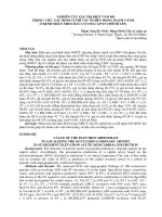
1. bệnh nhân nữ 45 tuổi không triệu chứng
I
aVR
V1
V4
II
aVL
V2
V5
III
aVF
V3
V6
2. bệnh nhân nam 24 tuổi đau ngực sau tập tạ
3
3
ECGs FOR THE EMERGENCY PHYSICIAN
I
aVR
V1
V4
II
aVL
V2
V5
III
aVF
V3
V6
I
aVR
V1
V4
II
aVL
V2
V5
III
aVF
V3
V6
II
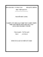
3. bệnh nhân nam 76 tuổi khó thở
II
4. bênh nhân nam 64 tuổi khơng triệu chứng
4
4
PART 1:ECGs
CASEFOR
HISTORIES
THE EMERGENCY PHYSICIAN
I
aVR
V1
V4
II
aVL
V2
V5
III
aVF
V3
V6
II
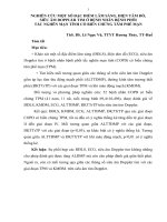
5. bệnh nhân nữ 48 tuổi đau đầu nhẹ khi đi bộ, gần đây mới dùng thuốc điều trị THA
I
aVR
V1
V4
II
aVL
V2
V5
III
aVF
V3
V6
II
6. bn nam 79 tuổi sau điều trị tiêu huyết khối do nmct cấp được 45 phút, hiện tại vẫn đang đau ngực
5
5
PART 1:ECGs
CASEFOR
HISTORIES
THE EMERGENCY PHYSICIAN
I
aVR
V1
V4
II
aVL
V2
V5
III
aVF
V3
V6
7. bn nam 43 tuỏi không triệu chứng
I
aVR
V1
V4
II
aVL
V2
V5
III
aVF
V3
V6
II
8. bn nam 82 tuổi gần đây có tăng liều beta blocker hiện tại đang chóng mặt
6
6
PART 1:ECGs
CASEFOR
HISTORIES
THE EMERGENCY PHYSICIAN
I
aVR
V1
V4
II
aVL
V2
V5
III
aVF
V3
V6
II
9. bn nam 49 tuổi đau ngực từng cơn
I
aVR
V1
V4
II
aVL
V2
V5
III
aVF
V3
V6
10. bn nam 65 tuổi nghiện thuốc lá đang điều trị tràn khí màng phổi nặng
7
7
PART 1:ECGs
CASEFOR
HISTORIES
THE EMERGENCY PHYSICIAN
I
aVR
V1
V4
II
aVL
V2
V5
III
aVF
V3
V6
II
11. bn nữ 54 tuổi đau giữa ngực và chóng mặt
I
aVR
V1
V4
II
aVL
V2
V5
III
aVF
V3
V6
II
12. bn nữ 86 tuổi mệt mỏi toàn thân
8
8
PART 1:ECGs
CASEFOR
HISTORIES
THE EMERGENCY PHYSICIAN
I
aVR
V1
V4
II
aVL
V2
V5
III
aVF
V3
V6
II
13. bn nam 61 tuổi hồi hộp chóng mặt
I
aVR
V1
V4
II
aVL
V2
V5
III
aVF
V3
V6
II
14. bệnh nhân nữ 44 tuổi liên tục có cơn hồi hộp đ á n h t r ố n g n g ự c
9
9
PART 1:ECGs
CASEFOR
HISTORIES
THE EMERGENCY PHYSICIAN
I
aVR
V1
V4
II
aVL
V2
V5
III
aVF
V3
V6
II
15. bệnh nhân nữ 24 tuổi có thai, 3 ngày nay nôn liên tục
I
aVR
V1
V4
II
aVL
V2
V5
III
aVF
V3
V6
II
16. bn nam 37 tuổi đau màng phổi
1
0
10
PART 1:ECGs
CASEFOR
HISTORIES
THE EMERGENCY PHYSICIAN
I
aVR
V1
V4
II
aVL
V2
V5
III
aVF
V3
V6
II
17. bn nam 63 tuổi chóng mặt, hồi hộp
I
aVR
V1
V4
II
aVL
V2
V5
III
aVF
V3
V6
II
18. bn nam 33 tuổi béo phì, đau chói ở ngực và khó thở
1
1
11
PART 1:ECGs
CASEFOR
HISTORIES
THE EMERGENCY PHYSICIAN
I
aVR
V1
V4
II
aVL
V2
V5
III
aVF
V3
V6
II
19. bn nữ 81 tuổi hồi hộp, mệt mỏi tồn thân
I
aVR
V1
V4
II
aVL
V2
V5
III
aVF
V3
V6
20. bn nam 61 tuổi khơng triệu chứng
1
2
12
PART 1:ECGs
CASEFOR
HISTORIES
THE EMERGENCY PHYSICIAN
I
aVR
V1
V4
II
aVL
V2
V5
III
aVF
V3
V6
II
21. bn nữ 57 tuổi đau ngực nhẹ và hồi hộp
I
aVR
V1
V4
II
aVL
V2
V5
III
aVF
V3
V6
II
22. bn nam 75 tuổi ho, khó thở và thở khò khè
1
3
13