Advances in Biomimetics Part 11 pot
Bạn đang xem bản rút gọn của tài liệu. Xem và tải ngay bản đầy đủ của tài liệu tại đây (8.98 MB, 35 trang )
Advances in Biomimetics
342
Integrating twice
And given the no slip condition at the boundaries
And
Adding equations to solve for C
2
Substituting to solve for C
1
Biomimetics in Bone Cell Mechanotransduction:
Understanding Bone’s Response to Mechanical Loading
343
The equation takes the form
The volume flow rate (Q) may be determined by integrating the velocity (u) over the flow
chamber’s cross-sectional area
Since wall shear stress is defined as
Advances in Biomimetics
344
Upon substituting back
2. References
Ajubi NE, Klein-Nulend J, Nijweide PJ, Vrijheidlammers T, Albas MJ, Burger EH (1996)
Pulsating fluid flow increases prostaglandin production by cultured chicken
osteocytes: a cytoskeleton-dependent process. Biochem Biophy Res Commun
225:62-68.
Balls MM (1976) Organ culture in biomedical research: festschrift for Dame Honor Fell,
London: Cambridge University Press.
Barrett LA, Trump BF (1978) Maintaining human aortas in long-term organ culture. Meth
Cell Sci 4(13):861-862.
Beno T, Yoon Y, Cowin SC, Fritton SP (2006) Estimation of bone permeability using accurate
microstructural measurements. J Biomech 39(13):2378-2387.
Billiau A, Edy VG, Heremans H, Van Damme J, Desmyter J, georgiades JA, De Somer P
(1977) Human interferon: mass production in a newly established cell line, MG-63.
Antimicrob Agents Chemother 12:11-15.
Bonewald LF (2007) Osteocytes as dynamic multifunctional cells. Ann NY Acad Sci
1116:281-290.
Botchwey EA, Dupree MA, Pollack SR, Levine EM, Laurencin CT (2003a) Tissue engineered
bone: measurement of nutrient transport in three-dimensional matrices. J Biomed
Mater Res, 67A(1): 357-367.
Botchwey EA, Pollack SR, El-Amin S, Levine EM, Tuan RS, Laurencin CT (2003b) Human
osteoblast-like cells in three-dimensional culture with fluid flow. Biorheology
40:299-306.
Bottlang M, Simnacher M, Schmidt H, Brand RA, Claes L (1997) A cell strain system for
small homogenous strain applications. Biomedizinische Technik 42:305-309.
Boyd JD (2005) Embryology in war-time Britain. Anat Rec 87(1):91-97.
Biomimetics in Bone Cell Mechanotransduction:
Understanding Bone’s Response to Mechanical Loading
345
Brown TD, Bottlang M, Pedersen DR, Banes AJ (1998) Loading paradigms – intentional and
unintentional – for cell culture mechanostimulus. Amer J Med Sci 316:162-
168.;1359-1364.
Brown TD (2000) Techniques for mechanical stimulation of cells in vitro: a review. J Biomech
33(1):3-14.
Buhl KM, Jacobs CR, Turner RT, Evans GL, Farrell PA, Donahue HJ (2001) Aged bone
displays an increased responsiveness to low-intensity resistance exercise. J Appl
Physiol 90
Chan ME, Lu XL, Huo B, Baik AD, Chiang V, Guldberg RE, Lu HH, Guo XE (2009) A
trabecular bone explants model of osteocyte-osteoblast co-culture for bone
mechanobiology. Cell Molec Bioeng 2(3):405-415.
Cowin SC (1999) Bone poroelasticity. J Biomech 32(3):217-238.
Currey JD (2009) Measurement of the mechanical properties of bone. A recent history, Clin
Orthop Relat Res 467:1948-1954.
Dallas SL, Zaman G, Pead MJ, Lanyon LE (1993) Early strain-related changes in cultures
embryonic chick tibiotarsi parallel those associated with adaptive modeling in vivo.
J Bone Miner Res 8(3):251-259.
Davies CM, Jones DB, Stoddart MJ, Koller K, Smith E, Archer CW, Richards RG (2006)
Mechanically loaded ex vivo bone culture system ‘Zetos’: systems and culture
preparation. Eur Cell Mater 11:57-75.
Del Rizzo DF, Moon MC, Werner JP, Zahradka P (2001) A novel organ culture method to
study intimal hyperplasia at the site of a coronary artery bypass. Ann Thorac Surg
71:1273-1279.
Ewart JL, Cohen MF, Meyer RA, Huang GY, Wessels A, Gourdie RG, Chin AJ, Park SMJ,
Lazatin BO, Villabon S, Lo CW (1997) Heart and neural tube defects in transgenic
mice overexpressing the Cx43 gap junction gene. Development 124:1281-1292.
Fell HB (1972) Tissue culture and its contribution to biology and medicine. J Exper Biol 57:1-
13.
Fell HB, Balls M, Monnickendam MA (1976) Organ culture in biomedical research.
Cambridge University Press, Cambridge.
Forrest SM, NG KW, Findlay DM, Michelangeli VP, Livesey SA, Partridge NC, Zajac JD,
Martin TJ (1985) Characterizqation of an osteoblast-like clonal cell line which
responds to both parathyroid hormone and calcitonin. Calcif Tissue Int 37:51-56.
Frangos JA, Eskin SG, McIntire LV, Ives CL (1985) Flow effects on prostacyclin production
by cultured human endothelial cells. Science 227:1477-1479.
Frangos JA, McIntire LV, Eskin SG (1988) Shear stress induced stimulation of mammalian
cell metabolism. Biotechnol Bioeng 32:1053-1060.
Garrett R (2003) Assessing bone formation using mouse calvarial organ cultures. In:Helfrich
MH, Ralston SH (eds) Bone research protocols, chap 14, Humana Press, Totowa.
Gay CV (1991) Avian osteoclasts. Calcif Tissue Int 49:153-154.
Glucksmann A (1942) The role of mechanical stresses in bone formation in vitro. J Anat
76:231-239.
Gross TS, Srinivasan S, Liu CC, Clemens TL, Bain SD (2002) Non-invasive loading of the
murine tibia: an in vivo model for the study of mechanotransduction. J Bone Miner
Res 17(3):493-501.
Advances in Biomimetics
346
Hagino H, Kuraoka M, Kameyama Y, Okano T, Teshima R (2005) Effect of a selective
agonist for prostaglandin E receptor subtype EP4 (ONO-4819) on the cortical bone
response to mechanical loading. Bone 36(3):444-453.
Hillam RA, Skerry TM (1995) Inhibition of bone resorption and stimulation of formation by
mechanical loading of the modeling rat ulna in vivo. J Bone Miner Res 10(5):683-689.
Hung CT, Pollack SR, Reilly TM, Brighton CT (1995) Real-time calcium response of cultured
bone cells to fluid flow. Clin Orthop Relat Res 313:256-269.
Hung CT, Allen FD, Pollack SR, Brighton CT (1996) Intracellular Ca2+ stores and
extracellular Ca2+ are required in the real-time Ca2+ response of bone cells
experiencing fluid flow. J Biomech 29:1411-1417.
Imamura K, Ozawa H, Hiraide T, Takahashi N, Shibasaki Y, Fukuhara T, Suda T (1990)
Continuously applied compressive pressure induces bone resorption by a
mechanism involving prostaglandin E2 synthesis. J Cell Physiol 144:222-228.
Ishizeki K, Takigawa M, Harada Y, Suzuki F, Nawa T (1995) Meckel’s cartilage
chondrocytes in organ culture synthesize bone-type proteins accompanying
osteocytic phenotype expression. Anat Embryol 185:421-430.
Jacobs CR, Yellowley CE, Davis BR, Zhou Z, Donahue HJ (1998) Differential effect of steady
versus oscillating flow on bone cells. J Biomech 31:969-976.
Jee WS, Ueno K, Deng YP, Woodbury DM (1985) The effects of prostaglandin E2 in growing
rats: increased metaphyseal hard tissue and cortico-endosteal bone formation.
Calcif Tissue Int 37:148-157.
Jones DB, Broeckmann E, Pohl T, Smith EL (2003) Development of a mechanical testing and
loading system for trabecular bone studies for long term culture. Eur Cell Mater
5:48-60.
Jubb RW (1979) Effect of hyperoxia on articular tissues in organ culture. Ann Rheum Dis
38(3):279-286.
Kato Y, Windle JJ, Koop BA, Mundy GR, Bonewald LF (1997) Establishment of an osteocyte-
like cell line, MLO-Y4. J Bone Miner Res 12:2014-2023.
Klein-Nulend J, Burger EH, Semeins CM, Raisz LG, Pilbeam CC (1997) Pulsating fluid flow
stimulates prostaglandin release and inducible prostaglandin G/H synthase
mRNA expression in primary mouse bone cells. J Bone Miner Res 12:45-51.
Lauritzen C, Munro IR, Ross RB (1985) Classification and treatment of hemifacial
microsomia. Scand J Plast Reconstr Surg 19:33–39.
Lyubimov EV, Gotleib AI (2004) Smooth muscle cell growth monolayer and aortic organ
culture is promoted by a nonheparin binding endothelial cell-derived soluble
factors. Cardiovas Pathol 13(3):139-145.
Meghji S, Hill PA, Harris M (1998) Bone organ cultures. In:Henderson B, Arnett T (eds)
Methods in bone biology, chap 4, Thomson Science, New York.
Merrick AF, Shewring LD, Cunningham SA, Gustafsson K, Fabre JW (1997) Organ culture of
arteries for experimental studies of vascular endothelium in situ. Transpl Immunol
5(1):3-7.
Merrilees MJ, Scott L (1982) Organ culture of rat carotid artery: maintenance of
morphological characteristics and of pattern of matrix synthesis. In vitro 18(11):900-
910.
Biomimetics in Bone Cell Mechanotransduction:
Understanding Bone’s Response to Mechanical Loading
347
Mikic B, Battaglia TC, Taylor EA, Clark RT (2002) The effect of growth/differentiation
factor-5 deficiency on femoral composition and mechanical behavior in mice. Bone
30(5):733-737.
Murray JE, Mulliken JB, Kaban LB, Belfer M (1979) Twenty-year experience in
maxillocraniofacial surgery. An evaluation of early surgery and growth, function
and body image. Ann Surg 190:320–331.
Murrills RJ (1996) In vitro bone resorption assays. In: Bilezekian JP, Raisz LG, Rodan GA
(eds) Principles of bone biology, chap 90, Academic Press, San Diego.
Nicholson GC, Moseley JM, Sexton PM, Martin TJ (1987) Chicken osteoclasts do not possess
calcitonin receptors. J Bone Miner Res 2(1):53-9.
Owan I, Burr DB, Turner CH, Qui J, Tu Y, Onyia JE, Duncan RL (1997)
Mechanotransduction in bone: osteoblasts are more responsive to fluid forces than
mechanical strain. Amer J Physiol Cell Physiol 273 (3 Pt 1), C810-C815.
Piekarski K, Munro M (1977) Transport mechanism operating between blood supply and
osteocytes in long bones. Nature 269:80-82.
Ponten J, Saksela E (1967) Two established in vitro cell lines from human mesenchymal
tumours. Int J Cancer 2:434-447.
Pruzansky S (1969) Not all dwarfed mandibles are alike. Birth Defects 1:120.
Raisz L (1965) Bone resorption in tissue culture. Factors influencing the response to
parathyroid hormone. J Clin Inv 44:103-116.
Raisz L, Niemann I (1967) Early effects of PTH and thyrocalcitonin in bone organ culture.
Nature 214:486-488.
Reich KM, Gay CV, Frangos JA (1990) Fluid shear stress as a mediator of osteoblast cyclic
adenosine monophosphate production. J Cell Physiol 143:100-104.
Reynolds JJ (1976) Organ cultures of bone: studies on the physiology and pathology of
resorption. In: Balls M, Monnickendam M (eds) Organ culture in biomedical
research. Cambridge University Press, Cambridge, pp 355-366.
Rubin CT, Lanyon LE (1984) Regulation of bone formation by applied dynamic loads. J Bone
Joint Surg 66A:397-402.
Rubin CT, Lanyon LE (1985) Regulation of bone mass by mechanical strain magnitude.
Calcif Tissue Int 37:411-417.
Saunders MM, You J, Trosko JE, Yamasaki H, Donahue HJ, Jacobs CR (2001) Gap junctions
and fluid flow in MC3T3-E1 cells. Am J Physiol Cell Physiol 281(6):1917-1925.
Saunders MM, You J, Zhou Z, Li Z, Yellowley CE, Kunze E, Jacobs CR, Donahue HJ (2003)
Fluid-flow induced prostaglandin E2 response of osteoblastic ROS 17/2.8 cells is
gap junction-mediated and independent of cytosolic calcium. Bone 32(4):350-356.
Saunders MM, Donahue HJ (2004) Development of a cost-effective loading machine for
biomechanical evaluation of mouse transgenic models. Med Eng Phys 26:595-603.
Saunders MM, Taylor AF, Du C, Zhou Z, Pellegrini VD Jr, Donahue HJ (2006) Mechanical
stimulation effects on functional end effectors in osteoblastic MG-63 cells. J
Biomech 39(8):1419-1427.
Saunders MM, Simmerman LA, Reed GL, Sharkey NA, Taylor AF (2010) Biomimetic bone
mechanotransduction modeling in neonatal rat femur organ cultures: Structural
verification of proof of concept. Biomech Model Mechanobiol 9:539-550.
Sidman JD, Sampson D, Templeton B (2001) DO of the mandible for airway obstruction in
children. Laryngoscope 111(7):1137-1146.
Advances in Biomimetics
348
Sorkin AM, Dee KC, Knothe Tate ML (2004) “Culture shock” from the bone cell’s
perspective: emulating physiological conditions for mechanobiological
investigations. Am J Cell Physiol Cell Physiol 287:C1527-C1536.
Stepita-Klauco M, Dolezalova H (1968) Organ culture of skeletal muscle subjected to
intermittent activity. Biomed Lif Sci 24(9):971
Swanson N, Javed Q, Hogrefe K, Gershlick A (2002) Human internal artery organ culture
model of coronary stenting: a novel investigation of smooth muscle cell response to
drug-eluting stents. Clin Sci 103(4):347-353.
Takahashi M, Chernin MI, Yamamoto O, Tonzetich J, Kinsey CG, Novak JF (2002)
Transformation of MC3T3-E1 cells following stress and transfection with pSV2neo
plasmid. Anticancer Res 22(2A):585-598.
Takai E, Mauck RL, Hung CT, Guo XE (2004) Osteocyte viability and regulation of
osteoblast function in a 3D trabecular bone explants under dynamic hydrostatic
pressure. J Bone Miner Res 19(9):1403-1410.
Takezawa T, Inoue M, Aoki S, Sekiguchi M, Wada K, Anazawa H, Hanai N (2000) Concept
of organ engineering: a reconstruction method of rat liver for in vitro culture. Tiss
Eng 6(6):641-650.
Taylor AF, Saunders MM, Shingle D, Cimbala JM, Zhou Z, Donahue HJ (2007) Osteocytes
communicate fluid flow-mediated effects to osteoblasts altering phenotype. Am J
Physiol Cell Physiol 292:C545-C552.
Turner CH, Akhter MP, Raab DM, Kimmel DB, Recker RR (1991) A noninvasive in vivo
model for studying strain adaptive bone remodeling. Bone 12:73-79.
Voisard R, v Eicken J, Baur R, Gschwend JE, Wendroth U, Kleinschmidt K, Hombach V,
Hoher M (1999) A human arterial organ culture model of postangioplasty
restenosis: results up to 56 days after ballooning. Atherosclerosis 144(1):123-134.
Wang L, Ciani C, Doty SB, Fritton SP (2004) Delineating bone's interstitial fluid pathway in
vivo. Bone 34(3):499-509.
Weinbaum S, Cowin SC, Zheng YA (1994) A model for the excitation of osteocytes by
mechanical loading induced bone fluid shear stresses. J Biomech 27:339-360.
Weiss A, Livne E, von der Mark K, Heinegard D, Silbermann M (1988) Growth and repair of
cartilage: organ culture system utilizing chondroprogenitos cells of condylar
cartilage in newborn mice. J Bone Miner Res 3(1):93-100.
Wetzel DM, Salpeter MM (1991) Fibrillation and accelerated A Ch R degradation in long-
term muscle organ culture. Muscle Nerve 14(10):1003-1012.
Wong SY, Dunstan CR, Evans RA, Hills E (1982) The determination of bone viability: a
histochemical method for identification of lactate dehydrogenase activity in
osteocytes in fresh calcified and decalcified sections of human bone. Pathology
14(4):439-442.
You J, Yellowley CE, Donahue HJ, Zhang Y, Chen Q, Jacobs CR (2000) Substrate deformation
levels associated with routine physical activity are less stimulatory to bone cells
relative to loading-induced oscillatory fluid flow. J Biomech Eng 122:387-393.
Zaman G, Dallas SL, Lanyon LE (1992) Cultured embryonic bone shafts show osteogenic
responses to mechanical loading. Calcif Tissue Int 51(2):132-136.
Ziambaras K, Lecanda F, Steinberg TH, Civitelli R (1998) Cyclic stretch enhances gap
junctional communication between osteoblastic cells. J Bone Miner Res 13:218-228.
17
Novel Biomaterials with Parallel Aligned
Pore Channels by Directed Ionotropic
Gelation of Alginate: Mimicking the
Anisotropic Structure of Bone Tissue
Florian Despang
1
, Rosemarie Dittrich
2
and Michael Gelinsky
1
1
Max Bergmann Center of Biomaterials and Institute for Materials
Science, Technische Universität Dresden, 01062 Dresden
2
Institut für Elektronik- und Sensormaterialien,
TU Bergakademie Freiberg, 09596 Freiberg
Germany
1. Introduction
Regenerative medicine intends to restore lost functionality by healing tissues defects. For this
novel types of biodegradable implants have to be used that first foster healing and later take
part in the natural remodelling cycle of the body. In this way, patient’s cells can reconstruct
and adapt the tissue according to the local situation and needs. Ideally, the implant should
mimic the desired tissue. That means that the biomaterial should resemble the extracellular
matrix (ECM) which is expressed by specific cells and acts as the biological scaffold of living
tissues. The closer an artificial scaffold material mimics the pattern the easier it can be involved
in the natural healing and remodelling processes, which is why more and more researchers try
to establish biomimetic approaches for the development of tissue engineering scaffolds.
Biological materials are seldom isotropic and for many tissue engineering applications distinct
anisotropic materials are needed. E. g. compact bone exhibits a honeycomb-like structure with
overlapping, cylindrical units (osteons) with the so-called Haversian canal in the centre.
Scaffolds with parallel aligned pores, mimicking the osteon structure of compact bone can be
synthesised by directed ionotropic gelation of the naturally occurring polysaccharide alginate.
The parallel channels are formed via a sol-gel-process when di- or multivalent cations diffuse
into the sol in broad front, forming an alginate hydrogel. The pore size and pore alignment of
such gels is influenced by the starting materials (e.g. concentrations, additives like powders or
polymers) and the preparation process (e.g. temperature, drying process). The phenomenon
was discovered already in the 50
th
of the last century but the biomedical potential of alginate
scaffolds with parallel aligned pores structured by ionotropic gelation has been explored for
osteoblasts, stem cell based tissue engineering, axon guiding or co-culture of vascular and
muscle cells only in the past few years.
2. Biomimetic approaches for biomaterials and Tissue Engineering (TE)
In natural tissues, cells are embedded in three dimensional, fibrous environments – the so
called extracellular matrix (ECM). General task of the ECM is to act as a scaffold for cell
Advances in Biomimetics
350
adhesion, to provide certain mechanical stability and elasticity, to protect the cells and to
facilitate the development of the proper cell morphology. In addition, ECM is the space of
nutrient and oxygen supply, of intercellular communication and it is relevant for storage of
water and soluble substances. Each ECM is perfectly adapted to the special needs of a
distinct tissue and its dedicated cells.
When developing artificial tissues in terms of tissue engineering a biomaterial called scaffold
has to take over the basic functions of the natural ECM, at least until the construct has been
fully integrated and remodelled by the host tissue after implantation. It is obvious that it is
difficult to design artificial materials which meet all the requirements described above.
Therefore many researchers started to mimic the natural ECM with their scaffold material,
either concerning chemical composition, micro- or nanostructure or special properties like
anisotropy which is also an important feature of most tissues (Ma, 2008). Biomimetic strategies
can include the utilisation of ECM components like natural biopolymers (e. g. collagen),
material synthesis under physiological conditions (37°C, pH of 7.4, buffered aqueous solutions
etc.) or the creation of structural features similar to those of extracellular matrices.
The better an artificial scaffold material mimics its biological model, the faster it will be
integrated by the host tissue after implantation and the easier it will be included in the
remodelling cycle, leading finally to a complete degradation and healing of the defect.
3. Bone tissue: a natural, highly anisotropic nanocomposite material
In humans (general in mammals), different types of bone exist or are formed intermediately
during development or healing, mainly cortical (compact), spongy (trabecular) and woven
bone (Weiner & Wagner, 1998). Their organisation is highly hierarchical, but at the lowest
level all consist of the same nanocomposite, made of fibrillar collagen type I and the calcium
phosphate phase hydroxyapatite (HAP). Collagen is produced by bone cells called
osteoblasts, which also express the enzyme alkaline phosphatise (ALP), necessary for
calcium phosphate mineral formation. A variety of non-collagenous proteins, also
synthesised by osteoblasts, are responsible for control of the matrix formation and
mineralisation processes, but the molecular mechanisms are not completely understood yet.
With the exception of woven bone, collagen fibrils are deposited in an alternating, sheet-like
manner and with a parallel fibre alignment (called “lamellae”) into the free space, created by
resorbing osteoclasts during bone remodelling. Lamellae form osteons in compact bone –
always aligned parallel to the bone axis – and trabecules in spongy bone (Rho et al., 1998).
These structure elements are responsible for the outstanding mechanical properties of bone
tissue and its perfect adaptation to the local force distribution.
Compact bone has only pores with diameters in the micrometer range, filled either with
blood capillaries (Haversian canals, located in the centre of the osteons) or osteocytes
(lacunae – interconnected by the canaliculi pore system). In contrast, the trabecules in spongy
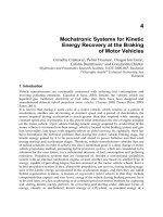
bone form a highly open porous structure with pore widths of up to a few millimetres. Fig. 1
shows the hierarchical organisation of (cortical) bone tissue – from the macroscopic organ
down to the nanometre scale.
4. Directed ionotropic gelation of alginate – a biomimetic method for
generating anisotropic materials
Alginate is the structural saccharid of brown algae. Being a co-polymer, it consists of
mannuronic (M) and guluronic acid (G) monosaccharide units, possessing identical
Novel Biomaterials with Parallel Aligned Pore Channels by Directed Ionotropic
Gelation of Alginate: Mimicking the Anisotropic Structure of Bone Tissue
351
Fig. 1. Hierarchical organisation of cortical bone tissue from the centimetre to the nanometre
scale (taken from Roh et al. (1998) with permission)
carboxylic and hydroxyl functional groups but differing in their configuration. These
functional groups coordinate multivalent cations and build intermolecular complexes which
results in the formation of a stable hydrogel. Straight MM-sequences do not exhibit sites for
specific binding of cations (Braccini et al., 1999); the interaction takes place between GG-
sequences leading to so-called egg-box motifs (Grant et al., 1973; Braccini & Perez, 2001).
Alternating MG-sequences may also contribute but to a much lower extent (Donati et
al., 2005). The composition of the alginates derived from different algae varies; the flexible
stipes of algae, growing next to the sea surface, contain M-rich alginate whereas those
exposed to strong flow exhibit high G-content (Zimmermann et al., 2007).
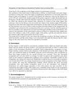
If an alginate sol gets into contact with gelling ions (electrolyte), the molecules gel
immediately by covering the sol with a dense skin or membrane. Microbeads are produced
by dropping small volumes into electrolyte solutions whereas the skin is trapping the sol
which gets radially transformed into a gel by the diffusing ions. Anisotropic gels with
channel-like pores develop when cations diffuse in broad front from one direction into an
alginate sol whereas the saccharide molecules get arranged and complexed. Together with
the gelation parallel aligned, channel-like pores are formed which can run through the
whole length of the gel (Fig. 2).
4.1 Theoretical models for the phenomenon
The discoverer of the phenomenon, the German colloid scientist Heinrich Thiele, proposed
the phase separation mechanism of droplet segregation. The gelation process
Sol + Electrolyte (A) ↔ Gel + Electrolyte (B) + Water (1)
is accompanied by dehydration. The finely distributed drops of water are trapped within
the zone of sol-gel-transition. Further delivered water molecules will accumulate and are
Advances in Biomimetics
352
Fig. 2. Sketch of the process of ionotropic gelation of alginate. The scheme in the middle was
adapted from Wenger (1998)
pushed by the gelation front towards the sol creating electrolyte containing and alginate free
pore channels (Thiele & Hallich, 1957; Thiele, 1967b). Khairou and co-workers described the
sol-gel-formation as diffusion controlled process which one step of primary membran
formation and further growth of the anisotropic gel (Khairou et al., 2002).
In a series of 5 articles, Kohler and his group developed the theory of chemically fixed
dissipative structure formation from the first idea (Kohler & Thumbs, 1995) until the
summary of the work (Treml et al., 2003). Based on the observation, that there was a
movement in the sol next to already gelled alginate visualized by tiny glass beads, they
assumed a coupled mechanism of convection and diffusion. The alginate chains are subject
to a conformational change during the complexation by the cations. If the sol exhibits an
adequate viscosity, this contraction will induce a movement of the sol which resembles to
pattern of the Rayleigh-Benard-Konvection. This pattern gets fixed by the sol-gel-transition.
For a stable reaction, a sufficient mass transport is needed to ensure a certain contraction
velocity of the alginate molecules. The mathematical description consists of the Navier-
Stokes equation for the hydro-dynamical model (Kohler & Thumbs, 1995; Thumbs & Kohler,
1996), Fick’s law for the diffusional macroscopic part (Treml & Kohler, 2000) and the results
from random walk simulations of a phantom chain (Woelki & Kohler, 2003). The
phenomenon of capillary creation due to the ionotropic gelation was postulated as
chemically fixed dissipative formation, which is based on the concentration of the alginate
sol and gel as well as the electrolyte, the diffusion coefficients of the reactants, the degree of
polymerization, length and number of rigid segments of the alginate chain and the gelation
rate constant (a fitting parameter obeying to boundary conditions) (Treml et al., 2003).
So far about growth but what about the initiation of the pores? Thiele and Hallich
postulated periodic water droplets which segregate by the dehydration during gelation
(Thiele & Hallich, 1957). The contraction of the alginate causes accumulations and lower
Novel Biomaterials with Parallel Aligned Pore Channels by Directed Ionotropic
Gelation of Alginate: Mimicking the Anisotropic Structure of Bone Tissue
353
concentrated areas as nucleation seeds (Purz, 1972). Lateral variations in chain mass fraction
and composition were also considered which would laterally vary the contraction capacity
(Thumbs & Kohler, 1996). The origin of first segregation and pore creation was tried to
identify by Purz and coworkers by electron microscopy – interestingly not with alginate but
cellulose xanthate (Purz, 1972; Purz et. al., 1985). The ionotropic gelation is not specific for
alginate but can occur also with other polymers (e.g. pectin, cellulose) and even inorganic
anisometric colloids (e. g. V
2
O
5
) get oriented by the flux of counter ions.
4.2 History of ionotropic gelation
The phenomenon of ionotropic gelation was discovered by Heinrich Thiele, professor at the
chemical department of Kiel University, Germany. Initially he studied in- and organic
anisometric colloids which were oriented by diffusing ions. He created the term ionotropy
(ionos = ion, trepein = turn) (Thiele, 1964) as a special case of gelation (Higdon, 1958). The
properties of the gels were birefringence, anisotropic swelling and reversible ion exchange.
He was fascinated by the similarity between structures of biological origin and the
artificially created anisotropic gels (Thiele & Andersen, 1953). In his pioneering work, Thiele
intensively studied parameters which influence the structure formation and different
methods to characterise the oriented colloids (Thiele, 1967b). He restlessly compared the
structure of ionotropic gels with those of tissues or other biological specimens and found a
variety of similarities (Thiele, 1954b; Thiele, 1967a). Based on this comparison, he predicted
a model for the principle of biological structure formation – especially supported by studies
on dissolution and re-constitution of an eye lens (Thiele et al., 1964). His last publication on
ionotropic gelation was dealing with mineralisation of the gels especially with calcium
phosphates (Thiele & Awad, 1969).
More than 25 years later, the phenomenon was theoretically investigated with a new vision
on the mechanism (Kohler & Thumbs, 1995) as well as towards the kinetics of ionotropic
gelation (Khairou et al., 2002) – and finally, the capillary formation could be described by a
mathematical model (Treml et al., 2003). At the same time, the idea re-emerged to use the
membranes, produced by ionotropic gelation, as filters with adjustable pore diameter. Not
only the hydrogels could be utilised for this application (Thiele & Hallich, 1959; Moll, 1963),
but also sintered ceramics, derived by structuring slurries of alginate mixed with ceramic
powders like e.g. Al
2
O
3
(Weber et al., 1997) or even with the mineral phase of bone,
hydroxyapatite (HAP) (Dittrich et al., 2002). The pore distribution and run was
characterized by µCT in ceramic (Goebbels et al., 2002) or composite (Despang et al., 2005b)
state. Since 2005/6, the anisotropic structures have been subject of research in the area of
tissue engineering with human cells for hard tissue (Despang et al., 2005a, Dittrich et
al., 2006) and vascularisation (Yamamoto et al., 2010), in in vitro and in vivo studies in rats
for nerve regeneration (Prang et al., 2006) and with murine embryonic stem cells opening
opportunities for the formation of many types of tissue (Willenberg et al., 2006). A more
detailed and chronological list of scientific contributions to the field with short summaries of
their content follows (Table 1).
4.3 Anisotropic hydrogels
The phenomenon of ionotropic gelation was discovered for alginate leading to a hydrogel
with parallel aligned, channel-like pores. At the early beginning, the gelation was carried
out solely with Cu
2+
which needs to be replaced in case of medical applications by acidic
exchange or ion substitution for a biocompatible one such as Ca
2+
. Since 2005, hydrogels
Advances in Biomimetics
354
Author(s) Year Content
Thiele, 1947
[in German]
Alignment and gelation of anisometric particles in colloidal solutions
(thin layer), resulting in birefringence pattern in polarized light
Thiele & Micke,
1948 [German]
First full article on alignment and gelation of anisometric particles in
colloidal solutions, but not yet about capillary formation
Thiele & Kienast,
1952 [German]
Dependence of alignment of anisometric particles on type and
concentration of ions of electrolyte including electron microscopy
images of sol and thixotropic gel
Thiele & Ander-
sen, 1953 [Ger.]
Identical structure and pattern of decalcified femur (collagen) and
ionotropic gel (Cu
2+
gelled pectin) observed in polarised light
Thiele, 1954a
[German]
Change in experimental set-up: diffusion of electrolyte from outside
into the sol, from thin layer of sol to beads and cylinders, direction of
ion diffusion from radial to broad front
Thiele, 1954b
English summary of previous work; differentiation of ionotropic gels
from other structures, claim on model for some biological patterns:
bone (collagen), see weed (alginate) and ripe fruits (pectin)
Thiele & Ander-
sen, 1955a
[German]
Transition from inorganic to organic colloids for ionotropic gelation
(alginate, pectin); first thoughts on theory of droplet demixing;
swelling and birefringence antipodal
Thiele & Ander-
sen, 1955b [Ger.]
Effect of chain length of alginate and pectin on ionotropic gelation
(viscosity); first images of radial pore channels in multiphasic gels
Schuur, 1955 [Ge.] Structure formation of ionotropic gels through material flux
Thiele & Kroenke,
1955 [German]
Reversible Pb-based mineralisation of ionotropic gels (cellulose
glyconat) within the cavities or pore walls of gel
Thiele & Hallich,
1957 [German ]
Channel-like pores in 3D gels of alginate through ionotr. gelation
including images and theory of droplet demixing; influence of type
and concentration of cations and sol on pore channel diameter
Thiele & Hallich,
1959 [German]
Application of capillary structure of ionotropic alginate gels as filters:
void volume, permeability (water, gas), pore size distribution
Thiele et al., 1962
[German]
Distinction between 5 zones of ionotropic gels with parallel aligned
pores; focus on primary membrane and diffusion induced membrane
potential; ion exchange after cross-linking with DIC
Moll, 1963
[German]
Application of Al-alginate gels with channel-like pores as reversible
filter for bacteria and viruses, filtering of a 5 nm gold sol
Thiele, 1964
[German]
Diverting overview about ionotropic gelation (theory, helices,
mineralisation) as model of biological pattern formation
Thiele et al., 1964
[German]
Ionotropic gelation as principle of biological pattern formation based
on similarities to natural tissues in appearance (osteons in bone, layers
of pearl) and reversible gelation of eye lens and cornea etc.
Thiele & Cordes,
1967 [German]
Influence of counter ions on gel formation; ligand field theory
Thiele, 1967
[German]
Short summary of principles of structure formation: bone, eye lens,
cornea
Novel Biomaterials with Parallel Aligned Pore Channels by Directed Ionotropic
Gelation of Alginate: Mimicking the Anisotropic Structure of Bone Tissue
355
Author(s) Year Content
Thiele, 1967b
[German book]
Exhaustive summary and overview on ionotropic gelation (book)
Thiele, 1967c
[German]
Ionotropic gels as template for oriented intra- or intercapillary
mineralisation in native and cross-linked gels by ion waves
Thiele & Awad,
1969
Mineralisation of alginate hydrogels with parallel aligned pores with
calcium phosphate phase brushit by ion waves followed by
conversion to hydroxyapatite
Purz, 1972
Anisotropic hydrogels based on cellulose-xanthate structured via
ionotropic gelation by thallium or zinc ions; SEM investigations
El-Cheik & Awad,
1976
Conductance of ions-free-washed metal alginate inversely
proportional to polarisability of gelling cations
Awad et al., 1980
Kinetic of ionotropic gel formation in two steps (quick
membrane formation, slow gel growth) evaluated by change in
concentration of electrolyte and description as diffusion controlled
process
Purz et al., 1985
[German]
Morphology of anisotropic cellulose-derivate gels structured by ions
of Tl, Pb, Zn, La and combinations studied by electron microscopy
Hassan et al., 1989
Latest of 3 similar articles on kinetics of sol-gel-transformation of
alginate with different ions (nickel, copper and cobalt)
Heinze et al., 1990
[German]
Structure and application of carboxy-containig polysaccharides,
especially anisotropic alginate hydrogels for cell immobilisation, drug
release; rheological investigations
Hassan et al., 1991
Structure formation of alginate by interaction of cations with two
carboxylic and two hydroxy groups
Hassan, 1991
Kinetics of acidic ion exchange of cations (Ni
2+
, Co
2+
, Cu
2+
) in
anisotropic alginate hydrogels by conductimetry
Hassan, 1993
Kinetics of anisotropic Ni-alginate gels: idea for application on
separation of ion mixtures and capture of isotopes based on selective
alginate binding
Kohler & Thumbs,
1995
[German]
New idea on theory of capillary development by ionotropic gelation
of alginate as chemically fixed dissipative structure: contraction of
alginate during gelling yields a movement of sol next to gelation front
which was visualised by adding 0.3 µm glass beads
Thumbs & Kohler,
1996
Mathematical description of ionotropic gelation similar to Rayleigh-
Benard convection by Navier-Stokes equation and introduction of
critical convection velocity
Weber et al., 1997
Al
2
O
3
membranes with capillaries produced by Cu
2+
-gelled
alginate-Al
2
O
3
-slurries and change in volume by drying procedures
Treml & Kohler,
2000
Mathematical description of diffusive mass transport of alginate and
gelling ions: correlation of convective transport to bulk concentrations
Dittrich et al., 2002
Synthesis of ceramic membrans (Al
2
O
3
, TiO
2
, HAP) by ionotropic
gelation of alginate/ceramic powder-slurries (drying process,
Advances in Biomimetics
356
Author(s) Year Content
influence of sintering temperature on density, macro-structure)
Goebbels et al.,
2002
Non-destructive analysis (µCT) of pore structure of ceramic
membranes (Al
2
O
3
, TiO
2
, HAP), synthesised by ionotropic gelation
Khairou et al., 2002
Kinetic study of ionotropic gelation induced by heavy metal ions and
interpretation of change of electrolyte concentration: influence of ionic
radius and electrolyte density; model of intra- and intermolecular
binding of cations to alginate chains
Woelki & Kohler,
2003
Modelling of the integration of alginate chains to the growing gel by
conformational changes/degree of contraction (length of chain,
velocity of gelation front, velocity of cross-linking reaction)
Treml et al., 2003
Summary of new theory on capillary formation as chemically fixed
dissipative structure depending on bulk concentrations, diffusion
constants, properties of alginate chain (number, length of Kuhn
segments), rate constant of gelation reaction
Despang et al.,
2005a
Ca-alginate hydrogels and composites of alginate/HAP for bone TE:
addition of HAP powder or synchronous mineralisation in situ
Despang et al.,
2005b
µCT-evaluation of composites of alginate-gelatine, reinforced with
HAP (powder and synchronous mineralisation)
Renzo et al., 2005 Pore channels in Cu-alginate microbeads and mineralisation
Dittrich et al., 2006
Alginate-gelatine-composites reinforced with HAP or ß-TCP
mimicking composition of bone (70:30 in- : organic) and biocompa-
tibility test by cultivation of osteogenically induced hMSC
Willenberg et al.,
2006
Cu-gelled alginate scaffold as polyelectrolyte with chitosan as
matrix for TE with murine embryonic stem cells: structure and in
vitro experiment for 4 days
Prang et al., 2006
Oriented axonal regrowth on isocyanate cross-linked, Cu-gelled
alginate hydrogels with in vitro (entorhinal-hippocampal slice
culture) & in vivo (spinal cord) experiments in rats
Mueller et al., 2006
Axonal regrowth on Cu
2+
-, Ni
2+
- or Ba
2+
-alginate hydrogels (after ion
exchange) with in vitro & in vivo experiments in rats
Eljaouhari et al.,
2006
Al
2
O
3
membrans based on Cu
2+
- or Ca
2+
-alginate-slurries including
optimized drying procedure, consolidation and permeability data
Dittrich et al., 2007
Influence of processing parameters on pore structure of Ca
2+
-alginate-
HAP-slurries (drying process, pore run (µCT), influence of media on
softening, hMSC in vitro culture)
Gelinsky et al.,
2007
Biphasic but monolithic scaffolds for therapy of osteochondral defect
with 2 layers (alginate/hyaluronate and alginate/HAP)
Despang et al.,
2008
Scaffolds for bone TE produced by ceramic processing chain;
composite, brown-body & ceramic: change of microstructure and
biocompatibility of hMSC
Bernhardt et al.,
2009
Biocompatibility of alginate-gelatine-HAP-scaffolds evaluated with
osteogenically induced human mesenchymal stem cells (hMSC) over 4
Novel Biomaterials with Parallel Aligned Pore Channels by Directed Ionotropic
Gelation of Alginate: Mimicking the Anisotropic Structure of Bone Tissue
357
Author(s) Year Content
weeks (incl. mechanical testing)
Mueller et al.,
2009a
Axonal regrowth on Ba- or Ni-gelled alginate with more and longer
linear axon ingrowth in dorsal ganglion in vitro culture with 10 µm
than 120 µm pore diameters
Mueller et al.,
2009b
Summary on axonal regrowth guided by anisotropic alginate
hydrogels
Khan et al., 2009
Alginate or polyelectrolyte dextran/alginate w/o particle
reinforcement of Au, TiO
2
and
Fe
3
O
4
Yamamoto et al.,
2010
Co-culture of HUVEC w/o smooth muscle cells seeded onto Ca-
alginate hydrogel for revascularization – static and perfusion cultures
Table 1. Chronology of scientific publications on ionotropic gelation leading to structures
with parallel aligned pores (excluding PhD theses and patents); milestones highlighted bold.
Abbreviations: DIC - diisocyanate, hMSC - human mesenchymal stem cells, HUVEC -
human umbilical vein endothelial cells, HAP – hydroxyapatite.
with channel-like pores created by ionotropic gelation of alginate were in focus for tissue
engineering. The idea of creating a tube-like template for capillary tissue structures e. g. for
blood vessels (Yamamoto et al., 2010) is fascinating. Depending on the needs, the pore
diameter can be adjusted between 30-460 µm by the processing conditions, meanly type and
concentration of alginate and electrolyte (Table 2). The swollen hydrogels exhibit a macro-
porosity of approx. 30% due to the pore channel diameter but the walls consist of an alginate
network with a high nano-porosity. The pore density was found to be 530/mm
2
and the
mean pore diameter around 30 µm for Cu
2+
as cation (Willenberg et al., 2006; Prang et al.,
2006).
Interestingly, using a different type of alginate gelled with Cu
2+
, we found a pore
density of 124/mm
2
with an mean pore diameter of only 20 µm. Anisotropic hydrogels
based on this type of alginate (ISP Manugel DMB) gelled by diffusion of Ca
2+
ions exhibited
a pore density of 77/mm
2
whereas ISP Manucol DM yields 5/mm
2
. The mean pore diameter
is inversely related to the pore density. Using Ba
2+
or Ni
2+
ions instead of Cu
2+
the pore
density was 960/mm
2
and 30/mm
2
, respectively, and the mean pore diameter 10 and 120
µm, respectively (Müller et al., 2008).
Target tissue
Dimension
(ØH or
LWH)
Pore-Ø
Alginate
concentr.
Mol.
weight
Electrolyte Reference
[mm] [µm] [Ma.%] [kDa] [M]
Bone
10x5 40-230 2 40-60 1 M CaCl
2
Despang et al.,
2005a
Embryonic
stem cells
7x5x3 30 2 12-80
0.5 M
CuSO
4
Willenberg et
al., 2006
Neuronal
tissue
0.5x0.5x3 27 2 100
1 M
Cu(NO
3
)
2
Prang et al.,
2006
Vascularisation
5x2 220-460 0.5-4 64-110
0.5-1.5 M
CaCl
2
Yamamoto et
al., 2010
Table 2. Alginate hydrogel scaffolds designed for different tissue engineering applications
Advances in Biomimetics
358
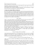
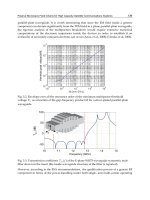
The pore diameter is also influenced by the pH value (Fig. 3), mainly, because the alginate
conformation (coiled or stretched) can be changed with the pH. Adjusting the pH value,
mostly HCl or NaOH is used which also changes the ion strength and therefore the
electrostatic conditions within the sol. To achieve a homogenous alginate sol, the aqueous
solution should be buffered because at low pH isotropic gelation can occur due to the ability
of H
+
ions to interact with the alginate molecules (Thiele & Hallich, 1957).
Fig. 3. Variation of pore diameter and pore distribution in the cross section of hydrogels,
prepared at different pH values (1% Alginate Manugel, ISP) – microstructure after freeze
drying
Up to distinct ranges, other biopolymers can be added to the alginate sol without preventing
the process of ionotropic gelation. This also allows to further stabilise the hydrogels by
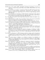
means of covalent cross-linking, e. g. applying carbodiimide chemistry. If a cationic polymer
like chitosan is chosen, polyelectrolytic hydrogels which means symplexes of two differently
charged polymers are formed (Fig. 4). For a biomimetic approach, we incorporated
successfully fibrillar collagen type I as the main component of most mammalian ECMs, but
only minor amounts could be used without disturbing the ionotropic gelation process. Also
addition of gelatine (thermally denaturised collagen) is possible but the mixture has than to
be kept above 30°C to prevent untimely gelation of gelatine.
The most stable (concerning degradation under cell culture conditions) polyelectrolytic
hydrogel was found while adding chitosan which additionally facilitated mineralisation by
immersion in simulated body fluids (SBF).
Novel Biomaterials with Parallel Aligned Pore Channels by Directed Ionotropic
Gelation of Alginate: Mimicking the Anisotropic Structure of Bone Tissue
359
Fig. 4. Polyelectrolytic hydrogels of the negatively charged alginate and positively charged
biopolymers (microstructure after air drying)
4.4 Anisotropic composites
Heading for regeneration of hard tissue, the mineral phase of bone, the calcium phosphate
hydroxyapatite (HAP), should be incorporated. Alginate-HAP-composites with parallel
aligned pores can be achieved following different strategies (Fig. 5), either by mineralisation
of the hydrogels after gelation or directly during the sol-gel-process. Possible routes are:
• Immersion of the structured gel in simulated body fluid (SBF) and heterogeneous
precipitation of HAP,
• Ion waves, i.e. diffusion of ions (alternating calcium and phosphate ions) in broad front
into the hydrogel in some runs creating initially brushit which can be transformed into
HAP (Thiele & Awad, 1969),
Advances in Biomimetics
360
• Synchronous mineralisation, i.e. precipitation of calcium phosphate during the sol-gel-
process (Despang et al., 2005),
• HAP powder, i.e. addition of HAP powder to the alginate sol and structuring of this
slurry via ionotropic gelation (Despang et al., 2005; Dittrich et al., 2006; Dittrich et al.,
2007; Bernhardt et al., 2009),
• Biphasic but monolithic scaffolds for the therapy of osteochondral defects can be
produced through deposition of sol layers differing in composition prior to the gelation
(Gelinsky et al., 2007).
Fig. 5. Strategies of mineralisation – which also can be used in combination
The mineral content of the composites, which was determined by ignition loss, varied
between the methods. A dried hydrogel, obtained from a 2% alginate sol without any
calcium phosphate phase exhibits approx. 11% of ash due to the gelling ions (Ca
2+
) and
reaction productes (CaCO
3
or CaO) during combustion (Despang et al., 2005). 5-9% more
mineral content was found for composites which were mineralised simultaneously during
the ionotropic gelation. In this case, the Ca
2+
ions not only orientated the alginate chains but
also reacted with the phosphate ions which had been added to the alginate sol before the
sol-gel-transition was initiated. Immersion in SBF increased the mineral content up to 11%.
Higher contents, mimicking the inorganic-to-organic-ratio of bone (aprox. 70:30), and even
more could only be realised by mixing HAP powder to the alginate sol. Thiele reached 50%
i.e. a little less than the ratio of bone ECM and each wave led to shrinkage of the structure
and therefore the pore diameter decreased (Thiele & Awad, 1969). Interestingly, the place of
mineralisation, either intracapillar or in the pore walls, could be adjusted by the processing
conditions as well as the shape of the precipitate was changed from round to needle-like by
addition of citrate. The different approaches of mineralisation are expressed in varying
microstructures (Fig. 6) and change the mechanical properties of the composite materials.
The high amount of HAP introduced by addition of ceramic powder results in improved
strength compared to the synchronously mineralised composites which was evaluated in
wet state (Despang et al., 2005; Bernhardt et al., 2009).
All changes in composition of the sol or slurry prior to ionotropic gelation will influence the
pore formation (diameter, length, density) during the sol-gel-process (Dittrich et al., 2007).
However, the gel or composite with parallel aligned pores can be influenced after gelation
Novel Biomaterials with Parallel Aligned Pore Channels by Directed Ionotropic
Gelation of Alginate: Mimicking the Anisotropic Structure of Bone Tissue
361
Fig. 6. Composite materials of biopolymer and mineral
by exposure to organic solvents benefiting of the different swelling behaviour. Other
strategies are exchange of the gelling ions or different drying procedures. Freeze, air and
supercritical drying were studied when the interest on ceramic membranes aroused (Weber
et al., 1997) and was further optimised (Dittrich et al., 2002; Eljaouhari et al., 2006).
Investigations by micro computer tomography (µCT) revealed that the pore structure was
destroyed by ice crystals during freeze drying whereas the structure remained intact when
water was exchanged against tert. butanol. Following the run of pore channels, this non
destructive method also unveiled that pore channels can merge with distance from the
primary membrane (Dittrich et al., 2007).
A mineral gradient in the direction of the long axis of the pore channels can be obtained by
carefully covering layers of alginate sol on top of each other which differ in composition
(Gelinsky et al., 2007). Bi-phasic but monolithic scaffolds consisting of a hydrogel-part and a
Advances in Biomimetics
362
mineralised part were under current investigations for regeneration of osteochondral defect.
Both parts contain additional components of the respective ECM, i.e. hyaluronic acid for
articular cartilage and hydroxyapatite for bone. Furthermore, living chondrocytes were
successfully embedded into the cartilage portion and stayed alive within 2 weeks of in vitro
culture. Incorporating living cells into the process of ionotropic gelation demands of course
work under sterile conditions and with sterile components during all process steps.
For medical applications, scaffolds need to be sterile but alginate (like other biopolymers) is
affected by all common sterilisation methods (Despang et al., 2008b). Since tissue
engineering comprises the degradation of the scaffold after implantation, the sterilisation
method enables to adapt this kinetic to the tissue or application of interest. For use in hard
tissue regeneration, the type of calcium phosphate powder incorporated in the composites
de- or accelerates the degradation because HAP and tricalcium phosphate (TCP) possess
different solubility (Dittrich et al., 2006). In vitro studies of the degradation kinetics should
be carried out under conditions as close as possible to those in vivo, i.e. in the incubator at
37°C/5%CO
2
and in cell culture medium (Bernhardt et al., 2009). Also the mechanical
stability over time is differently affected by cell culture medium compared to water or PBS
(Dittrich et al., 2007).
The biocompatibility of alginate-gelatine-HAP composites with a pore diameter of approx.
90 µm was evaluated by human mesenchymal stem cells (hMSC) which were osteogenically
induced. The seeding efficiency was 10-34% and cell number increased by a factor of 4-7
within 4 weeks (Dittrich et al., 2006; Bernhardt et al., 2009). Osteogenic differentiation was
confirmed by reverse transcriptase-PCR by gene expression of ALP and BSPII which were
not present at day 1 but were found clearly at day 21 (Bernhardt et al., 2009). A clear
difference between osteogenically induced and non-induced cells was observed, too. Cells
adhere at the face surface but were also found inside the channel-like pores visualised by
confocal laser scanning microscopy (Bernhardt et al., 2009).
4.5 Anisotropic ceramics
Two observations paved the road for the synthesis of anisotropic inorganic materials. First
of all, the channel-like structure was conserved in the ash after burning the organic part of
mineralised alginate which was intended to determine the mineral content (Thiele, 1967c).
Additionally, impurities or additives are not segregated like in the case of crystallisation but
incorporated into the hydrogel (Thiele, 1964). Only Weber et al. (1997) described the
synthesis of ceramic membranes based on structuring via sol-gel-process of ionotropic
gelation of alginate/powder-slurries followed by calcination.
Ceramic processing for membrane manufacturing was studied with Al
2
O
3
or TiO
2
including
development of adapted drying regimes for the wet composites applying method inherent
shrinkage, followed by heat treatment to obtain a sintered ceramic without cracks (Weber et
al., 1997; Dittrich et al., 2002; Eljaouhari et al., 2006). Dittrich et al. (2002) for the first time
synthesised such ceramics consisting of the mineral phase of bone, hydroxyapatite, with
parallel aligned pores and investigated their structure by µCT in cooperation with Goebbels
et al. (2002). The pore size and wall thickness was adjusted by the ratio of alginate-to-HAP
powder (Dittrich et al., 2002).
Anisotropic Al
2
O
3
ceramics with a pore diameter of 19 µm (Dittrich et al., 2002),
approximately 70 µm (Eljaouhari et al., 2006) or even 250-320 µm (Weber et al. 1997) were
manufactured. For TiO
2
ceramics a range of 10-30 µm was reported (Goebbels et al. 2002).
Novel Biomaterials with Parallel Aligned Pore Channels by Directed Ionotropic
Gelation of Alginate: Mimicking the Anisotropic Structure of Bone Tissue
363
Lower sintering temperatures result in less shrinkage and larger pore diameters (Dittrich et
al., 2002). For tissue engineering of bone, pores in the range of 100-300 µm are demanded.
Additionally, HAP which was exposed to temperatures of more than ca. 1000°C is no longer
resorbable in vivo by osteoclasts (the bone degrading cells). Therefore heat treatment at a
lower temperature is required to achieve biodegradable implant materials. Sintering is
performed for consolidating ceramic materials. A first HAP ceramic without organic
components can be derived as intermediate state after bisquit firing, leading to a material
called brown body which normally is consolidated in a further sintering step (Fig. 7).
Fig. 7. Sketch of ceramic processing using sol-gel-technique of ionotropic gelation of alginate
slurries with ceramic powders
Mechanical tests of HAP brown bodies with parallel aligned pores revealed a compressive
strength of 4.5 MPa. This value is quite comparable to that of cancellous bone of human
origin (5.9 MPa) tested at the same instrument under similar conditions (Rauh et al., 2009).
The brown body exhibited a crystallite size of 41 nm compared to 238 nm of the sintered
ceramic after treatment at 1200°C (Despang et al., 2008). Biocompatibility was evaluated by
proliferation and differentiation of human mesenchymal stem cells (hMSC). The typical
maximum of the specific alkaline phosphatase (ALP) activity was observed at day 14 after
seeding. Cell number increase by a factor of 2.5 within 3 weeks for osteogenically induced
hMSC cultivated on brown body samples as well as on sintered ceramics (Despang et al.,
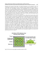
2008). Osteogenically induced hMSC adhered at the face surface as well as inside the
channel-like pores and grew to a confluent layer (Fig. 8).
The pore diameter (40-165 µm) was larger for the brown body than for the sintered ceramics
with approximately 30-115 µm (Fig. 9), depending on the type of alginate used for structure
formation. The pore density varied between 20-90 pores/mm
2
for the brown bodies and 50-
100 pores/mm
2
for the sintered ceramics. Largest samples prepared were 11x8 mm (ØxH).
Since alginate is gelled by many cations, anisotropic ceramics could also be synthesised by
different complexing metal ions. Within the non-toxic elements, Zn
2+
generates hydrogels
with pores larger than those derived from Ca
2+
(Thiele, 1967b). The more the cations orient
the alginates molecules, the smaller the pore diameter of the gelled structure which also
leads to an increase of the mechanical strength (Thiele & Hallich, 1957). But this does not
primarily apply for the ceramics because the fibrous organic part was burnt. Even so using
Zn
2+
for gelling alginate-HAP-slurries led to larger pore channels than Ca
2+
(Fig. 10), but the
biocompatibility was poor i.e. no cell proliferation was observed within 4 weeks on
composite material – in vitro studies on HAP bioceramics based on Zn-alginate-HAP-slurry
still need to be accomplished.
Advances in Biomimetics
364
Beside the actual pore size, the specific surface is an important parameter for scaffolds in
tissue engineering by regulating protein and growth factor adsorption. The specific surface
(BET) was measured for all states of the ceramic processing including the starting HAP
powder, green body (composite of alginate and HAP after drying), brown body (thermal
removal of organic phase) and consolidated ceramic (Fig. 11). Highest value is reached for
the initial powder whereas in the composite material, the alginate is occupying some space
and therefore the specific surface decreased. During heat treatment, the organic phase was
removed and the consolidation started by sintering. Nano-sized pores of the walls were
filled during sintering but the macro-porosity as relevant parameter for cell ingrowth
remained unaffected.
Face surface
Longitudinal section
Fig. 8. Osteogenically induced hMSC after 14 days of in vitro cultivation on nano-crystalline
HAP scaffolds in the state as brown body (Ca
2+
gelled slurry) – SEM (200x) after
supercritical drying
Fig. 9. Hydroxyapatite bioceramic based on Ca-alginate-HAP-slurry (top: state after thermal
treatment at 650°C) with channel-like pores (bottom: sintered ceramic)
Novel Biomaterials with Parallel Aligned Pore Channels by Directed Ionotropic
Gelation of Alginate: Mimicking the Anisotropic Structure of Bone Tissue
365
Fig. 10. Ceramics based on Zn-alginate-HAP-slurries (top: brown body, bottom: sintered)
Fig. 11. Change of the specific surface (BET) during ceramic processing through thermal loss
of alginate and due to sintering effects
5. Similarities between natural tissues and materials, generated by ionotropic
gelation of alginate
In section 3, the highly hierarchical organisation of bone tissue has been described. Many
attempts has been undertaken up to now to develop biomimetic materials which resemble