Advances in Biomimetics Part 12 pot
Bạn đang xem bản rút gọn của tài liệu. Xem và tải ngay bản đầy đủ của tài liệu tại đây (1.61 MB, 35 trang )
Advances in Biomimetics
376
mass and is responsible for its rigidity and load bearing capacity lies between these layers; it
undergoes the maximum mineralization and therefore it contains the maximum inorganic
content in the entire skeletal system. Due to their proximity with the bonemass the
periosteum and the endosteum make up the two distinct orthopedic interfaces in long
bones. They respectively play key roles in the formation and degeneration of the bone
tissue. The cellular and biochemical organization of these two orthopedic interfaces along
with the large mass of mineralised bone tissue that lies between them are the main targets of
biomimetic designing and manufacturing products for the human skeletal system.
Structurally the periosteum is a vascularised membranous layer that covers the entire outer
surface of all bones and functionally it acts as the regenerative orthopedic interface for the
entire diaphysial region of the bone,. Externally it combines with the fibers and ligaments of
the skeletal muscles and internally it provides attachment to the flattened osteoprogenitor
cells which divide by mitosis and differentiate into osteoblasts and then osteocytes. The
existence of the periosteum is essential for the regeneration of the bone after trauma injury.
The endosteum, which makes the degenerative interface of the bonemass, lines the inner
side of the mineralized cortical bone and has two surfaces - one which faces the outer
mineralized side of the bone mass and another which faces the inner non mineralized
sinusoidal bone marrow. The inner surface of endosteum makes several endosteal niches
which harbor multipotent stem cells that generate hematopoietic, muscular, adipose and
mesenchymal cell precursors in the marrow region. The outer surface of endosteum acts as
the site for producing differentiated osteoclast cells that migrate into the mineralized bone
matrix, between the periosteum and endosteum, and participate in its breakdown.
Osteoclasts also remove the dead osteocytes that lie embedded in the matrix. The
endosteum thus plays a key role in the bone remodeling by actively assisting the bone
resorption process through osteoclasts.
2.2 Histological and biochemical organization
In general the bone tissue exhibits a unique histological organization, it exhibits the general
properties of vertebrate connective tissues, but its matrix is uniquely dense, semi-rigid,
porous and highly calcified because it is made up of an organic matrix and an inorganic
mineral component. In a typical appendicular bone the matrix is composed of
approximately 30-35% organic and 65-70% inorganic components. The organic component is
called the osteoid which is composed of type I collagen and ground substances like
glycoproteins, proteoglycans, peptides, carbohydrates and lipids. Mineralization of the
osteoid, which can occur by several methods (see Section 3) constitutes the inorganic
components of the bone and these constituents include calcium phosphate- hydroxyapatite
Ca
10
(PO
4
)
6
(OH)
2
and calcium carbonate along with similar salts of magnesium, fluoride and
sodium in lesser quantity [Clarke 2008; Kalfas 2001].
The cellular component of bone tissue comprises three main cell types: osteoblasts,
osteocytes and the osteoclasts. As mentioned above osteoblasts line the periosteal layer and
they are cuboidal to flat in shape. They secrete the unmineralized organic matrix which later
mineralizes and leads to increase in organic component of bone matrix. Osteoblasts, as they
migrate into the matrix or line the canaliculi the thin cylindrical spaces or canals seen in the
bone mass, differentiate into osteocytes, which possess long thin cytoplasmic processes
called the filopodia. The osteocyte lined canaliculi help in the passage of nutrients and
oxygen between the blood vessels and matrix localized osteocytes. Osteocytes also break
down the bone matrix by osteocytic osteolysis to release calcium for calcium homeostasis.
Bioinspired and Biomimetic Functional Hybrids as Tools for Regeneration of Orthopedic Interfaces
377
They also maintain extracellular phosphorus concentration. The third main category of cells in
the bone mass are the osteoclasts. These are bone resorbing cells which are multinucleated and
carry out the process of bone resorption. They are generated from the shallow depressions on
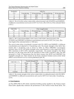
the inner side of the endosteum called howship lacunae. A schematic representation of the
cellular and inorganic organization of the bone mass is seen in Figure 3 below.
Fig. 3. A figurative description of the cellular organization in two orthopedic interfaces the
periosteum and the endosteum that surround the bone matrix in the hard cortical bone.
3. Biomimicry of bone components
The capacity of bone tissue components, both cellular and inorganic, to self-regenerate,
particularly after trauma related injuries, has attracted the interest of many scientists [Alves et
al., 2010]. During this regeneration process, we observe the recreation of mineral rich tissues of
different constitutions and hence this process is also referred to as biomineralization [Palmer,
2008]. Studying the process of biomineralization helps us in understanding the mechanisms by
which living organisms deposit mineralized crystals within matrix [Sarikaya, 1999]. Among
the approximately 40 different constituents found in the naturally formed biominerals,
carbonates, phosphates and silicates of calcium are the most common [Stephen, 1988]. These
salts have a significant role to play in determining the physiochemical properties and thermal
stability in hard bone tissue [Sarikaya, 1999; Cai & Tang, 2009].
In general terms, biomineralization process can be either biologically induced or biologically
controlled. In biologically induced mineralization (BIM) the shape and organization of the
Endosteum
Marrow
Osteoclast
Osteoc
y
te
Demineralization
Preosteoc
y
te
Mineralized Matrix
Osteoblast
Osteoid
Preosteoblast
Periosteum
Mineralization
Advances in Biomimetics
378
crystals is not directly under cellular control and it is determined entirely by inorganic
processes. As a result of this the shape and organization of the inorganic compounds made
by BIM is of a low order. In contrast to this biologically controlled mineralization (BCM) is
cell dependent and it shows a well balanced organization of the mineralizing salts with the
organic molecules resulting in well defined crystals of uniform shape, size and orientation
[Khaner, 2007; Weiner & Addadi, 1997]. During post trauma osteo-regeneration both types
of biomineralization processes are observed however the involvement of BCM is more
dominant. Features common to bone mineralization are also seen in the biomineralization of
many non skeletal tissues and cells and an examination of those properties helps in
understanding the mechanism behind skeletal tissue mineralization.
3.1 Non-skeletal biomineralization
The biomineralization process in non skeletal cells and tissues generates very complex,
diverse and interesting mineral forms and this process can be observed in almost in all
organisms [Ozawa & Hoshki 2008; Veiss, 2005]. An evolutionary break through about this
process was achieved in a report on the formation of magnetites in magnetotactic bacteria
which indicated the commonality of biomineralization mechanisms in different biological
forms and it also highlighted that this process is regulated by highly complex control
systems that are operational even in simple organisms. Several examples of non skeletal
biomineralization in multicellular organisms are observed in nature along with the more
common unicellular mineral producers. Some of these include silica spicule producing
sponges, diatoms and actinopoda; synthesis of amorphous calcium carbonate in ascidians
and formation of layered aragonite platelets in the nacreous layer of mollusk shells,few of
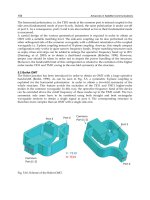
such examples has been shown in Figure 4 below. [Sarikaya, 1999].
Fig. 4. Biologically controlled mineralization of hierarchical structures observed in A)
magnetospirullum magnetium bacteria B) TEM of organic lattice of nacreous shell found in
atrina C) finely organized enamel rod structures of mouse tooth D) ordered structures in
siliceous skeleton lattice.[Atsushi et al., 2008; Yael et al.,2001;Sarikaya, 1999; James et al.,
2007]
3.2 Biomineralization in skeletal tissue
As indicated above, the biomineralization process in the bone tissue is different from what is
exhibited by nonskeletal cells and tissues, because in skeletal cells it is primarily cell
dependent i.e. it is controlled by BCM mechanisms. At the sub-cellular level
biomineralization in bones is mediated by the formation of matrix vesicles (MV) which are
membrane encased vesicles of size 20-200nm that are formed by a special exocytic membrane
Bioinspired and Biomimetic Functional Hybrids as Tools for Regeneration of Orthopedic Interfaces
379
budding process in polarized and differentiatiating osteoblasts/osteocytes of the long bones
and also in the hypertrophic chondrocytes of the cartilage and odontoblasts of the growing
teeth [Anderson, 2003]. After being secreted out of the cell, the MVs begin to deposit
calcium phosphate/apatite crystals within the lumen of the vesicle itself or are specifically
transported through the vesicular membrane into the matrix and they mineralize in
conjunction with matrix collagen [Ciancaglini, 2006]. This process can thus be divided into 2
phases - in phase I intra-luminal deposition of amorphous calcium phosphate, octa-calcium
phosphates and HAp crystals is seen and in phase II seepage of HAp crystals occurs
through the MV membrane into extracellular fluid resulting in nucleation of the crystals
within collagen fibrils as calcified nodules [Guido & Isabelle, 2004; Kazuhiko et al, 2009].
Type-1 collagen acts as a template for initiating the crystallization of secreted calcium
hydroxyapatite crystals [Vincet, 2008] which subsequently gets associated with other ECM
components such as proteins, polysaccharides, proteolipids and proteoglycans to support
activities such as cell adhesion, transport of ionic molecules, cell signaling etc.
Understanding the steps of matrix biomineralization and its degeneration is therefore
necessary in order to develop synthetic analogs that would mimic the matrix components
that aid in the regeneration of new tissue [Joshua et al., 2009; Alves et al., 2010; Veiss, 2005].
3.3 Steps in bone modeling and remodeling
As mentioned earlier and shown in Figure 3 the process of bone modeling and remodeling
is a homeostatic process where the bone formation and resorption processes are observed
simultaneously. The two processes are regulated by independent but related controls but
since basic steps are very different from one another they need to be understood sperately in
order to design materials to replace this integral component of the bone tissue.
3.3.1 Bone modeling
As mentioned above the bone modeling process in long bones is dependent mainly upon the
calcification of the collagenous matrix of the bone mass. This process of physiological
mineralization of collagen is controlled by the balance of enzymes, such as
metalloproteinases, transporters, such as type III Na/Pi co-transporter, and channels, such
as the annexin channels, which together aid to efficiently export the mineralizing molecules
from the MVs into the matrix. In a recent study, using proteo-liposomal vesicles, it has been
shown how to reconstruct a model that would mimic the MV microenvironment and would
help us in better understanding the MV microenvironment [Simao et al., 2010]. In addition
to the MV associated enzymes, transporters and channels some other molecules in the
matrix such as tissue nonspecific alkaline phosphatase (TNAP), the group of docking
proteins ankyrins and nucleotide associated inorganic phosphate, that influence the
transport of MV pyrophosphate into the matrix and thereby regulate its calcification [Ellis,
2009, Robert, 2001]. These matrix associated molecules exert their effects by directly
controlling the amount of free inorganic phosphate in the ECM which in turns determines
the transport PPi from the MVs [Ellis, 2009]. The effective role of matrix associated TNAP in
controlling vesicle mineralization is highlighted in a disease named hypophosphatasia
where TNAP activity is decreased because of a mutation in this gene the mobility of PPi
from MVs to the matrix is very high [Robert, 2001]. Mineralization initiation in matrix
vesicles is a function of several inhibitors, promoters that needs a proper balance between
the elements that maintain them.
Advances in Biomimetics
380
In addition to Type I collagen there are some other proteins in the matrix that also associate
with the mineralized collagen and then further enhance or inhibit the mineralization
process. Some of these proteins observed in bones and teeth are shown in Table 1.
Osteopontin[OPN] and Bone Sialoprotein[BSP] are acidic proteins with high affinity for Ca
2+
ions are localized within the collageneous matrix found adjacent to mineralization front that
are involved in determining calcification. BSP are found to be initiator of mineralization
whereas OPN affinity for apatite crystal founds to inhibit the crystal maturation process
[Hunter et al ., 1996; Bernards et al., 2008].
Bone Dentin Enamel
Osteocalcin (OC) dentin matrix protein 1
Enamelin
Osteopontin (OPN)
dentin sialo-phospho
protein
Matrix extracellular phospho-
glycoprotein (MEPE)
Osteonectin (ON) - -
Bone sialoprotein (BSP) - -
Table 1. Major non-collageneous proteins that associate with mineralized ECM in different
bone tissues
3.3.2 Bone remodeling
In contrast to the matrix modeling process the remodeling of the mineralized matrix is more
complex because it can be controlled by many different mechanisms. In the case of normal
bone homeostasis we observe a balance between the calcification and decalcification reactions
in the bone matrix where the decalcification of the matrix is facilitated by the removal of the
dead osteocytes and discharged MVs from the matrix. This process is primarily carried out by
osteoclasts which arise from the endosteum. However, the decalcification process can be
disturbed due to several reasons which could be either related to blockages or total stoppage
of the calcification process or due to pathological changes in the tissue such as migration of
cancer metastatic cells, activation of osteoporotic reactions etc.
The modeling and remodeling of the matrix thus represent the two orthopedic interfaces of
the bone which are generated at periosteum and endosteum respectively and their
mineralizing and de-mineralizing functions overlap in the matrix as shown in Figure 3.
4. Materials and methods for the mimicry of bone components
Based upon the details of the natural processes that lead to mineralized bone formation and
its degradation, as described above, there are several reports in the literature that describe
strategies to generate materials in vitro that are similar to the in vivo physicochemical and/or
biological properties of the bone components. In fact bone biomimetism remains as one of
the most actively pursued and financially a very rewarding area of human tissue
engineering. A brief summary describing the different types of materials and processes that
are currently in use to generate bone like materials, for their use as bone implants or
substitutes, is provided here.
Bioinspired and Biomimetic Functional Hybrids as Tools for Regeneration of Orthopedic Interfaces
381
4.1 Materials useful as substrates or modifiers in bone implants and/or bone
substitutes
The choice of materials that can be used to repair or replace a damaged or deformed bone is
very wide. An overriding factor in choosing a base material for this purpose is its bioactivity
and biocompatibility in vivo.
Materials References
Metals
Stainless steel AISI 316L, Co–Cr–Mo
alloy
Ti and its alloys
Ti6Al4V, TNZT alloys (Ti–Nb–Zr–Ta),
Ni Ti, TiNbZr
Ceramics and Bioglass
α-Al
2
O
3,
high alumina ceramics
,
PSZ
(partially stabilized zirconia)
,
45S5 BG
,
S45P7
Polymers
Polyethylene (PE), Polymethacrylic
acid (PM MA), polyglycolic acid (PGA),
poly lactic acid (PLA), polycarbonate
(PC), polypropylene(PP)
Composites
Mg–Zn–Zr, HA-PEEK poly (aryl-ether-
ether-ketone), Polyphospha zenes, BG-
COL-HYA-PS (glass-collagen
hyaluronic acid-phosphatidylserine)
Yeung et al.,2007; Aksakal et al., 2008; Seligson et
al.,1997;
Marti 2000
Aksakal et al., 2008; Chakraborty et al., 2009;
Yeung et al.,2007; Banerjee et al., 2004; Banerjee et
al., 2006; Niinomi 2003; Ning et al., 2010; Seligson
et al.,1997
Kapoor et al., 2010; Christel et al., 1988;
Gorustovich et al., 2010; Yuan et al., 2001
Andersson et al., 2004; Reis et al., 2010; Oral et al.,
2007; Butler et al., 2001; Athanasiou et al., 1998;
Smith et al.,2007; Geary et al., 2008; Shalumon et
al., 2009; Jayabalan et al., 2001
Ye et al.,2010; Kurtz et al., 2007; Sethuraman et al.,
2010; Xu et al., 2010
Table 2. A list of materials in use as base/substrate material in bone implants
Since there is no material available that can per se become a bone substitute, several
modifications on the original material are required to make it biocompatible. The aim to do
these modifications is that the new material should be nontoxic and biologically inert but yet
it should show orthopedic bioactivity and its production should be cost effective. The
biocompatibility of the material is also dependent upon certain host factors such as general
health, age, tissue perfusion and immunological factors [Wooley et al., 2001] and therefore
only certain types of materials have been used so far for this purpose. A list of such
materials currently in use is given in Table 2.
Each of the listed materials in the Table has some unique quality that qualifies it to be used
as the base material or the substrate of an orthopedic implant. Cationic metals for example
can form ionic bonds with non-metals and can be easily converted into alloys which have
good ductile properties and heavy load bearing strength. Among the nonmetals, ceramics
are interesting because their inter-atomic bonds are either totally ionic or predominantly
Advances in Biomimetics
382
ionic and they can be covalently bonded to a number of compounds including proteins.
Among the polymers for orthopedic use, plastics and elastomers have been the main choice
but because of their limited weight bearing capacities their use is restricted. The composites
are useful because they can combine the properties of two or more compounds making it a
more versatile material to get a functional hierarchy of substances needed to make a bone
like substance.
Besides the substances which are used as substrates for making biocompatible materials,
there are many other unique elements of bone structure which lend themselves to be
mimicked by manmade materials as functionalizing compounds of the substrates. One of
the most commonly mimicked biomaterial for this purpose is apatite which is the most
abundant phosphate mineral on earth found in mineralizing vertebrates. Among all the
calcium phosphate minerals available hydroxyapatite (HAp) is found to be the most
thermodynamically stable bioceramic material at physiological environment which helps in
faster osteointegration. Hence the most sought after properties that material scientists and bone
tissue engineers look for in their apatite are bone bonding ability and osteo-conductivity in
addition to their general biocompatibility and bioactivity. The starting compounds used for
making HAp is generally calcium phosphate and based on some solution parameters like
super saturation, other ionic products and pH we can get many other apatite phases apart
from HAp. These non-naturally occurring apatite phases can be more useful than naturally
occurring ones.
MINERAL NAME Ca/P ratio Abbreviation
Monocalcium phosphate monohydrate 0.5 MCPM
Monocalcium phosphate:dihydrate 0.5 MCPD
Dicalcium phosphate: dehydrate mineral brushite 1.0 (DCPD)
Anhydride mineral monetite 1.0 (DCPA)
Octacalcium phosphate 1.33 (OCP)
α-tricalcium phosphate 1.5 (αTCP)
β-tricalcium phosphate 1.5 (β-TCP)
Whitelock mineral 1.29
Hydroxyapatite O- HAp 1.67 OHAp
Calcium-deficient hydroxyapatite 1.5-1.67 (CDHA)
Fluorapatite 1.67 (FAp)
Chloroapatite 1.67 (ClAp)
Carbonated apatite TYPE A 1.67 (CO3Ap)
Tetracalcium phosphate, mineral hilgenstokite
2.0
(TTKP or
tetcp)
Table 3. Different types of calcium phosphates obtained during preparation of HAp
A list of the various types of apatite phase that can be obtained from different calcium
phosphates is given in Table3. Besides using calcium phosphate, a combination of various
salts is also used to generate HAp. This process is more close to the natural process because
the constituents of starting material are based upon the constituents of the natural body
fluid such as blood plasma. The solution that most represents the similarity with blood
plasma is referred to as simulated body fluid or SBF and its many constituents have been
described elsewhere Tadashi and Hiroaki 2006 and Jalota et al 2006.
Bioinspired and Biomimetic Functional Hybrids as Tools for Regeneration of Orthopedic Interfaces
383
Na
+
K
+
Ca
2+
Mg
2+
HCO3
-
Cl
-
HPO4
2-
SO4
2-
Ca/P Ph
Blood Plasma
142 5 2.5 1.5 27 103 1 0.5 2.5 7.4
SBF Range
127-
734
5-10
2.5-
12.5
1.5-
7.5
4.2-35 111-724 1-5 0.05-1 0-2.5 7.25-7.4
TYPE-1
142 5 2.5 1.5 4.2 148 1.8
1.4 7.25
TYPE-2
142 5 2.5 1.5 27 147.8 1 0.5 2.5 7.4
TYPE-3
c-SBF2
c-SBF3
SBF-
J
L1
SBF-JL2
142
142
142
142
5
5
2.5
2.5
2.5
-
1.5
1.5
-
-
4.2
35.23
34.9
34.88
147.96
117.62
111
109.9
1
1
1
1.39
0.5
0.5
-
-
2.5
2.5
2.5
0
7.4
TYPE-4
SBF
d-SBF
142
142
5
5
2.5
1.6
1.5
0.7
4. 2
4.2
147.8
144.1
1
1
0.5
0.5
2.5
1.6
7.25
7.25
TYPE-5
142 5 2.5 1.5 4.2 148 1 0.5 2.5 7.4
TYPE-6
SBF
5XSBF
142
714.8
5
2.5
12.5
1.5
7.5
4.2
21
147.8
723.8
1
5
0.5
2.5
2.5
7.4
7.6
TYPE-7
127 10 12.5 3 35 123 5
2.5
7.4
TYPE-8
142 5 2.5 1.5 4.2 147.8 1 0.05 2.5 7.4
TYPE-9
SBF-1
5XSBF
SBF-2
142
213
142
5
7.5
5
2.5
3.8
2.5
1.5
2.3
1.5
4.2
6.3
4.2
148
223
148.8
1
1.5
1
0.5
0.75
0.5
2.5
2.53
2.5
7.4
TYPE
10
SBF-a
SBF-b
714.8
704.2
12.5
12.5
7.5
1.5
21
10.5
723.8
711.8
5
5
-
-
2.5
2.5
7.4
TYPE-11
142 5 2.5 1.5 4.2 148.8 1 0.5 2.5 7.4
TYPE-12
142 5 2.05 1.5 4.2 148 1 2.05 7.4
TYPE-13
142 5 2.5 1.5 4.2 148.5 1 0.5 2.5 7.4
TYPE-
14
1XSBF
3CaP
SBF
142
109.5
5
6
2.5
7.5
1.5
1.5
4.2
17.5
147.8
110
1
3
0.5
-
2.5
2.5
7.5
TYPE-
15
SBF(N)
SBF(O)
142
142
5
5
2.5
2.5
1.5
-
27
-
123
123
1
1
0.5
0.5
2.5
2.5
7.2
TYPE-16
109.5 6 7.5 1.5 17.5 110 3 0
6.65-6.71
6.55-6.65
6.24-6.42
Table 4. Recipes for making different types of Simulated Body Fluids for biomimetic
preparation of Apatite
[Reference for the above Table are a-Liu et al.,1998; b-Kokubo & Kim, 2004; c-Marc &
Jacques,2009; d-Chikara et al., 2007; e-Kokubo,1996; f-Bharati et al.,2005; g-Qu & Mei,2008; h-
De Medeiros et al., 2008; i-Tsai et al.,2008; j-Habibovic et al.,2002; k-Hyun et al.,1996; l-Silvia
et al.,2006; m-Xin et al.,2007; n-Yajing et al.,2009; o-Kapoor et al.,2010; p-Haibo & Mei 2008]
Over the years the constitution of SBF has undergone so many modifications that would
be compiled into a list of different SBFs that can used to obtain bone like apatite for bone
remodeling purposes. This compilation is shown in Table 4. The original SBF was
intended to study mainly the bone-bonding ability of the apatite and it lacked in sulfate
Advances in Biomimetics
384
ions in relation to original plasma constituents. The SBF constitution was later upgraded
with major variations done in chlorine and bicarbonate compositions and to a lesser
extent in sulphate ions. SBF with higher Cl
-
and lower HCO3- concentrations and
variations in buffer systems and pH are found to be in equilibrium with the blood plasma.
The physiological pH is maintained in this in vitro system using tris (hydroxymethyl)
amino methane (Tris)/HCl.
4.2 Methods for preparing substrates and modifier materials
While the base substrate materials are prepared by conventional metallurgical methods,
their bioactivity is induced by functionalizing them with many modifier materials. The
modifier materials include proteins, enzymes and most importantly the different types of
apatites. There is an endless list of techniques by which apatite deposition can be carried
out on orthopedically selected substrates, but the successful methods are those which give
high bone bonding ability and good osseointegration. Among the different available
techniques, plasma spray, sol-gel synthesis and biomimetic methods are the most
successful. Some salient features of the first two and details of the biomimetic approaches
are provided here.
4.2.1 Plasma spray
Plasma spray coatings on to metal substrates have gained interest during the past decades
due to its high deposition rate and its large scale efficiency. This method is compatible with
various platforms including ceramic composites apart from metals. Numerous studies have
been carried out on the bone bonding behavior of these coatings with the substrates. The
thickness of the coating is of few microns size. The precursor is mainly fed in the form of
powder which is released into a plasma gun. A high voltage argon gas generates plasma
where the powder gets partly melted and is directed towards the substrate followed by
rapid cooling further impelling the substrate thus depositing a coat. This method has been
used to deposit different functionalized materials on either metal or non-metal surfaces.
[Chen et al., 2008; Chen et al., 2006; Culha et al., 2010]
But the major concerns regarding this process a) is the instability of the coatings therefore poor
binding of the coating with the substrate or implant .This necessitates them for further
processing to increase the mechanical interlocking of the coating-substrate system. b) High
processing temperatures involved lead to changes in CaP phases resulting in the formation of
less stable phases thereby reducing the bonding strength between the substrate and the
coating. c) These coatings are largely amorphous with less homogeneity over the entire
substrate resulting in structures of low crystallinity which signifies that the substrates are not
bioactive enough to induce the required bone attachment. Many functionalized scaffolds have
been developed by this technique and there biocompatibility was checked in-vivo so that these
implants can be used for various orthopedic applications [Heimann et al., 2004; Wu et al., 2009]
4.2.2 Sol-gel synthesis
This technique is one of the oldest in developing thin film coating having varied
applications like protective coatings, passivation layers, sensors and membranes. The
methodology involves the fabrication of materials by using a chemical solution (sol) which
acts as the precursor for a specialized integrated network (gel) of either particles or network
oligomers/polymers. The unique property of this method is that the kinetics of the reaction
Bioinspired and Biomimetic Functional Hybrids as Tools for Regeneration of Orthopedic Interfaces
385
can be controlled by monitoring the particle size, porosity and thickness of coating. Hence
the fabricated materials can be obtained in the form of films, powders, fibers, processed at a
lower temperature which differentiates it form the conventional processing strategies
[Podbielska and Ulatowska-arza 2005].
The starting materials used are inorganic or metal-organic precursors (alkoxides). The
chemistry of this process involves basically two reactions like hydrolysis and
polycondensation. When metal- alkoxides are used the alkoxide is dissolved in alcohol and
hydrolyzed by the addition of water, whereas in case of metalloids, acid or base catalyst is
added which replaces the alkoxide ligands with hydroxyl groups. In case of inorganic
precursors like salts, hydrolysis proceeds by the removal of a proton to form a hydroxo (-
OH) or oxo (=O) ligand. Therefore subsequent condensation reactions in case or organic and
inorganic produces oligomers or polymers composed of M-O-M or M-µ(OH)-M bonds.
The coating is generally done by depositing the precursor on to the substrate either by dip
coating or spin coating, later the samples are dried at high temperature which results in
shrinkage and also increases the density of the deposited precursors. The coating thickness
is a function of withdrawal speed, concentration and viscosity of the solution hence the
porosity of the gel is dependent on the rate at which the solvent is removed. The simplicity
of this procedure develops uniform coatings of high homogeneity [Klein, 1988].Many
biocompatible, bioactive and stable metals/non-metals and bioglass scaffolds are deveopled
by this technique by depositing HAp, various bioactive proteins in the form of thin films
and nanoparticles[Weng et al., 2003; Wang etal.,2008; Vijayalakshmi et al.,2008] for hard and
soft tissue replacement[Kim et al.,2005; Nguyen et al.,2004; Sepulveda et al.,2002; Zheng et
al.,2009].
4.2.3 Biomimetic process
Since the theory of biomimetic process proposed by Kokubo, the study of bioactivity using
SBF has been reviewed by many research groups all these years. Why these studies are at a
faster pace and what makes this process so challenging from other technologies in
predicting bone bioactivity in vivo. This process aims at mimicking the blood plasma
compositions in acellular conditions using SBF [Tadashi & Hiroaki, 2006]. For natural bone
to bond with the implants there must be specific appropriate response which it feels that it
can be accepted, is mainly achieved by depositing apatite on to these surfaces termed as
bioactivity/bone-bonding ability. Bones ability to deposit calcium phosphate defines its
characteristic property as a hard connective tissue. Several results have been obtained using
this procedure and they have been summarized in Table 6.
Bio-mimetic Coating Method used to Functionalize Ti-6Al-4V and α-Al
2
O
3
Our lab is also developing functionalized scaffolds which can be in long run used for bone
engineering applications.
We are working with metal (Ti and its alloys like (Ti-6Al-4V, TiZr, and TiNb), non-metals
(Ceramic like α-Al
2
O
3
) and glass, functionalizing them in order to check the cell behavior in
vitro and also check there bio-compatibility properties in vivo.
There are many methods to functionalize the metal/non-metal surface by using
HAp/calcium phosphate which can be done by various methods like plasma spray method,
sol-gel coating method, dip coating methods but the most easy and efficient way to mimic
the natural component of bone is by Biomimetic coating method, hence we have utilized this
process to develop an even, functionalized HAp coating on a titanium alloy (Ti-6Al-4V) and
Advances in Biomimetics
386
Cell culture studies
and materials used
Objectives Results References
Apatite and apatite/
collagen composite
coatings on PLLA
using Saos-2
osteoblast like cells
Cell attachment,
proliferation and
differentiation
Biomimetic apatite/collagen
coating found to exhibit
higher proliferation and
differentiation in comparison
to apatite coatings
Chen et al.,
2008
Biomimetic and
electrolyti -cally
deposited carbonate
apatite on Ti alloy
using MC3T3-E1
cells.
Cellular
proliferation and
differentiation
Higher proliferation and OC
and BSP mRNA expression
on biomimetically coated
substrates than
electrolytically deposited
method.
Jiawei et al.,
2009
Chemically
pretreated CP Ti
immersed in SBF for
2 and 14 days and
tested using human
osteoblasts (MG-63)
cells.
Cell spreading,
proliferation and
differentiation
A well spread morphology
was observed both
functionalized surfaces. TiCT
and TiHCA surfaces
rendered increased
expression of collagen 1 and
ALP at 7 and 14 days.
Barbara et al
., 2008
HA deposition on
negatively charged
SAM coated glass
cover slips by
culturing human
mature OC of bone
cell tumor for 24hrs
Osteoclastic
activity through
F-Actin ring
formation,
calcium release
and formation of
resorption pits
Osteoclast were able to attach
and resorb on coated glass
cover slips
Asiri et al.,
2009
Biomimetic apatite
deposition on
hyaluronic acid
(HA)-based polymer
scaffold
Osteogenic
induction of
mesenchymal
stromal cells (h-
MSCs)
At higher mineralization on
HA-based scaffold.
Cristina et al
., 2010
Incorporation of
bisphosph -onate
sodium clondrate
into biomimetically
coated apatite on to
starch based scaffold
using human
osteoblast-like cell
line (SaOs-2)
Effect of BP on
osteoclastic
activity and cell
morphology,
attachment and
proliferation
Osteoblastic activity was
simulated with
bisphopshonates at dose
dependent concentration of
0.32mg/ml by enhanced cell
viability
Oliveira et
al., 2010
BMP-2 into
biomimetic apatite
coatings using Rat
bone marrow
stromal cells for
8days on Ti implants
Osteogenic
activity
Protein incorporated CaP
coatings enhanced the
alkaline phophatase activity
Yuelian et
al., 2004
Table 6. Cellular responses to biomimetically prepared substrates and coatings
Bioinspired and Biomimetic Functional Hybrids as Tools for Regeneration of Orthopedic Interfaces
387
on a bioinert ceramic substrate (α-Al
2
O
3
). In our method, the metal /ceramic substrates were
incubated in simulated body fluid (SBF) at 25°C for different time points with prior
treatment with globular protein BSA (bovine serum albumin) [Chakraborty et al., 2009;
Kapoor et al., 2010]. This process leads to the formation of HAp coating exhibiting bone like
apatite growth on the surface. It may further be noted that bone, a natural composite
comprises non stoichiometric calcium hydroxyapatite (HAp) precipitated in a controlled
reaction environment of a highly aligned, anisotropic organic template. It differs from
stoichiometric hydroxyapatite (HA) in composition, crystallinity and other physical and
mechanical properties developed artificially through various methods.
The surface treatment and coating of these materials had shown a better cellular response in
vitro and also a good biocompatibility property in vivo when compared with untreated and
uncoated materials. The surface treatment by globular protein i.e., BSA might provide a
functionalized template comprising of charged amino-acids which resulted in more
nucleation sites [Chakraborty et al., 2009] hence led to the even coverage of HAp (about 280-
300µm) by immersion of the materials in SBF at desired temperature of 25°C between the
pH range of 5-7, which resulted in the formation 30-40 nm albumin globules, under
specified conditions, on both ceramic and Ti-6Al-4V alloy substrates. In comparison with the
untreated substrates the coverage of HAp was very much poor(less than 200µm), hence BSA
treatment has led to the development of nano-sized globules after HAp coating which have
led to the better cellular-activity in-vitro which is due to “cooperativity” reaction
[Chakraborty et al., 2009] between protein molecules and the charged surface of HAp,
depending on the concentration of the protein molecules in the coating [SBF] solutions.
We have done a comparative study of biological properties of the unique coating of HAp
developed on both metal and non-metal which is less reported. Based on the methodology
of functionalizing these materials we have generated many substrates of Ti and Ceramic
which showed a different structural variation and these specific morphological structures of
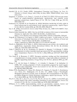
protein and HAp has led to good fibroblast [NIH-3T3] cell response. The Ti-6Al-4V which is
BSA treated and coated for 4 days has shown a nano-sized globules (as indicated by arrows)
due to globular protein treatment has shown a better in-vitro and in-vivo activity which can
be seen in Figure 1 panel c in comparison with the bare Ti-6Al-4V panel a, BSA treated Ti-
6Al-4V panel b and coated Ti-6Al-4V for 4 days without prior treatment with BSA panel d
which did not show nano-sized HAp globules.
The unique structural property of HAp coating on Ti-6Al-4V treated with BSA and coated
for 4 days is shown in Figure 2 where panel a shows the inter and intra connection of HAp
fibers into plates which can be seen in higher magnification in panel b. Panel c shows the
femur bone like growth of HAp fibers [Kapoor et al., 2010] which represents the unique
methodology in mimicking the bone like components by generating a highly functionalized
scaffold for in-vivo applications.
On the contrary, micron sized globules of HAp [Figure. 3(c)] were observed on the BSA
treated and coated for 2days ceramic substrate surface. This may be attributed to the
enhanced hydrophilicity of the BSA treated ceramic substrate (it already has intrinsic
hydrophilicity) that accumulates –OH groups throughout the mechanically roughened (grit
blasted) surface, on immersion in simulated body fluid (SBF), aqueous medium. These act as
nucleation sites and induce Ca
2+
ions from SBF to be coordinated to the above –OH groups
on the substrate, by electrostatic force of attraction. Hence nucleation of a large number of
HAp globules takes place and they grow fast into micron sized globules owing to the high
surface energy as mentioned, resulting in a dense coverage of substrate surface. Hence due
Advances in Biomimetics
388
to large deposition of micron-sized HAp globules the NIH-3T3 cellular response was much
better on this ceramic substrate in comparison to the bare ceramic (panel a), BSA treated
ceramic(panel b) and untreated and coated for 2 days panel d which showed a much bigger
HAp deposition.
Fig. 4. SEM images of different Ti-6Al-4V where (a)Bare Ti-6Al-4V (b)BSA Treated Ti-6Al-4V
(c)BSA Treated and Coated for 4 days Ti-6Al-4V (d) Coated for 4 days Ti-6Al-4V.( Image
generated from Kapoor et al., 2010).
Fig. 5. SEM Images of Ti-6Al-4V substrate which is BSA treated and coated for 4 days where
(a) Inter- and intraconnection of the HAp fiber in the crystal plates of 4-day coated substrate.
(b) Higher-magnification image of B showing the fiber merges into the crystal plates of the
HAp coating. (c) Femur bone-like structure obtained in B4. (Image generated from
Chakraborty et al., 2009).
Fig. 6. SEM images of α-Al
2
O
3
where (a)Bare α-Al
2
O
3
(b)BSA Treated α-Al
2
O
3
(c)BSA Treated
and Coated for 2 days α-Al
2
O
3
(d) Coated for 2 days α-Al
2
O
3
.( Image generated from
Kapoor et al., 2010).
Our in vivo experiments also proven that metal/nonmetal implants which are protein
treated and coated are more bioactive as they showed no negative response in term of any
kind of inflammatory responses.
Bioinspired and Biomimetic Functional Hybrids as Tools for Regeneration of Orthopedic Interfaces
389
This comparative assessment of metal/non-metals structural and biological properties
showed that metal when treated with protein and biomimtically coated for HAp can be
used as a scaffold for many biomedical applications especially for osteoconduction. In
modification for the method proposed, many biologically active molecules like osteogenic
agents and growth factors can be co-precipitated with apatite crystals onto metal implants
for the better osteogenic behavior as this biomimetic coating can be readily absorbed in-
vivo.
5. Orthopedic challenges
As new methodologies for making functional components of human tissues to rectify a
deformity or for developing new treatments of disease and trauma get developed we realize
the limitations of the techniques and principles of biomimetic tissue engineering in facing
up the real challenges of this approach. While many new methodologies have become
available for the management of orthopedic disease and trauma, the computability of the
manmade materials in this area is far from ideal. We describe here some of the unmet
challenges of this field.
5.1 Biocompatibility and stability of in-vivo scaffolds
One of the most important aims of biomimetic design and production of materials for bone
implants is to make them stable and compatible to the local bone tissue. Since there is
considerable diversity in the details of local anatomies of specific bones the presently
available general implant materials are prone to infection, extensive inflammation, and poor
osteointegration. Besides their life span is less than 15 years which clearly shows the
inability to mimic the longetivity of the molecular components of bone [Harold 2006, Porter
2009]. The implant failure is mainly attributed to acute complications, host responses,
prosthesis dislocations and surgery failures seen at initial stages after surgery, and also after
several years post surgery when implant loosening, osteolysis, implant wear and tear,
instability, infection and fractures are observed.
In order to increase implant life it would be advisable to seed them with young osteoblasts
which would sustain the production of bone mass on the implants (Xynos et al., 2001). It
would also be useful to use bioactive agents in the coatings that would activate pathways
related to cell survival, proliferation and differentiation. Thus it is clear that in order to
increase the life of the implanted material it would be advisable to shift the focus of material
production from a purely material science outlook to a cell biological and molecular
biological approach.
5.2 Materials for osteoporotic applications
Osteoporosis a major health threat to bone degenerations due to decreased bone quality, are
characterized by reduction in bone mass and disordered skeletal micro-architecture and are
susceptible to fracture risks at sites of hip, spine and wrist [Borges & Bilezikian, 2006]. Much
of the concerns regarding this are found in older populations where treatment becomes
possible to an extent through regular controlled diet activities. Since the loss in bone mass
can be directly attributed to the abnormal remodeling process therefore biomimetic tissue
engineering approaches could offer alternate approaches to reduce the hyperactive bone
resorption process. One of the targets for this could be the receptor for nuclear factor kappa
B which seems to be involved in osteoblast–osteoclast coupling mechanisms.
Advances in Biomimetics
390
6. Conclusion
We have shown in this chapter how one can use biomimetic approaches to simulate the
osteoregenerative (periosteal surface) and osteo-degenerative (endosteal surface) interfaces
of appendicular bones. These processes include novel tissue engineering strategies that
combine developments in the field of material science with the cell and molecular biological
pathways that are seen in the natural differentiation of osteoblast and osteoclast. We hope
that some of these strategies would lead to the better management of trauma and age related
degeneration of bone tissues.
7. Acknowlegments
This work is supported by grant Nos. GAP 0311, GAP022 and CMM002 to GP from the
Council of Scientific and Industrial Research and Department of Science and Technology
Government of India New Delhi. SR is supported with DBT-Indo Australian Biotech Grant
(GAP0311) and RK is supported by grant No.GAP220 from the Department of Science and
Technology, Government of India.
8. References
Aksakal, B. & Hanyaloglu, C. (2008). Bioceramic dip-coating on Ti–6Al–4V and 316L SS
implant Materials. Mater Sci: Mater Med, Vol.19, No. , pp. 2097–2104
Anderson, H. (2003). Matrix Vesicles and Calcification. Curr Opin Rheumatol, Vol.5, pp.222–
226
Andersson,
O.H.; Rosenqvist, J. & Karlsson, K.H. (2004).Dissolution, leaching, and Al
2
O
3
enrichment at the surface of bioactive glasses studied by solution analysis. J.
Biomed. Mater. Res, Vol.27, pp. 941–948
Alves,M.; Leonor,B.; Azevedo,H.; Reis,L & Mano,F. (2010).Designing biomaterials based on
biomineralization of of bone. Mater. Chem, Vol.20, pp. 2911–2921
Asiri, K., Wijenayaka, C., Colby, G. & Atkins, P. (2009).Biomimetic hydroxyapatite coating
on glass coverslips for the assay of osteoclast activity in vitro. J Mater Sci: Mater
Med, Vol.20, pp.1467–1473
Athanasiou, K.A.; Agrawal, C.M.; Barber, F.A. & Burkhart, S.S. (1998). Orthopaedic
applications for PLA-PGA biodegradable polymers. Arthroscopy, Vol.14, No.7,
pp.726-737
Atsushi, A.; Hidekazu, N.; Michiko, N.; Tetsushi ,M. & Tadashi, M.(2008). Formation of
magnetite by bacteria and its application. J. R. Soc. Interface, Vol.5, pp.977-999
Balasundaram, G. & Webster, T.J. (2007). An Overview of Nano-Polymers for Orthopedic
applications. Macromol Biosci, Vol.7, pp.635–642
Banerjee, R.;Nag, S.;Samuel, S. & Fraser, H.L. (2004).Strengthening mechanisms in Ti–Nb–
Zr–Ta and Ti–Mo–Zr–Fe orthopaedic alloys.Biomaterials, Vol. 25, No.17, pp. 3413-
3419
Banerjee, R.;Nag, S.;Samuel, S. & Fraser, H.L. (2006).Laser-deposited Ti-Nb-Zr-Ta orthopedic
alloys. J Biomed Mater Res,Vol. 78A, pp.298–305
Barbara, J.; Lenka, M.; Frank, L.; Andrea, E.; Claudia, B.; Egle, C. & Frank, A. (2008).
Osteoblast response to biomimetically altered titanium surfaces. Acta Biomaterialia,
Vol.4, No.6, pp.1985-1995
Bioinspired and Biomimetic Functional Hybrids as Tools for Regeneration of Orthopedic Interfaces
391
Bernards, M.T.;Qin, C. & Jiang, S.(2008). MC3T3-E1 cell adhesion to hydroxyapatite with
adsorbed bone sialoprotein, bone osteopontin, and bovine serum albumin. Colloids
Surf B Biointerfaces, Vol.64, No.2, pp.236-247
Bharati, S.; sinha, m. k & basu,D.(2005).Hydroxyapatite coating by biomimetic method on
titanium alloy using concentrated sbf. Bull. Mater. Sci, Vol. 28, pp. 617–621
Butler, K.; Benghuzzi, H. & Tucci, S. (2001).Tissue-implant response following soft tissue
implantation of poly-L-lysine coated UHMW-polyethylene into adult male rats.
Biomed Sci Instrum, Vol.37, pp.19-24
Cai,Y. &Tang,R.(2009). Towards understanding biomineralization: calcium phosphate in a
biomimetic mineralization process. Front. Mater. Sci. China,Vol.3,No.2,pp.124–131
Ciancaglini,P.; Simão, S.; Camolezi,L.; Millán,J. & Pizauro,M. (2006). Contribution of matrix
vesicles and alkaline phosphatase to ectopic bone formation. Braz J Med Biol Res,
Vol.39, No.5, pp.603-610
Chakraborty, J.; Mazaj, M.; Kapoor, R.; Gouri, S.P.; Daneu, N.; Sinha, M.K.; Pande, G . &
Basu, D. (2009).Bone-like growth of hydroxyapatite in the biomimetic coating of Ti-
6Al-4V alloy pretreated with protein at 25°C. J Mat Res,Vol.24,pp.2145-2153
Chen, C.C.; Huang, T.H.; Kao, C.T. & Ding, S.J. (2006).Characterization of Functionally
Graded Hydroxyapatite/Titanium Composite Coatings Plasma-Sprayed on Ti
Alloys. J Biomed Mater Res Part B: Appl Biomater, Vol.78B, pp. 146–152
Chen, D.; Jordan, E.H. & We, M.G.M. (2008).Apatite formation on alkaline-treated dense
TiO2 coatings deposited using the solution precursor plasma spray process. Acta
Biomaterialia,Vol. 4,pp.553-559
Chen, Y., Mak,A., Wang, M., Li, J. & Wong, J. (2008). In vitro behavior of osteoblast-like
cells on PLLA films with a biomimetic apatite or apatite/collagen composite
coating. J Mater Sci: Mater Med, Vol.19, pp.2261–2268
Chikara, O.; Masanobu, K & Toshiki, M. (2007).Coating bone-like apatite onto organic
substrates using solutions mimicking body fluid. J Tissue Eng Regen Med, Vol.1, pp.
33–38
Christel, P.; Meunier, A.; Dorlot, J.M.; Crolet, J.M.; Witvoet, J.; Sedel, L. & Boutin,
P.(1988).Biomechanical Compatibility and Design of Ceramic Implants for
Orthopedic Surgery. Ann. N. Y. Acad. Sci, Vol. 523, pp. 234–256
Clarke, B. (2008).Normal Bone Anatomy and Physiology. Clin J Am Soc Nephrol,Vol. 3,
pp.S131–S139
Cristina, M., Vincenzo, G., Nicoletta, Z., Maria, G., Andrea, F., Francesco, G., Elena, G.,
Stefano, S., Andrea, F., Luigi, A. & Gina, L. (2010). Mineralization behavior with
mesenchymal stromal cells in a biomimetic hyaluronic acid-based scaffold.
Biomaterials, Vol.31, No.14, pp.3986-3996
Culha, O.; Tekmen, C.; Toparli, M. & Tsunekawa, Y. (2010).Mechanical properties of in situ
Al2O3 formed Al–Si composite coating via atmospheric plasma spraying. Materials
& Design, Vol.31, pp.533-544
De Medeiros, W.S.; De Oliveira, M. V.; Pereira, L. C & De Andrade, M. C.(2008).Bioactive
Porous Titanium: An Alternative to Surgical Implants . Artif Organs, Vol. 32, pp.
277–
282
Dreinhofer,K.; Feron, J.; Herrera, A.; Hube, R.; Johnell, O.; Lidgren, L.; Miles,K.; Panarella,
L.; Simpson, H. & Wallace, A. (2004).Orthopaedic surgeons and fragility fractures. J
Bone Joint Surg [Br], Vol.86-B, pp.958-961
Dixon, R.A. (2005).Engineering of plant natural product pathways. Curr Opin Plant,Vol. 8,
pp.329–336
Advances in Biomimetics
392
Elliott, J.C.; Wilson, R.M. & Dowker, S.E.P. (2002).Apatite structures. Adv X Ray
Anal,Vol.45, pp.172-181
Ellis, E. (2009). Role of matrix vesicles in biomineralization. Biochimica et Biophysica Acta
1790, pp.1592–1598
Feng, L.; Song, Y.; Zhai, J.; Liu, B.; Xu, J.; Jiang, L. & Zhu, D. (2003).Creation of a
Superhydrophobic Surface from an Amphiphilic Polymer. Angew. Chem. Int. Ed,
Vol.42, No. 7, pp.800-802
Feng, X. & Jiang, L. (2006).Design and Creation of Superwetting/Antiwetting Surfaces.
Adv.Mater,Vol. 18, pp.3063-3078
Geary, C.; Birkinshaw, C. & Jones, E. (2008). Characterisation of Bionate polycarbonate
polyurethanes for orthopaedic applications. J Mater Sci: Mater Med,Vol. 19, pp.3355–
3363
Gorustovich, A.A.; Steimetz, T.; Cabrini, R.L. & Lopez, J.M.P. (2010).Osteoconductivity of
strontium-doped bioactive glass particles: A histomorphometric study in rats. J
Biomed Mater Res,Vol. 92A, pp. 232–237
Guido, M. & Isabelle, J. ( 2004). Cells, tissues, and disease: principles of general pathology, Oxford
University Press, 0-19-514090-7, New York
Habibovic, P.; Florence, B.; Clemens, A.B.; De Groot, K & Layrolle, P. (2002). Biomimetic
Hydroxyapatite Coating on Metal Implants. J. Am. Ceram. Soc, Vol. 85, pp. 517–522
Haibo, Qu & Mei, W. (2008). Effect of temperature and intital ph on biomimetic apatite
coating. J Biomed Mater Res Part B: Appl Biomater 87B, pp. 204–212
Harold, C.;Slavkin,P. & Bartold, M. (2006).Challenges and potential in tissue engineering.
Periodontol 2000, Vol. 41, pp.9–15
Heimann, B.R.;Suhurmann, N. & Muller, T.R. (2004).In vitro and in vivo performance of
Ti6Al4V implants with plasma sprayed osteoconductive hydroxyapaptite-bioinert
titania bond coat “duplex” systems: an experimental study in sheep.J Mater
Sci:Mater Med,Vol.15, pp.1045-1052.
Hunter, K.; Hauschka,V.; Poole, A.; Rosenberg,C. & Harvey, A. (1996). Nucleation and
inhibition of hydroxyapatite formation by mineralized tissue proteins. International
Review of Cytology, Vol.242, pp.121-156.
Hyun,M.K.; Fumiaki, M.; Tadashi, K & Takashi, N.(1996).Preparation of bioactive Ti and its
alloys via simple chemical surface treatment. Journal of Biomedical Materials Research,
Vol. 32, pp. 409-417
James, C.; Joanna, A.; Georg, E.; David, K.; Alexander, W.; Peter, A.; Kirk, F.; Michael, J.;
Frank, W.; Paul, K.; Peter, F. & Daniel, E. (2007)Hierarchical assembly of the
siliceous skeletal lattice of the hexactinellid sponge Euplectella aspergillum. Journal
of Structural Biology, Vol.158, pp.93–106
Jayabalan, M.; Thomas, V. & Rajesh, P.N. (2001).Polypropylene fumarate/phloroglucinol
triglycidyl methacrylate blend for use as partially biodegradable orthopaedic
cement. Biomaterials, Vol.22, No.20, pp.2749-57
Jiawei, W., Jan de, B. & Klaas de, G. (2009). Proliferation and differentiation of osteoblast-
like MC3T3-E1 cells on biomimetically and electrolytically deposited calcium
phosphate coatings. J Biomed Mater Res,Vol. 90A, pp
.664–670
Joshua, R.; Timothy, T.; Popat, R.K.(2009). Bone Tissue Engineering: A Review in Bone
Biomimetics and Drug Delivery Strategies. Biotechnol. Prog, Vol.25, No.6, pp.1539–
1560
Kalfas, I.H. (2001).Principles of bone healing. Neurosurg Focus, Vol.10, No. 4, pp.7-10
Bioinspired and Biomimetic Functional Hybrids as Tools for Regeneration of Orthopedic Interfaces
393
Kamino, K. (2008). Underwater Adhesive of Marine Organisms as the Vital Link between
Biological Science and Material Science. Mar Biotechnol, Vol.10, pp.111–121
Kapoor, R.; Gouri, S.P.; Kumar, J.M.; Raj, A. T.; Srinivas, G.; Chakraborty, J.;Sinha, M.
K,.;Basu, D. & Pande, G. (2010).Comparative assessment of structural and
biological properties of biomimetically coated hydroxyapatite on alumina (α-
Al2O3) and titanium (Ti-6Al-4V) alloy substrates. J Biomed Mat Res Part A, Vol.94A,
pp.913–926
Kazuhiko, K.;Buchanan,V.& Weiss,M.( 2009). Biomineralization in Humans: Making the
Hard Choices in Life. Annu. Rev. Genet, Vol.43,pp.119–142
Keselowsky, B. G.; Collard, D. M. & Garcia, A. J. (2005).Integrin binding specificity regulates
biomaterial surface chemistry effects on cell differentiation. PNAS, Vol. 102,
pp.5953–5957
Khaner, O. (2007). Evolutionary innovations of the vertebrates. Integrative Zoology, Vol.2,
pp.60-67
Kim, H.W.; Kim, H.E.; Salih, V. & Knowles, J.C. (2005).Hydroxyapatite and Titania Sol–Gel
Composite Coatings on Titanium for Hard Tissue Implants; Mechanical and In
Vitro Biological Performance. J Biomed Mater Res Part B: Appl Biomater, Vol.72B:
pp.1– 8
Klein, L.C. (1988) Sol-Gel Technology for Thin Films, Fibers, Preforms, Electronics and
Specialty Shapes, Noyes Publications, 0-8155-1154,USA
Kokubo,T.(1996). Formation of biologically active bone like apatite on metals and polymers
by a biomimetic process. Thermochemica acta, Vol. 280/281, pp.479-490
Kokubo, T.; Kim, H.M.; Kawashita,M.; Nakamura,T.(2004).Bioactive Metals: Preparation and
Properties. J Mater Sci Mater Med, Vol.15, pp.99-107
Kurtz, S.M. & Devine, J.N. PEEK. (2007).Biomaterials in Trauma, Orthopedic, and Spinal
Implants. Biomaterials, Vol.28,No.32, pp. 4845–4869
Lawrence, G. (2005).Pathogenesis of osteoporosis: concepts, conflicts, and prospects. J. Clin.
Invest, Vol.115, pp. 3318–3325
Le, M.H.; Ducheyne, P.; Lynch, L.; Boettiger, D. & Composto, R. J. (2006). Effect of
biomaterial surface properties on fibronectin–α5β1 integrin interaction and cellular
attachment. Biomaterials, Vol.27, pp.1907-1916
Lee, H.; Lee, B.P. & Messersmith P.B. (2007).A reversible wet/dry adhesive inspired by
mussels and geckos. Nature, Vol.448, pp.338-341
Liu, Y.; Huang, B.; Ruan, J. & He, Y. (1998). Behaviour of Composite Ca/P Bioceramics in
Stimulated Body Fluid. J. Mater.Sci.Technol, Vol.14, pp. 533-537
Liu, K.; Yao, X. & Jiang, L. (2010).Recent developments in bio-inspired special wettability.
Chem. Soc. Rev, (In press)
Marc, B & Jacques, L. (2009).Can bioactivity be tested in vitro with SBF solution?
Biomaterials, Vo
l. 30, pp. 2175–2179
Marti, A. (2000).Cobalt-base alloys used in bone surgery. Injury, Vol. 31, No. 4, pp.18-21
Meek, K.M. & Fullwood, N.J. (2001).Corneal and scleral collagens - a microscopist’s
perspective. Micron, Vol.32, pp.261–272
Mueller, G. & Russell, R. (2003). Osteoporosis: pathogenesis and clinical intervention.
Biochem. Syst. Ecol, Vol. 31, No. 2, pp.462-464
Nair, M.B.; Varma, H.K.; Menon, K.V.; Shenoy, S.J. & John, A. (2009).Tissue regeneration
and repair of goat segmental femur defect with bioactive triphasic ceramic-coated
hydroxyapatite scaffold. J Biomed Mater Res, Vol. 91A, pp.855–865
Advances in Biomimetics
394
Nguyen, H.Q.; Deporter, D.A.; Pilliar, R.M.;Valiquette, N. & Yakubovich, R. (2004).The effect
of sol–gel-formed calcium phosphate coatings on bone ingrowth and
osteoconductivity of porous-surfaced Ti alloy implants. Biomaterials,Vol.25, pp.865-
876
Niinomi, M. (2003).Fatigue performance and cyto-toxicity of low rigidity titanium alloy, Ti–
29Nb–13Ta–4.6Zr.Biomaterials, Vol.24, No.16, pp.2673-2683
Ning, C.; Ding, D.;Dai, K.; Zhai, W. & Chen, L. (2010).The effect of Zr content on the
microstructure, mechanical properties and cell attachment of Ti–35Nb–xZr alloys.
Biomed Mater, (In press)
Nystrom, D.; Lindqvist, J.;Ostmark, E.; Hult, A. & Malmstrom, E. (2006).Superhydrophobic
bio-fibre surfaces via tailored grafting architecture. Chem. Commun, pp.3594–3596
Nystrom, D.; Malmstrom, E.; Hult, A.; Blakey, I.; Boyer, C.;Davis, T.P. & Whittaker, M.R.
(2010).Biomimetic Surface Modification of Honeycomb Films via a “Grafting From”
Approach. Langmuir, (In press)
Oliveira,A.; Pedro,A.; Saiz,C.; Mano,J.; Rodriguez,G.; San, J. & Reis,R. (2010). Biomimetic
Ca-P Coatings Incorporating Bisphosphonates Produced on Starch-Based
Degradable Biomaterials. J Biomed Mater Res Part B: Appl Biomater, Vol.92B, pp.55–
67
Oral, E. & Muratoglu, O.K. (2007).Radiation cross-linking in ultra-high molecular weight
polyethylene for orthopaedic applications. Nucl Instrum Methods Phys Res B,
Vol.265, No.1, pp. 18–22
Ozawa,H.;Hoshi,K. & Amizuka,N.(2008).Current concepts of bone mineralization. . J.Oral
Biosci, Vol.50, No.1, pp.1-14
Palmer,C.;Christina,J.;Kaltz,R. & Spoerke,D.(2008). Biomimetic Systems for Hydroxyapatite
Mineralization Inspired By Bone and Enamel. Chem. Rev, Vol.108.pp. 4754–4783
Podbielska, H. & Ulatowska-jarza,A. (2005). Sol-gel technology for biomedical engineering.
Bull. Pol. Ac.: Tech. Vol.53, No.3, pp.261-271
Porter, J.R.; Ruckh, T.T. & Popat, K.C. (2009).Bone Tissue Engineering: A Review in Bone
Biomimetics and Drug Delivery Strategies. Biotechnol. Prog, Vol.25, pp.1539–1560
Puleo, D.A. & Nanci, A. (1999).Understanding and controlling the bone-implant interface.
Biomaterials, Vol. 20, pp. 2311- 2321
Qu, H & Mei, W. (2008). Improvement of Bonding Strength between Biomimetic Apatite
Coating and Substrate. J Biomed Mater Res Part B: Appl Biomater, Vol.84B, pp. 436–
443
Reis, J.; Kanagaraj S.; Fonseca, A.; Mathew, M.T.; Capela-Silva, F, Potes J, Pereira A, Oliveira,
M.S, Simoes, J.A. (2010).In vitro studies of multiwalled carbon nanotube/ultrahigh
molecular weight polyethylene nanocomposites with osteoblast-like MG63 cells.
Braz J Med Biol Res. Vol.43,No.5, pp.476-82
Rigo, E.C.S.; Boschi, A.O.; Yoshimoto, M.; Allegrini, S.;Konig, B. & Carbonari, M.J.
(2004).Evaluation in vitro and in vivo of biomimetic hydroxyapatite coated on
titanium dental implants. Mater Sci and Eng:C, Vol. 24, pp.647–651
Robert, M.; David, F.; Dorothy, A. & Clifford, J. (2001). Osteoporosis. Third edition-Volume 1,
Eli Lilly & Company, 978-0-12-370545-7,London,UK
Sarikaya, M. (1999).Biomimetic fabrication of apatite related biomaterials.PNAS, Vol.96,
No.25, pp.14183–14185
Sato, M. & Webster, T.J. (2004).Nanobiotechnology: implications for the future of
nanotechnology in orthopedic applications. Expert Rev. Medical devices, Vol.1,
pp.105-114
Bioinspired and Biomimetic Functional Hybrids as Tools for Regeneration of Orthopedic Interfaces
395
Seligson, D.; Mehta, S.; Mishra, A.K.; FitzGerald, T.J.; Castleman, D.W.; James, A.H.; Voor,
M.J.; Been, J. & Nawab, A. (1997). In vivo study of stainless steel and Ti-13Nb-13Zr
bone plates in a sheep model. Clin Orthop Relat Res, Vol.343, pp.213-23
Sepulveda, P.; Jones, J.R. & Hench, L.L. (2002).Bioactive sol-gel foams for tissue repair. J
Biomed Mater Res, Vol.59, pp. 340–348
Sethuraman, S.; Nair, L.S.; El-Amin, S.; Nguyen, M.T.; Singh, A.; Greish, Y.E.; Allcock, H.R.;
Brown, P.W. & Laurencin C.T.(2010).Development and Characterization of
Biodegradable Nanocomposite Injectables for Orthopaedic Applications Based on
Polyphosphazenes.J Biomater Sci Polym Ed, (In press)
Shalumon, K.T. & Jayabalan, M. (2009). Studies on biodegradation of crosslinked hydroxyl
terminated-poly (proplyene fumarate) and formation of scaffold for orthopedic
applications. J Mater Sci: Mater Med, Vol. 20, pp.S161–S171
Silvia, F.; Sally, J. M.; Eduardo, S.; Antoni, P. T. & Grayson, W. M. (2006). Functionally
graded bioactive coatings: Reproducibility and stability of the coating under cell
culture conditions. Acta Biomaterialia,Vol. 2, pp. 133–142
Simao, S.; Yadav,C.; Ciancaglini, P. & Millan, L.(2010). Proteoliposomes as matrix vesicles
biomimetics to study the initiation of skeletal mineralization. Braz J Med Biol Res,
Vol.43, No.3, pp.234-241
Smith, L.J.; Swaim, J.S.; Yao, C.; Haberstroh, K.M.; Nauman, E.A. & Webster,
T.J.(2007).Increased osteoblast cell density on nanostructured PLGA-coated
nanostructured titanium for orthopedic applications. Int J Nanomedicine, Vol.2,
No.3, pp.493–499
Stephen,M.(1988). Molecular recongnistion in biomimeralization. Nature, Vol.332, pp.119 –
124.
Stevens, B.; Yang, Y.; Mohandas, A.; Stucker, B. & Nguyen, K.T. (2008). A Review of
Materials, Fabrication Methods, and Strategies Used to Enhance Bone Regeneration
in Engineered Bone Tissues. J Biomed Mater Res Part B: Appl Biomater, Vol. 85B,
pp.573–582
Tadashi, K. & Hiroaki, T. (2006). How useful is SBF in predicting in vivo bone bioactivity.
Biomaterials, Vol.27, pp.2907–2915.
Tsai, S.W.; Hsu, F.Y & Chen, P.L. (2008). Beads of collagen–nanohydroxyapatite composites
prepared by a biomimetic process and the effects of their surface texture on cellular
behavior in MG63 osteoblast like cells. Acta Biomater, Vol. 4, pp. 1332-41
Veiss,A.(2005). A Window on Biomineralization. Science, Vol.4, No.307 (5714), pp.1419-1420.
Vijayalakshmi, U.; Prabakaran, K. & Rajeswari, S. (2008).Preparation and characterization of
sol–gel hydroxyapatite and its electrochemical evaluation for biomedical
applications. J Biomed Mater Res. Vol. 87A, pp.739–749
Vincent, J.F.V. (2003). Biomimetic modeling. Phil. Trans. R. Soc. Lond. B, Vol.358, pp.1597–
1603.
Wang, D.; Chen, C.; He, T. & Lei, T. (2008). Hydroxyapatite coating on Ti6Al4V alloy by a
sol–gel method. J Mater Sci: Mater Med, Vol.19, pp.2281–2286
Weiner,S .& Addadi,L. (1997). Design strategies in mineralized biological materials. J. Mater.
Chem, Vol.7, No.5, pp. 689–702
Weiner, S. & Wagner, H.D. (1998).The material bone: structure-mechanical function
relations. Annu
. Rev. Mater. Sci, Vol.28, pp.271–298
Weng W.; Zhang, S.; Cheng, K.; Qu, H.; Du, P.; Shen,G.; Yuan, J. & Han, G. (2003). Sol–gel
preparation of bioactive apatite films Surface and Coatings Technology, Vol.167,
pp.292–296
Advances in Biomimetics
396
Wooley, P.H.; Morren, R.; Andary, J.; Sud, S.; Yang, S.Y.; Mayton, L.; Markel, D.; Sieving, A.
& Nasser, S. (2002).Inflammatory responses to orthopaedic biomaterials in the
murine air pouch. Biomaterials, Vol.23, No.2, pp.517-526
Wu, C.; Ramaswamy, Y.; Liu, X.; Wang, G. & Zreiqat, H. (2009).Plasma-sprayed CaTiSiO5
ceramic coating on Ti-6Al-4V with excellent bonding strength, stability and cellular
bioactivity. J. R. Soc. Interface, Vol. 6, pp.159–168
Xin,Y.; Liu,C.; Zhang,X.; Tang, G.; Tian,X. & Chu, P. K.(2007) Corrosion behavior of
biomedical AZ91 magnesium alloy in simulated body fluid. J. Mater. Res, Vol. 22,
No. 7, pp.2004-2011
Xu,
C.; Su, P.; Wang, Y.; Chen, X.; Meng, Y.; Liu, C.; Yu, X.; Yang, X.; Yu, W.; Zhang, X. &
Xiang,
A.P. (2010).A novel biomimetic composite scaffold hybridized with
mesenchymal stem cells in repair of rat bone defects models. J Biomed Mater Res
Part A, (In Press)
Xynos, I.D.; Edgar, A.J.; Buttery, L.D.K.; Hench, L.L. & Polak, J.M. (2001). Gene-expression
profiling of human osteoblasts following treatment with the ionic products of
Bioglass 45S5 dissolution. J. Biomed. Mater.Res, Vol. 55A, pp. 151–157
Yael, K.; Giuseppe, F.; Lia, A. & Steve, W. (2001). Structure of the Nacreous Organic Matrix
of a Bivalve Mollusk Shell Examined in the Hydrated State Using Cryo-TEM.
Journal of Structural Biology, Vol.135, pp.8–17
Yajing, Z.; Guozhi, Z. & Mei, Wei. (2009)Controlling the Biodegradation Rate of Magnesium
Using biomimetic apatite coating. J Biomed Mater Res Part B: Appl Biomater 89B, pp.
408–414
Ye, X.; Chen, M.; Yang, M.; Wei, J. & Liu, D. (2010).In vitro corrosion resistance and
cytocompatibility of nano-hydroxyapatite reinforced Mg–Zn–Zr composites. J
Mater Sci: Mater Med, Vol.2, pp.1321–1328
Yeung, K.W.K.; Poon, R.W.Y.; Chu, P.K.; Chung, C.Y.; Liu, X.Y.; Lu, W.W.; Chan, D.; Chan,
S.C.W.; Luk, K.D.K. & Cheung, K.M.C. (2007).Surface mechanical properties,
corrosion resistance, and cytocompatibility of nitrogen plasma-implanted nickel–
titanium alloys: A comparative study with commonly used medical grade
materials. J Biomed Mater Res, Vol.82A, pp.403–414
Yuan, H.; De Bruijn, J.D.; Zhang, X.; van Blitterswijk, C.A. & de Groot K. (2001).Bone
Induction by Porous Glass Ceramic Made from BioglassT (45S5). J Biomed Mater Res
(Appl Biomater), Vol.58, No.3, pp.270–276
Yuelian, L., Ernst, B., Pierre L., Joost, D., De, B. & Klaas, D.(2004). Tissue Engineering Bone
Morphogenetic Protein 2 Incorporated into Biomimetic Coatings Retains Its
Biological Activity. Tissue Engineering, Vol.10, No.1-2, pp.101-108
Zheng, Y.; Lv, H.; Wang, Y.; Lu, H.; Qing, L. & Xi, T. (2009).Performance of novel bioactive
hybrid hydrogels in vitro and in vivo used for artificial cartilage. Biomed Mater,
Vol.4, No.1, pp.015015
19
Advances in Biomimetic Apatite
Coating on Metal Implants
C.Y. Zhao, H.S. Fan and X.D. Zhang
National Engineering Research Center for Biomaterials,
Sichuan University, Sichuan, Chengdu 610064
China
1. Introduction
Artificial implants are generally encapsulated by a fibrous tissue when implanted into bone
defects. However, Hench et al. showed that bioglass directly bonded to living bone via a
biologically active bone-like apatite layer instead of the formation of surrounding fibrous
tissue(Hench et al., 1971). Meanwhile, with the mineral compositional resemblance with the
inorganic phase of human bone, calcium phosphate ceramics possessed excellent
biocompatibility and osteoconductivity, and it also showed bone-bonding ability via a
biologically active bone-like apatite layer(W.P. Cao & Hench, 1996; Hench, 1998).
Nowadays, they are both extensively used as hard tissue repair or substitution materials in
clinic. However, these materials cannot be used under load-bearing conditions such as
femur, tibia and spinal interbody, because they are usually very stiff and brittle, and have
low impact resistance and relatively low tensile strength(Rezwan et al., 2006).
Titanium and its alloys are widely used as orthopaedic implants due to their superior
mechanical properties and excellent biocompatibility( X.Y. Liu et al., 2004; Ratner, 2001).
However, their bioactivity are not as good as that of calcium phosphate ceramics and during
implantation they can only form osteointegration at the interface of titanium and bone
tissue, instead of bone-bonding(Feng et al., 2002). To overcome these disadvantages, various
methods of coating the titanium surface have been developed to combine the mechanical
properties of metals with bone-bonding ability of bioactive ceramics, such as ion-beam(Ong
et al., 1992) or radiofrequency magnetron sputter deposition(Wolke et al., 1998), sol–gel
method (Brendel et al., 1992; Weng & Baptista, 1999) et al, with plasma spraying being the
most popular(Y. Cao et al., 1996; J. Chen et al., 1994). However, each of them has its own
technical limitations, for example, the inability to coat those complex-shaped implants with
internal cavities or porous implants and incorporate biologically active agents. Therefore, an
optimal technique for apatite coatings on complex-shaped or porous implants still has to be
developed.
One alternative method is the so-called biomimetic apatite coating, which consists of
mimicking the bone mineralization process by immersing implants in simulated body fluid
(SBF) that mimics the inorganic composition, pH, and temperature of human blood
plasma(Abe et al., 1990). As a result of the low temperature conditions of this technique,
diverse Ca-P phases such as amorphous calcium phosphate (ACP), octacalcium phosphate
(OCP) or carbonated apatite (CA), some of which are stable only at low temperatures, can be
Advances in Biomimetics
398
deposited on the metal implants(Barrère et al., 1999, 2001, 2002a, b; Habibovic et al., 2002).
Compared with the above mentioned techniques, biomimetic technique might have the
following advantages: (1) it is expected to endow the materials with high bioactivity, and the
properties of the coating such as phase composition, crystallinity and dissolution can be
adjusted by controlling the process parameters to meet specific clinic needs, (2) it is a low-
temperature process, free of adverse heat effect on substrates, and even heat-sensitive
substrates including polymers can be coated, (3) it can be used to produce biomimetic apatite
coating on/or even into porous or complex-shaped implants, (4) it can incorporate biologically
active agents or drugs into biomimetic apatite coating through coprecipitation rather than
merely absorb on the surface. The degradation of these coatings would result in a gradual
release of biologically active agents or drugs rather than in a single rapid burst, (5) it is a
simple and cost-effective way(Habibovic et al., 2004a; Wen et al., 1998). Two conditions,
however, must be met in order to insure an effective biomimetic apatite deposition: (1)
pretreatments of the metal surface, and (2) supersaturation of calcium and phosphate in the
solution(Narayanan et al., 2008; Q.Y. Zhang & Leng, 2005). Regarding the pretreatments,
surface morphology of metal implants such as surface roughness can affect the nucleation and
growth of apatite coating from the simulated body fluid, and the surface chemistry such as
hydroxyl groups on the titanium surface is beneficial for the chemical bonding with calcium
and phosphate ions (Barrère et al., 2004; Leitão et al., 1997). Regarding the degree of
supersaturation in the solution, it influences the calcification ability of metal implants(Barrère
et al., 2004). Both factors determine its in vitro and in vivo biological effects. In this chapter, the
effects of both factors including pretreatments of the metal surface and the simulated body
fluid on biomimetic coating as well as the possibility to incorporate biologically active agents
and drugs into the biomimetic apatite coatings are introduced. The in vitro and in vivo
biological performances of these biomimetic apatite coatings are also described.
2. Effect of pretreatments on biomimetic apatite coating
During the biomimetic deposition process, the heterogeneous nucleation ability of Ca
2+
and
PO
4
3-
ions are directly dependent on the activation of metal surface in the pretreatment
process. The purpose of the pretreatments is mainly to modify the surface topography,
and/or modify the chemical composition or structure of the oxide layer or form a new
surface layer. The solvent cleaning to remove the surface contaminants such as oils, greases
is not included in this chapter as a pretreatment(Lausmaa, 2001). The main pretreatments
are summarized as follows:
2.1 Physical methods to modify the surface topography
The metal surface becomes coarse and porous through special treatments, such as grit
blasting or other methods to form porous structure(Barrère et al., 2003a; Habibovic et al.,
2002; Ryan et al., 2006). After immersion in supersaturated calcium phosphate solution, the
Ca
2+
and PO
4
3-
ions adhere to these coarse and/or porous surfaces through mechanical
interlocking. Regarding the effects of surface topography on biomimetic apatite coating,
previous study showed that the nucleation and morphology of apatite coating could be
affected by the surface roughness of the substrate after immersion in Hank’s balanced salt
solution (HBSS)(Leitão et al., 1997). Furthermore, the adhesion strength of the biomimetic
apatite coating was dependent on the mechanical interlock between biomimetic coating and
implant surface(Leitão et al., 1997). There were many types of methods for the fabrication of
Advances in Biomimetic Apatite Coating on Metal Implants
399
coarse surface or porous structure. For example, the rough surface on Ti6Al4V plates was
obtained via grit blasting by using alumina particles and an average surface roughness of 3.5
μm was required for an optimal apatite coating(Habibovic et al., 2002). The porous implants,
such as porous tantalum implants manufactured by chemical vapor infiltration to deposit
pure tantalum onto vitreous carbon foams, porous Ti6Al4V implants produced by a positive
replica technique, were in favor of apatite deposition(Barrère et al., 2003a; Habibovic et al.,
2005). OCP or CA coating was successfully deposited on or into the porous tantalum or
porous Ti6Al4V implants by immersion into a highly concentrated simulated body
fluid(Barrère et al., 2003a; Habibovic et al., 2005).
2.2 Chemical and electrochemical methods
Chemical and electrochemical treatments of titanium and its alloys, such as acid treatment,
alkali or alkali-heat treatment, acid-alkali treatment, hydrogen peroxide (H
2
O
2
) treatment,
anodic (microarc) oxidation, are mainly based on chemical or electrochemical reactions
occurring at the interface between titanium and a solution, and a porous sodium titanate gel
or titania-based film forms on the substrate(Lausmaa, 2001). After immersion in SBF, a bone-
like apatite coating spontaneously deposits on its surface. The mechanism of apatite
formation can be interpreted as the electrostatic interaction between Ti-OH functional
groups on the film and Ca
2+
, PO
4
3-
ions in the simulated body fluid and/ or the matching of
crystal structure between the titania and apatite(Kim H.M. et al., 1996; Wang et al., 2000).
Chemical methods are substantially biomimetic in nature.
2.2.1 Acid treatment
Acid treatment is often used to remove surface oxide and contamination in order to obtain
clean, uniform and rough surface finishes. The mixed acid of 10–30 vol% of HNO
3
and 1–3
vol%of HF in distilled water is the most commonly used and is recommended to be a standard
solution as a pre-treatment. The ratio of nitric acid to hydrofluoric acid at 10:1 is preferred to
minimize the formation of free hydrogen. The free hydrogen results from the reaction between
titanium and hydrofluoric acid and can adsorb on the titanium surface to cause embrittlement
of the surface layer( ASTM standard B600, 1997; Lausmaa, 2001). The acid etching of titanium
in HCl under inert atmosphere as a pretreatment was used to obtain a uniform initial micro-
roughened surface before alkali treatment, which provided an improved condition for a
homogenous hydroxycarbonated apatite precipitation after exposition in SBF(Jonášová et al.,
2004). Nitric acid passivation was also used as a pretreatment before alkaline treatment to form
a microporous surface on NiTi alloy( M.F. Chen et al., 2003). Wen et al. employed a mixture of
100ml HCl(18mass%) and 100ml H
2
SO
4
(48 mass%) before alkaline treatment to obtain a
microporous surface(Wen et al., 1997, 1998). However, titanium with only acid treatment
could not spontaneously induce apatite deposition in SBF. Lately, Lu et al firstly revealed that
titanium with nitric acid treatment could induce biomimetic apatite coatings formation in SBF.
They confirmed that nitric acid treatment did not increase the oxide thickness on the Ti
substrates, but the increase of the nitric acid treatment temperature and duration helped to
improve its apatite-forming ability(Lu et al., 2007).
2.2.2 Alkaline treatment
Kim et al established a simple chemical treatment, i.e. an alkali and heat(AH) treatment
process, for spontaneously inducing a uniform bonelike apatite layer on titanium surface in
Advances in Biomimetics
400
SBF (H.M. Kim et al., 1996). Treatment of titanium in 5-10M NaOH or KOH solution at 60°C
for 24h produced a microporous and graded alkali titanate hydrogel layer(H.M. Kim et al.,
1996, 1998,1999; Kokubo et al., 1996). The as-formed gel layer, however, was mechanically
unstable. Subsequent heat treatment of the alkali-treated Ti at 600°C for 1h made the
hydrogel layer dehydrated and densified to form a cystalline alkali titanate layer. The heat
treatment considerably increased the mechanical strength of the surface gel layer to its
substrates, but it slightly lowered the bioactivity of the alkali titanate gel layer on NaOH-
treated Ti surfaces. That’s to say, it would take a little longer time to induce the apatite
formation on the titanium surface in SBF(H.M. Kim et al., 1997). The mechanism of apatite
formation on AH treated titanium in SBF is as follows: When AH-treated titanium is
exposed to SBF, the alkali ions are released from the alkali titanate layer and hydronium
ions enter into the surface layer via ions exchange, which result in the formation of
negatively charged Ti-OH groups in the surface. At the same time, the released Na
+
ions
increase the degree of supersaturation with respect to apatite by increasing pH. Because of
the electrostatic interaction, the negatively charged Ti-OH groups combine selectively with
the positively charged Ca
2+
in the fluid to form calcium titanate. Calcium titanate takes the
phosphate ions as well as the calcium ions in the fluid to form the apatite nuclei. Once the
apatite nuclei are formed, they spontaneously grow by consuming the calcium and
phosphate ions from SBF(H.M. Kim et al., 1996; Takadama et al., 2001a, b). The order of
calcium and phosphate ion deposition on AH-treated titanium surface is that the
precipitation of Ca ions is prior to that of phosphate ions(B.C.Yang et al., 1999).
AH treatment is a simple and economical method. It affects only the top 1 μm of the surface
and its effects can extend all over the irregular surface of the implant, which is especially
important for porous and porous-coated implants. The AH treatment can provide porous
and porous-coated implants with bioactive surface while does not reduce the pore space
available for bone ingrowth(Nishiguchi et al., 2001; Takemoto et al., 2005a).
Based on the AH treatment to improve the bioactivity of titanium and its alloys, many
researchers have further optimized the treatment process for better bioactivity. Wei et al
optimised the bioactivity of alkaline-treated titanium alloy by changing a variety of
conditions for the AH treatments of Ti6Al4V alloy, and found that the rate of apatite
formation on AH-treated titanium alloy could be significantly accelerated(M. Wei et al.,
2002). Uchida et al conjoined the hot water and heat treatments after alkali treatment
(Water-AH) to convert the sodium titanate gel into anatase, which significantly improved
the apatite-forming ability of the metal in SBF(Uchida et al., 2002). Some researchers have
successfully applied these techniques to porous or porous-coated metal implants, and these
treated implants all showed apatite-forming ability in SBF(Fujibayashi et al., 2004;
Nishiguchi et al., 2001; Takemoto et al., 2005a, 2006). Takemoto et al developed a dilute
hydrochloric acid (HCl) treatment between alkali treatment and heat treatment (HCl-AH)
for porous titanium implants, which could remove sodium from the alkali-treated porous
titanium more effectively than conventional hot water treatment, and the subsequent heat
treatment converted titania into anatase. The surface of HCl-AH implants possessed a more
complex porous structure than the others, which showed a combination of large and small
microporous structures. Both water-AH treated and HCl-AH treated porous titanium
showed high apatite-forming ability after immersion in SBF. Island-like apatite deposits
could be recognized on the surface of both implants within 1 day. There was larger size of
the apatite deposits in the HCl-AH treated implants and higher number of spherulites in the
Water-AH group(Fujibayashi et al., 2004; Takemoto et al., 2006).