Bio-MEMS Technologies and Applications - Wang and Soper (Eds) Part 16 pptx
Bạn đang xem bản rút gọn của tài liệu. Xem và tải ngay bản đầy đủ của tài liệu tại đây (306.65 KB, 12 trang )
Pharmaceutical Analysis Using Bio-MEMS 453
TABLE 16.1 (continued)
Clinical and Bioanalytical Applications
Analyte Significance Device/Technique Notes
NO Dilates blood vessels;
modulates synaptic signal
transmission
Cells grown in channels of
microfluidic chip with
amperometric detection
83
Bovine pulmonary artery endothelial cells
stimulated to release NO, detected with C
ink electrode coated with nafion to block
interfering species.
Cell culture, enzyme reactions, and
detection by a colorimetric reaction
and laser-induced thermal lens
microscopy
50
Indirect detection via Griess reagent of NO
released from mouse macrophages
Cytokine Cellular proteins that
regulate immune response
Immunoaffinity capture, dye-
labeling, electrophoresis, and LIF
detection
31
Immobilized antibodies in injection
port–captured cytokines in serum and CSF
of head-trauma patients
Botulinum toxin Bacterial toxin used in
biological warfare
ELISA, including sample prep of
blood, on-chip
47
Filtration, mixing, incubation coupled to
microfluidic channels, valves, filters, and
enzymatic reaction for colorimetric
detection
Cortisol Stress hormone Competitive immunoassay,
electrophoretic separation of bound
and free labeled antigen, and LIF
detection on-chip
84
Determination of cortisol in blood serum in
clinical range without extraction or other
sample prep (separation in less than 30 s)
DK532X_book.fm Page 453 Friday, November 10, 2006 3:31 PM
© 2007 by Taylor & Francis Group, LLC
454 Bio-MEMS: Technologies and Applications
electrochemical detection. First, the total amount of NO
2
was determined by
zone electrophoresis with amperometric detection at a carbon electrode.
Second, NO
3
was reduced to NO
2
–
on-chip with copper-coated cadmium
granules, separated, and detected. The total concentration of NO
3
–
was cal-
culated by subtracting the first run (NO
2
–
) from the second (NO
2
–
+ NO
2
–
from reduction of NO
3
–
).
49
A different microchip-based bioassay was developed for the detection of
nitric oxide release from macrophage cells stimulated by lipopolysaccharide
(LPS).
50
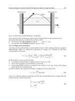
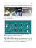
A diagram of the microchip is shown in Figure 16.3. Cells were
cultured on-chip in a microchamber and incubated at 37°C by a Peltier
element-based temperature control device. In order to stimulate NO produc-
tion, LPS in medium was introduced through a reservoir upstream from the
cells. The NO quickly degraded to generate NO
2
–
and NO
3
–
. Nitrate reductase
was introduced through another reservoir to reduce the nitrate to nitrite. The
resulting nitrite (from both NO
2
–
and NO
3
–
) was then reacted with sulfanil-
amide and N-1-naphthylethylendiamine to form a colored product that was
detected using a thermal lens microscope.
Another application of microchips to clinical analysis is immunoassays.
These are frequently used to detect the presence of certain proteins or anti-
bodies in blood or other tissues. Examples of microchip assays of this type
include those for simple protein analytes such as bovine serum albumin
(BSA)
51,52
or IgG.
51,53,54
Other on-chip assays are more complex. One example
is a chip that is designed to aid in the diagnosis of Duchene muscular
dystrophy.
55
In this assay, genomic DNA was extracted from whole blood
and amplified using an on-chip IR-mediated polymerase chain reaction
FIGURE 16.3
Microsyringe pumps, microchip, and temperature control device for bioassay of NO release
from macrophages. (From Goto, M. et al., Anal. Chem., 77, 2125, 2005. With permission.)
Switching valve
Cells
Medium
LPS in medium
Nitrate reductase
Sulranilamide
N-1-Naphtylethylendiamine
TLM detection
Waste
Te mperature
control device
37°C
50°C
20°C
DK532X_book.fm Page 454 Friday, November 10, 2006 3:31 PM
© 2007 by Taylor & Francis Group, LLC
Pharmaceutical Analysis Using Bio-MEMS 455
(PCR). The resulting DNA related to the disease was detected following
electrophoretic separation.
Wang et al. have coupled an enzymatic bioassay with an electrophoretic
separation on-chip for the measurement of the renal markers creatine, cre-
atinine, p-aminohippuric acid, and uric acid.
56
These markers are routinely
monitored to assess kidney function. Sample and a mixture of creatinase,
creatininase, and sarcosine oxidase were combined and allowed to react.
The enzyme reactions produced hydrogen peroxide, which is neutral and
can be electrophoretically separated from other anionic analytes of interest,
urate, and p-aminohippuric acid. Amperometric detection was used for
quantitation.
In large clinical labs, most assays take place at a location that is far from
the patient. In these cases, the analysis can take a great deal of time and the
sample may be mishandled, mislabeled, or lost. Microfluidic devices offer
the opportunity for point-of-care analysis, giving both the patient and the
doctor instant feedback. These small, fast, and disposable devices offer the
potential for quick, less error-prone analyses—at the doctors’ office or at
of microchip-based clinical assays.
16.4.2 Therapeutic Drug Monitoring
Microchip-based assays have also been developed for therapeutic drug mon-
theophylline was developed by Chiem and Harrison.
51,85
Theophylline is an
antiasthmatic drug. Physicians can adjust the dosage for maximum efficacy
if serum theophylline levels are followed. In this study, the antibody, labeled
theophylline, and sample serum were mixed on-chip by electroosmotic
pumping. The antibodies were allowed to react with the antigen and then
the bound versus free fractions were separated by electrophoresis and
detected by fluorescence. Limits of detection for theophylline of 26 mg/L in
serum were achieved.
Vrouwe et al.
86
created a microchip analysis system for measurement of
Li
+
in whole blood. Li
+
is normally not present in the body; however, it is
used in the treatment of manic-depressive illnesses. The upper therapeutic
level of this drug is dangerously close to toxic concentrations; therefore,
careful monitoring of blood concentration is important. Vrouwe exploited
the fact that in an electric field, Li
+
ions move more quickly than the much
larger blood cells. The sample was injected electrokinetically, and Li
+
was
loaded into the separation channel before the blood cells had a chance to
enter the injection T. Glucose was also added to the run buffer to match the
osmotic strength of the run buffer to that of blood so that the cells did not
lyse and release contents that could interfere with Li
+
detection. Because
sodium concentrations are high and fairly constant in blood, sodium was
used as an internal standard. The Li
+
peak areas were normalized to Na
+
DK532X_book.fm Page 455 Friday, November 10, 2006 3:31 PM
home. Table 16.1 lists a majority of the current research toward development
itoring as Table 16.2 shows. An on-chip competitive immunoassay for serum
© 2007 by Taylor & Francis Group, LLC
Pharmaceutical Analysis Using Bio-MEMS 457
16.4.3 High-Throughput Screening
As mentioned previously, microchips and bio-MEMS are of great value to
the pharmaceutical industry for the purpose of high-throughput screening.
TABLE 16.2 (continued)
Therapeutic Drug Monitoring
Analyte Significance Device/Technique Notes
Lithium Used as treatment in
manic-depressive
illness, therapeutic
range dangerously
close to toxic level
Microchip
electrophoresis
separation of Li+,
K+, and Na+ with
AC conductivity
detection
86
Whole blood mixed with
anticoagulant, loaded
on-chip, channels
coated to resist
contamination by
proteins, voltage
controlled so that
sample entered
separation channel, yet
RBC did not
Phenytoin Anticonvulsant Competitive
immunoassay of
whole blood with
fluorescence
detection
91
T-sensor allows diffusion
of side-by-side streams
of antigen and antibody;
binding of antigen
(small and thus fast) to
antibody (larger thus
slower) slows their
diffusion; diffusion
profile changes
compared to profile of
freely diffusing antigen.
Albumin bound to
iophenoxate to decrease
binding assay
interference.
General
immunoassay
microfabrication
technique
Patterning
protein antigens
on-chip with
surface plasmon
resonance, then
probed antigens
with complement-
ary antibodies to
visualize
patterning
92
Fabrication of chip
microarray for
immunoassay
Theophylline Drug for respiratory
diseases
On-chip
competitive
immunoassay,
detection down to
1.25 ng/mL, linear
in therapeutic
range
51,85,93
Reagent and serum
samples mixed, reacted,
separated, and analyzed
all on one chip
DK532X_book.fm Page 457 Friday, November 10, 2006 3:31 PM
© 2007 by Taylor & Francis Group, LLC
458 Bio-MEMS: Technologies and Applications
Multiple channels and detectors on one chip greatly increase the number of
analyses that can be run. To that end, several research groups have developed
microchips to investigate high volumes of samples. This enables drug screen-
ing of molecular libraries to identify successful drug candidates.
94
Currently, many high-throughput screenings involve microarrays. Numer-
ous physiological processes involve protein–carbohydrate interactions, and
the ideal microanalysis tool to study these interactions in the high-through-
put format is the carbohydrate microarray.
95,96
These chips consist of carbo-
hydrates immobilized on glass slides. Detection is external, frequently
fluorescence detection. A high-throughput carbohydrate array microchip
was developed and used to determine the binding affinities between lectins
and carbohydrates.
97
A different carbohydrate microarray was used to screen
85 compounds to find inhibitors of fucosyltransferases.
98
Another form of high-throughput screening, multiplexed enzyme assay,
was developed to screen enzymatic activity of MAP, IR, and PKA kinases.
51,99
These assays were especially valuable because they were multiplexed; three
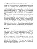
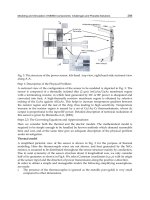
FIGURE 16.4
Electropherograms of separations of a mixture of (1) serotonin, (2) propranolol, (3) 3-phenoxy-
1,2-propandiol, and (4) tryptophan using different detection systems. (a) Conventional capillary
electrophoresis with UV absorbance detection. (b) Microchip electrophoresis with deep UV
fluorescence detection. (c) Commercial microchip electrophoresis system with UV absorbance
detection. (From Schulze, P. et al., Anal. Chem., 77, 1325, 2005. With permission.)
2
50 75
1
2
3
4
Absorbance
(a)
1
2
3
4
Fluorescence
(b)
t (S)t (S)
61830
(c)
1
2
3
4
5101520
x (mm)
Absorbance
DK532X_book.fm Page 458 Friday, November 10, 2006 3:31 PM
© 2007 by Taylor & Francis Group, LLC
Pharmaceutical Analysis Using Bio-MEMS 459
enzymes, each from a different kinase family, were assayed simultaneously
within each channel. Although sample preparation was conducted off-chip,
enzymes and product were separated in a double-T microchip design. Using
this device, drugs were screened for activity, cross-reactivity, specificity, and
potential side effects. In a separate high-throughput application, a 6-channel
microfluidic immunoreactor/immunoassay was developed for the simulta-
neous assay of ovalbumin and antiestradiol within 30 to 60 s.
51,100
Another
high-throughput immunoassay was used to screen affinity complexes of
phenobarbitol antibody and nine barbiturates, including phenobarbitol.
Sample loading, washing, and dissociation steps were performed on-chip,
and the device was then coupled to ESI-MS for detection.
101
Tabuchi et al. developed an integrated cell-culture chip that incorporated
protein separation and detection along with cell culture. Washing, stimula-
tion, and lysis could be accomplished on-chip and were coupled to a com-
mercial Agilent microelectrophoresis chip.
102
The culture chip contained 48
to 96 wells 5 to 6.5 mm in diameter, with a second layer that contained
molded cups that fit into the wells of the first chip. Jurkat cells were cultured
in the first row of wells in the strip of cups that fit into those wells. For a
medium change, the cups in which the cells were cultured were removed
and placed into the next row of wells, which were filled with fresh medium.
This process was repeated for each new step, that is, stimulation, lysis, and
protein extraction, until finally the cups were placed in wells fitted with the
wires for the electrophoretic separation chip. This Agilent chip has 12 chan-
nels; in this application, 11 samples from the cells and a protein ladder sample
were run and detected by LIF. The cell density remained constant in contrast
to conventional cell culture and CE analysis, where cells are consistently lost
to dilution, washing, medium change, and pipetting. Ultimately, this device
enabled analysis of extracted proteins without sample loss at a rate of 12
samples per minute.
DNA sequencing is the most popular application of microchips in the high-
cussion of this topic will be left to that chapter.
16.5 Conclusion
Although many examples of useful clinical and bioanalytical pharmaceutical
applications have been presented in this chapter, most of the bio-MEMS used
in these are prototypes. There is still much work to be done to improve the
limits of detection, reproducibility, and ruggedness of these devices. In addi-
tion, the integration and fabrication of several components onto a single chip
has been accomplished by only a few groups thus far. However, the potential
utility of these microchip devices for high-throughput and point-of-care
analyses makes this research well worth the effort.
DK532X_book.fm Page 459 Friday, November 10, 2006 3:31 PM
throughput world; however, Chapter 13 addresses DNA directly, and dis-
© 2007 by Taylor & Francis Group, LLC
460 Bio-MEMS: Technologies and Applications
References
1. Gawron, A.J., Martin, R.S., and Lunte, S.M., Microchip electrophoretic separa-
tion systems for biomedical and pharmaceutical analysis, Eur. J. Pharm. Sci.,
14, 1, 2001.
2. Kricka, L.J. and Wilding, P., Micromachining: a new direction for clinical ana-
lyzers, Pure Appl. Chem., 68, 1831, 1996.
3. Verpoorte, E., Microfluidic chips for clinical and forensic analysis, Electrophore-
sis, 23, 677, 2002.
4. Wang, J. et al., Single-channel microchip for fast screening and detailed iden-
tification of nitroaromatic explosives or organophosphate nerve agents, Anal.
Chem., 74, 1187, 2002.
5. Fang, Q., Xu, G.M., and Fang, Z.L., A high-throughput continuous sample
introduction interface for microfluidic chip-based capillary electrophoresis sys-
tems, Anal. Chem., 74, 1223, 2002.
6. Alarie, J.P., Jacobson, S.C., and Ramsey, J.M., Electrophoretic injection bias in
a microchip valving scheme, Electrophoresis, 22, 312, 2001.
7. Jacobson, S.C., Ermakov, S.V., and Ramsey, J.M., Minimizing the number of
voltage sources and fluid reservoirs for electrokinetic valving in microfluidic
devices, Anal. Chem., 71, 3273, 1999.
8. Roddy, E.S., Xu, H., and Ewing, A.G., Sample introduction techniques for
microfabricated separation devices, Electrophoresis, 25, 229, 2004.
9. Attiya, S. et al., Design of an interface to allow microfluidic electrophoresis
chips to drink from the fire hose of the external environment, Electrophoresis,
22, 318, 2001.
10. Chen, S.H. et al., Flow-through sampling for electrophoresis-based microchips
and their applications for protein analysis, Anal. Chem., 74, 5146, 2002.
11. Huynh, B.H. et al., On-line coupling of microdialysis sampling with microchip-
based capillary electrophoresis, Anal. Chem., 76, 6440, 2004.
12. Wilding, P. et al., Integrated cell isolation and polymerase chain reaction anal-
ysis using silicon microfilter chambers, Anal. Biochem., 257, 95, 1998.
13. Foote, R.S. et al., Preconcentration of proteins on microfluidic devices using
porous silica membranes, Anal. Chem., 77, 57, 2005.
14. Xu, N. et al., A microfabricated dialysis device for sample cleanup in electro-
spray ionization mass spectrometry, Anal. Chem., 70, 3553, 1998.
15. Yu, C. et al., Preparation of monolithic polymers with controlled porous prop-
erties for microfluidic chip applications using photoinitiated free-radical poly-
merization, J. Polym. Sci., Part A: Polym. Chem., 40, 755, 2002.
16. Kutter, J.P., Jacobson, S.C., and Ramsey, J.M., Solid phase extraction on microf-
luidic devices, J. Microcolumn Sep., 12, 93, 2000.
17. Oleschuk, R.D. et al., Trapping of bead-based reagents within microfluidic
systems: on-chip solid-phase extraction and electrochromatography, Anal.
Chem., 72, 585, 2000.
18. Delaunay-Bertonicini, N. and Hennion, M.C., Immunoaffinity solid-phase ex-
traction for pharmaceutical and biomedical trace-analysis-coupling with HPLC
and CE-perspectives, J. Pharm. Biomed. Anal., 34, 717, 2004.
19. Giordano, B. et al., Microchip laser-induced fluorescence detection of proteins
at submicrogram per milliliter levels mediated by dynamic labeling under
pseudonative conditions, Anal. Chem., 76, 4705, 2004.
DK532X_book.fm Page 460 Friday, November 10, 2006 3:31 PM
© 2007 by Taylor & Francis Group, LLC
Pharmaceutical Analysis Using Bio-MEMS 461
20. Gottschlich, N. et al., Integrated microchip-device for the digestion, separation
and postcolumn labeling of proteins and peptides, J. Chromatog. B, 745, 243,
2000.
21. Jacobson, S.C. et al., Microchip structures for submillisecond electrophoresis,
Anal. Chem., 70, 3476, 1998.
22. Veenstra, T.T. et al., Characterization method for a new diffusion mixer applicable
in micro flow injection analysis systems, J. Micromech. Microeng., 9, 199, 1999.
23. Eijkel, J.C.T. et al., Micromachined heated chemical reactor for pre-column
derivatisation, J. Chromatog. A, 815, 265, 1998.
24. Unger, M.A. et al., Monolithic microfabricated valves and pumps by multilayer
soft lithography, Science, 288, 113, 2000.
25. Jacobson, S.C., McKnight, T.E., and Ramsey, J.M., Microfluidic devices for elec-
trokinetically driven parallel and serial mixing, Anal. Chem., 71, 4455, 1999.
26. Weinberger, R., Practical Capillary Electrophoresis. 2nd ed., San Diego, CA: Aca-
demic Press, 2000.
27. Culbertson, C.T., Jacobson, S.C., and Ramsey, J.M., Microchip devices for high-
efficiency separations, Anal. Chem., 72, 5814, 2000.
28. Han, J. and Singh, A.K., Rapid protein separations in ultra-short microchannels:
microchip sodium dodecyl sulfate-polyacrylamide gel electrophoresis and iso-
electric focusing, J. Chromatog. A, 1049, 205, 2004.
29. Rodriguez, I., Lee, H.K., and Li, S.F., Microchannel electrophoretic separation
of biogenic amines by micellar electrokinetic chromatography, Electrophoresis,
20, 118, 1999.
30. Shackman, J.G. et al., Perfusion and chemical monitoring of living cells on a
microfluidic chip, Lab Chip, 5, 56, 2005.
31. Phillips, T.M., Rapid analysis of inflammatory cytokines in cerebrospinal fluid
using chip-based immunoaffinity electrophoresis, Electrophoresis, 25, 1652, 2004.
32. Wang, J., Ibanez, A., and Chatrathi, M.P., On-chip integration of enzyme and
immunoassays: simultaneous measurements of insulin and glucose, J. Am.
Chem. Soc., 125, 8444, 2003.
33. Wang, J., Ibanez, A., and Chatrathi, M.P., Microchip-based amperometric im-
munoassay using redox tracers, Electrophoresis, 23, 3744, 2002.
34. Wooley, A.T. et al., Capillary electrophoresis chips with integrated electrochem-
ical detection, Anal. Chem., 70, 684, 1998.
35. Wang, J., Tian, B.M., and Sahlin, E., Micromachined electrophoresis chips with
thick-film electrochemical detectors, Anal. Chem., 71, 5436, 1999.
36. Tantra, R. and Manz, A., Integrated potentiometric detector for use in chip-
based flow cells, Anal. Chem., 72, 2875, 2000.
37. Martin, R.S. et al., In-channel electrochemical detection for microchip capil-
lary electrophoresis using an electrically isolated potentiostat, Anal. Chem.,
74, 1136, 2002.
38. Lui, Y. et al., Electrophoretic separation of proteins on a microchip with non-
covalent, postcolumn labeling, Anal. Chem., 72, 4608, 2000.
39. Tamaki, E. et al., Single cell analysis by a scanning thermal lens microscope
with a microchip: direct monitoring of cytochrome-c distribution during apo-
ptosis process, Anal. Chem., 74, 1560, 2002.
40. Schulze, P. et al., Deep UV laser-induced fluorescence detection of unlabeled
drugs and proteins in microchip electrophoresis, Anal. Chem., 77, 1325, 2005.
41. Chau, L.K. et al., Microfabricated silicon flow-cell for optical monitoring of
biological fluids, Anal. Sci., 15, 721, 1999.
DK532X_book.fm Page 461 Friday, November 10, 2006 3:31 PM
© 2007 by Taylor & Francis Group, LLC
462 Bio-MEMS: Technologies and Applications
42. Deng, Y. et al., Chip-based capillary electrophoresis/mass spectrometry deter-
mination of carnitines in human urine, Anal. Chem., 73, 639, 2001.
43. Deng, Y., Zhang, H., and Henion, J., Chip-based quantitative capillary electro-
phoresis/mass spectrometry of drugs in human plasma, Anal. Chem., 73, 1432,
2001.
44. Li, J. et al., Integration of microfabricated devices to capillary electrophoresis-
electrospray mass spectrometry using a low dead volume connection: applica-
tion to rapid analyses of proteolytic digests, Anal. Chem., 71, 3036, 1999.
45. Zhang, B. et al., Microfabricated devices for capillary electrophoresis-electro-
spray mass spectrometry, Anal. Chem., 71, 3258, 1999.
46. Grayson, A.C.R. et al., A bioMEMS review: MEMS technology for physiologi-
cally integrated devices, IEEE, 2, 6, 2004.
47. Moorthy, J. et al., Microfluidic tectonics platform: a colorimetric, disposable
botulinum toxin enzyme-linked immunosorbant assay system, Electrophoresis,
25, 1705, 2004.
48. Pasas-Farmer, S.A., New analytical methods for the determination of homocys-
teine in human plasma, Ph.D. thesis, Pharmaceutical Chemistry, University of
Kansas, Lawrence, 2004.
49. Kikura-Hanajiri, R., Martin, R.S., and Lunte, S.M., Indirect measurement of
nitric oxide production by monitoring nitrate and nitrite using microchip elec-
trophoresis with electrochemical detection, Anal. Chem., 74, 6370, 2002.
50. Goto, M. et al., Development of a microchip-based bioassay system using
cultured cells, Anal. Chem., 77, 2125, 2005.
51. Guijt, R.M., Baltussen, E., and Van Dedem, G.W.K., Use of bioaffinity interac-
tions in electrokinetically controlled assays on microfabricated devices, Electro-
phoresis, 23, 823, 2002.
52. Qui, C.X. and Harrison, D.J., Integrated self-calibration via electrokinetic solvent
proportioning for microfluidic immunoassays, Electrophoresis, 22, 3949, 2001.
53. Martynova, L. et al., Fabrication of plastic microfluid channels by imprinting
methods, Anal. Chem., 69, 4783, 1997.
54. Dodge, A. et al., Electrokinetically driven microfluidic chips with surface-mod-
ified chambers for heterogeneous immunoassays, Anal. Chem., 73, 3400, 2001.
55. Ferrance, J.P. et al., Developments toward a complete micro-total analysis system
for Duchene muscular dystrophy diagnosis, Anal. Chim. Acta, 500, 223, 2003.
56. Wang, J. and Chatrathi, M.P., Microfabricated electrophoresis chip for bioassay
of renal markers, Anal. Chem., 75, 525, 2003.
57. Deng, Y., Zhang, H., and Henion, J., Chip-based quantitative capillary electro-
phoresis/mass spectrometry determination of drugs in human plasma, Anal.
Chem., 73, 1432, 2001.
58. Stefan, R. et al., Simultaneous determination of L- and D-carnitine using a
sequential injection analysis/amperometric biosensors system, J. Pharm.
Biomed. Anal., 33, 323, 2003.
59. Kameoka, J. et al., A polymeric microfluidic chip for CE/MS determination of
small molecules, Anal. Chem., 73, 1935, 2001.
60. Uhlig, A. et al., Miniaturised ion-selective sensor chip for potassium measure-
ment in a biomedical application, Sens. Actuators B, 34, 252, 1996.
61. Lichtenberg, J., De Rooij, N.F., and Verpoorte, E., A microchip electrophoresis
system with integrated in-plane electrodes for contactless conductivity detec-
tion, Electrophoresis, 23, 3769, 2002.
DK532X_book.fm Page 462 Friday, November 10, 2006 3:31 PM
© 2007 by Taylor & Francis Group, LLC
Pharmaceutical Analysis Using Bio-MEMS 463
62. Bruno, A.E. et al., All-solid-state miniaturized fluorescence sensor array for the
determination of critical gases and electrolytes in blood, Anal. Chem., 69, 507, 1997.
63. Wang, J. et al., Microfabricated electrophoresis chips for simultaneous bioas-
says of glucose, uric acid, ascorbic acid, and acetaminophen, Anal. Chem., 72,
2514, 2000.
64. Freaney, R. et al., Novel instrumentation for real-time monitoring using miniatur-
ized flow systems with integrated biosensors., Ann. Clin. Biochem., 34, 291, 1997.
65. Dempsey, E. et al., Design and development of a miniaturised total chemical
analysis system for on-line lactate and glucose monitoring in biological sam-
ples, Anal. Chim. Acta, 346, 341, 1997.
66. Nakamura, H. et al., A compactly integrated flow cell with a chemiluminescent
FIA system for determining lactate concentration in serum, Anal. Chem., 73,
373, 2001.
67. Bohm, S. et al., A flow-through amperometric sensor based on dialysis tubing
and free enzyme reactors, Biosens. Bioelectron., 16, 391, 2001.
68. Du, Y. et al., Direct electrochemical detection of glucose in human plasma on
capillary electrophoresis microchips, Electrophoresis, 25, 3853, 2004.
69. Munro, N.J. et al., Indirect fluorescence detection of amino acids on electro-
phoretic microchips, Anal. Chem., 72, 2765, 2000.
70. Von Heeren, F. et al., Micellar electrokinetic chromatography separations and
analyses of biological samples on a cyclic planar microstructure, Anal. Chem.,
68, 2044, 1996.
71. Kennedy, R.T. et al., In vivo neurochemical monitoring by microdialysis and
capillary separations, Curr. Opin. Chem. Biol., 6, 659, 2002.
72. Haskins, W.E. et al., Discovery and neurochemical screening of peptides in
brain extracellular fluid by chemical analysis of in vivo microdialysis samples,
Anal. Chem., 76, 5523, 2004.
73. Pasas, S.A. et al., Detection of homocysteine by conventional and microchip
capillary electrophoresis/electrochemistry, Electrophoresis, 23, 759, 2002.
74. Ceriotti, L. et al., Low-density lipoprotein analysis in microchip capillary elec-
trophoresis systems, Electrophoresis, 23, 3615, 2002.
75. Chen, Y. et al., Plastic microchip electrophoresis for analysis of PCR products
of hepatitis C virus, Clin. Chem., 45, 1938, 1999.
76. Price, A.K. et al., Deformation-induced release of ATP from erythrocytes in a
poly(dimethylsiloxane)-based microchip with channels that mimic resistance
vessels, Anal. Chem., 76, 4849, 2004.
77. Sorouraddin, H.M., Hibara, A., and Kitamori, T., Use of a thermal lens micro-
scope in integrated catecholamine determination on a microchip, Fresenius J.
Anal. Chem., 371, 91, 2001.
78. Sorouraddin, H.M. et al., Integrated FIA for the determination of ascorbic acid
and dehydroascorbic acid in a microfabricated glass-channel by thermal-lens
microscopy, Anal. Sci., 16, 1033, 2000.
79. Suzuki, S. et al., Rapid analysis of amino sugars by microchip electrophoresis
with laser-induced fluorescence detection, Electrophoresis, 22, 4023, 2001.
80. Fanguy, J.C. and Henry, C.S., The analysis of uric acid in urine using microchip
capillary electrophoresis with electrochemical detection, Electrophoresis, 23,
767, 2002.
81. Qin, J. et al., Simultaneous and ultrarapid determination of reactive oxygen
species and reduced glutathione in apoptotic leukemia cells by microchip elec-
trophoresis, Electrophoresis, 26, 1155, 2005.
DK532X_book.fm Page 463 Friday, November 10, 2006 3:31 PM
© 2007 by Taylor & Francis Group, LLC
464 Bio-MEMS: Technologies and Applications
82. Roper, M.G. et al., Microfluidic chip for continuous monitoring of hormone
secretion from live cells using an electrophoresis-based immunoassay, Anal.
Chem., 75, 4711, 2003.
83. Spence, M.D. et al., Amperometric determination of nitric oxide derived from
pulmonary artery endothelial cells immobilized in a microchip channel, Ana-
lyst, 129, 995, 2004.
84. Koutny, L.B. et al., Microchip electrophoretic immunoassay for serum cortisol,
Anal. Chem., 68, 18, 1996.
85. Chiem, N.H. and Harrison, D.J., Microchip systems for immunoassay: an in-
tegrated immunoreactor with electrophoretic separation for serum theophyl-
line determination, Clin. Chem., 44, 591, 1998.
86. Vrouwe, E.X., Luttge, R., and Van Den Berg, A., Direct measurement of lithium
in whole blood using microchip capillary electrophoresis with integrated con-
ductivity detection, Electrophoresis, 25, 1660, 2004.
87. Kriikku, P. et al., Isotachophoresis of β-blockers in a capillary and on a poly(me-
thyl methacrylate) chip, Electrophoresis, 25, 1687, 2004.
88. Tanyanyiwa, J. and Hauser, P.C., Capillary and microchip electrophoresis of
basic drugs with contactless conductivity detection, Electrophoresis, 25, 3010,
2004.
89. Ramseier, A., Von Heeren, F., and Thormann, W., Analysis of fluorescein isothio-
cyanate derivatized amphetamine and analogs in human urine by capillary
electrophoresis in chip-based and fused-silica capillary instrumentation, Elec-
trophoresis, 19, 2967, 1998.
90. Wallenborg, S.R. et al., On-chip chiral and achiral separation of amphetamine
and related compounds labeled with 4-fluoro-7-nitrobenzofurazane, Electro-
phoresis, 21, 3257, 2000.
91. Hatch, A. et al., A rapid diffusion immunoassay in a T-sensor, Nature Biotech.,
19, 461, 2001.
92. Kanda, V. et al., Label-free reading of microarray-based immunoassays with
surface plasmon resonance imaging, Anal. Chem., 76, 7257, 2004.
93. Chiem, N. and Harrison, D.J., Microchip-based capillary electrophoresis for
immunoassays: analysis of monoclonal antibodies and theophylline, Anal.
Chem., 69, 373, 1997.
94. Sundberg, S.A. et al., Microchip-based systems for target validation and HTS,
Drug Discov. Today, 5, 92, 2000.
95. Khan, I., Desai, D.V., and Kumar, A., Carbochips: a new energy for old bio-
builders, J. Biosci. Bioeng., 98, 331, 2004.
96. Wang, D., Carbohydrate microarrays, Proteomics, 3, 2167, 2003.
97. Park, S. et al., Carbohydrate chips for studying high-throughput carbohydrate-
protein interactions, J. Am. Chem. Soc., 126, 4812, 2004.
98. Bryan, M.C., Lee, L.V., and Wong, C.H., High-throughput identification of
fucosyltransferase inhibitors using carbohydrate microarrays, Bioorg. Med.
Chem. Lett., 14, 3185, 2004.
99. Xue, Q. et al., Multiplexed enzyme assays in capillary electrophoresis single-
use microfluidic devices, Electrophoresis, 22, 4000, 2001.
100. Cheng, S.B. et al., Development of a multichannel microfluidic analysis system
employing affinity capillary electrophoresis for immunoassay, Anal. Chem., 73,
1472, 2001.
DK532X_book.fm Page 464 Friday, November 10, 2006 3:31 PM
© 2007 by Taylor & Francis Group, LLC
Pharmaceutical Analysis Using Bio-MEMS 465
101. Jiang, Y. et al., Integrated plastic microfluidic devices with ESI-MS for drug
screening and residue analysis, Anal. Chem., 73, 2048, 2001.
102. Tabuchi, M. and Baba, Y., Self-contained on-chip cell culture and pretreatment
system, J. Proteom. Res., 3, 871, 2004.
DK532X_book.fm Page 465 Friday, November 10, 2006 3:31 PM
© 2007 by Taylor & Francis Group, LLC