Atlas of the Diabetic Foot - part 5 potx
Bạn đang xem bản rút gọn của tài liệu. Xem và tải ngay bản đầy đủ của tài liệu tại đây (905.31 KB, 22 trang )
90 Atlas of the Diabetic Foot
(a ‘painful–painless foot’) is a quite com-
mon feature of neuropathic diabetes.
Keywords: Neuropathic ulcer; granulating
tissue
NEUROPATHIC ULCER
OVER A COLLAPSED
MIDFOOT
A typical neuropathic ulcer under a bony
prominence in a patient with midfoot
collapse due to neuro-osteoarthropathy is
shown in Figure 5.4. Callus formation is
present at the margins of the ulcer, while
Figure 5.4 Neuropathic ulcer over a bony
prominence in a patient with neuro-osteoarth-
ropathy
its base is clean, covered by healthy gra-
nulating tissue.
Therapeutic footwear was prescribed
(extra depth shoes with an orthotic insole
and a window under the ulcerated area)
and the patient was advised to minimize his
activities. The ulcer healed in 3 months.
Ulcers in patients with midfoot collapse
recur very often. Prevention of new ulcers
over the same bony prominence is achieved
by prophylactic surgery (osteotomy of the
prominent bone). Preservation of plantar
ligaments is essential, since their extensive
resection may cause progression of the
rocker bottom deformity.
Keywords: Neuropathic ulcer; bony promi-
nence; prophylactic osteotomy
NEUROPATHIC ULCER
UNDER FOURTH
METATARSAL HEAD
A 74-year-old female patient with type 2
diabetes diagnosed at the age of 62 years,
was referred to the outpatient diabetic foot
clinic because of callus formation on her
right sole. She was being treated with
insulin and had a history of hypertension
and ischemic heart disease.
On examination she was found to have
severe peripheral neuropathy and normal
peripheral pulses. In addition, significant
muscle atrophy of her feet, claw toes
and a hemorrhagic callus on the fourth
metatarsal head of her right foot were found
(Figure 5.5). An impressive finding was the
palpation of her metatarsal heads just below
the skin as the fat pads had been displaced
anteriorly. After callus removal a super-
ficial ulcer was revealed (Figure 5.6). An
anteroposterior radiograph showed diffuse
Neuropathic Ulcers at Various Sites 91
Figure 5.5 Hemorrhagic callus under the
fourth metatarsal head. Claw toes and prominent
metatarsal heads are also present
Figure 5.6 A neuropathic ulcer in the same
patient whose foot is shown in Figure 5.5
demineralization of the foot and signifi-
cant widening with periosteal reaction at
the metatarsal heads (Figures 5.7 and 5.8).
The patient was advised to rest. Extra depth
Figure 5.7 Diffuse osteopenia and significant
widening with periosteal reaction on the meta-
tarsal heads can be seen in this X-ray of the foot
shown in Figure 5.5
shoes and orthotic insoles were prescribed
in order to accommodate her deformed toes
and relieve the load under the metatarsal
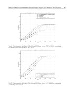
heads. Post-debridement in-shoe pressures
when she used her own shoes showed a
significant load under her metatarsal heads
(Figure 5.9 Panel A). The maximum pres-
sure in this area was 282 kPa; however,
after insertion of an orthotic insole the
maximum in-shoe pressure was reduced to
155 kPa (Figure 5.9 Panel B). The ulcer
healed in 8 weeks.
Reduced thickness of the fat pad is asso-
ciated with high plantar pressures. Although
some authors have suggested that thresh-
old pressures of 500–1000 kPa may lead
to the development of foot ulceration when
walking barefoot, it seems that each patient
has an individual threshold. In the present
case the maximum pressure was obviously
below this threshold. However, high plantar
92 Atlas of the Diabetic Foot
Figure 5.8 Significant widening with periosteal reaction of the first three metatarsal heads (same
patient whose foot is shown in Figures 5.5–5.7)
Figure 5.9 Plantar pressures before (A) and
after (B) orthotic insoles in the patient whose
foot is shown in Figures 5.5–5.7
pressures alone do not cause foot ulcera-
tion; a combination of different risk factors
(mentioned in Chapter 1) is necessary for
the development of ulceration.
Demineralization of the foot bones is
not common, but when this occurs it sig-
nifies an adequate circulation, which is a
prerequisite for bone resorption. Localized,
mature periosteal reaction and demineral-
ization involving metatarsal heads is com-
mon in diabetic patients with neuropathy. Its
etiology is poorly understood. Focal oste-
olysis of phalanges, metatarsal heads, and
other single foot bones, as well as stress frac-
tures of the metatarsal heads can also be seen
in neuropathic patients. Bone resorption at
the phalanges may be so extensive that a part
or even a whole phalanx may be resorbed.
Metatarsal resorption usually starts from the
metaphysis and extends to the epiphysis
sparing the diaphysis. Bones which have
become demineralized may have a pencil-
like appearance.
Keywords: Neuropathic ulcer; plantar pres-
sures, periosteal reaction
Neuropathic Ulcers at Various Sites 93
NEUROPATHIC ULCERS
UNDER PROMINENT
METATARSAL HEADS
This 32-year-old type 1 female diabetic
patient, diagnosed at the age of 16 years,
attended the outpatient diabetic foot clinic
for chronic neuropathic ulcers of her feet.
She was treated with intensive insulin treat-
ment. The patient had a renal transplant
at the age of 30 years, because of end-
stage renal failure due to diabetes, and
she had laser treatment on both eyes at
the age of 28 years. Soon after her trans-
plantation she noticed a bulla under her
last three left metatarsal heads which read-
ily ruptured and a superficial ulcer devel-
oped. She also reported an ulcer of 2 years’
duration under the third metatarsal head
of her right foot. She had never been
instructed in foot care and had never worn
Figure 5.10 Neuropathic ulcers under promi-
nent metatarsal heads and on the midsole. Claw
toes and dry skin are also apparent
the correct footwear. She had two small
children and had not been taking good care
of her f eet. The patient was being treated
with erythropoietin injections, cyclosporin,
methylprednisolone, mycofenolate mofetil
and furosemide.
On examination she was found to have
bounding pedal pulses, and severe dia-
betic neuropathy. The vibration perception
threshold was above 50 V in both feet
bilaterally.
A non-infected neuropathic ulcer was
noted under her left third, fourth and fifth
metatarsal heads. Its dimensions were 3.5 ×
4 × 0.4 cm, and it was surrounded by cal-
lus. A smaller neuropathic ulcer was also
observed under her midsole (Figure 5.10).
Claw toe deformity of her lesser toes, dry
skin and desquamation of the tip of her
third toe were also present. Under her
Figure 5.11 Neuropathic ulcer surrounded by
callus. Claw toe. Right foot of patient whose left
foot is shown in Figure 5.10
94 Atlas of the Diabetic Foot
Figure 5.12 Original in-shoe peak plantar pres-
sures on the left (upper panel) and right foot
(lower panel) of the patient whose feet are
illustrated in Figures 5.10 and 5.11
Figure 5.13 Healing neuropathic ulcers in the
patient whose feet are shown in Figures 5.10–
5.11. Note bunionette deformity at the right foot
right third metatarsal head a neuropathic
ulcer was noted in an area of gross callus
formation, in addition to claw toe defor-
mity (Figure 5.11). A callus was present
under her right fifth metatarsal head over
a bunionette deformity. Mild callus forma-
tion was observed on the heels of both
feet. Onychomycosis affecting a ll toes was
also present (discussed in Chapter 8, see
Figure 8.7).
A plain radiograph did not reveal osteo-
myelitis. Sharp debridement was performed
and therapeutic half shoes were prescribed.
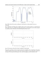
In-shoe peak pressure measurement showed
high pressures under both heels, metatarsal
heads, and halluxes when the patient wore
Figure 5.14 Effect of orthotic insoles and cor-
rect footwear on in-shoe peak plantar pressures
on the left (upper panel) and right foot (lower
panel) in the patient whose feet are illustrated in
Figures 5.10–5.11
Neuropathic Ulcers at Various Sites 95
her own shoes (Figure 5.12). She had
standard treatment on a weekly basis and
the ulcers began to heal slowly. Six months
after her first visit, an ulcer developed
under her left third metatarsal head and
a callus under her right fifth metatarsal
head (Figure 5.13). New shoes were pre-
scribed with orthotic insoles: the in-shoe
peak pressures were reduced from 33.3
to 16.83 N/cm
2
under her right, and from
37.42 to 20.13 N/cm
2
under her left foot
(Figure 5.14).
The patient continued visiting the out-
patient foot clinic almost every week, and
6 months after her first visit her ulcers had
healed.
Keywords: Neuropathic ulcer; peak plan-
tar pressures
ULCERS OVER A CHARCOT
FOOT
The following two figures (before and
after debridement) show the left foot of
a male patient of 62 years of age with
type 2 diabetes diagnosed at the age of
48 years and treated with insulin. A smoker
since the age of 18 years, the patient
had had an ulcer on the plantar aspect
of his left hallux which was complicated
by osteomyelitis and led to amputation
3 years previously. One year before his
first visit to the foot clinic the patient
developed an ulcer on the lateral aspect of
his left foot which resulted in osteomyelitis
and surgical debridement of the metatarsal
bone. After a femoral-popliteal bypass graft
in his left f oot, the patient developed
neuro-osteoarthropathy. He presented to
the outpatient clinic with two painless
ulcers under his first and third metatarsal
heads surrounded by hemorrhagic calluses.
Hyperkeratosis under his fifth metatarsal
head and a scar at the site of the surgical
debridement were noted (Figure 5.15).
The graft was functioning well and the
patient had no claudication. Debridement
of the ulcer under his fourth metatarsal
exposed the bone (Figure 5.16). Cultures
were obtained from the sloughy base of the
ulcer — a positive sign of infection — and
the patient was treated with an empiri-
cal combination of cotrimoxazole and clin-
damycin. The patient did not attend follow-
up, therefore no X-ray or any further studies
are available.
Charcot foot typically does not develop
in patients with peripheral vascular dis-
ease since increased blood supply to the
bone is needed for the osseous tissue to be
overmetabolized. Autonomic sympathetic
neuropathy leads to bone arteriovenous
Figure 5.15 Hallux disarticulation at the meta-
tarsophalangeal joint, callus under first and fifth
metatarsal heads, and deep infected neuropathic
ulcer under the third metatarsal head. Claw toes
96 Atlas of the Diabetic Foot
Figure 5.16 Foot shown in Fig-
ure 5.15 after sharp debridement.
Note bone exposure at the base of
the ulcer under the third metatarsal
head
shunting, hypervascularity and demineral-
ization. Some cases are reported to occur
after bypass surgery of the arteries.
Exposure of the bone denotes osteomy-
elitis and it should be treated accordingly.
Keywords: Neuropathic ulcers; Charcot
foot; osteomyelitis; amputation
A NEUROPATHIC ULCER
UNDER THE HEEL
A 51-year-old female patient with type
2 diabetes since the age of 38 years and
treated with insulin, was referred to the
outpatient diabetic foot clinic because of
a chronic non-healing ulcer under her
right heel. She had good diabetes control
(HBA
1c
: 7.2%). Four months before her
first visit she noticed a painless blister on
the right heel caused by a small stone in
her shoe; the blister ruptured and since the
patient did not feel any pain she did not give
her foot any attention. Some discharge was
present on her socks, but it was the patient’s
daughter who saw a superficial ulcer on the
right heel. The patient visited a primary
care clinic and was advised to clean the
ulcer with povidone iodide and apply clean
dressings every day. A 2-week course of
Neuropathic Ulcers at Various Sites 97
antibiotics was prescribed. She continued
her daily activities and after 4 months the
ulcer was still active.
On examination the patient was found
to have severe diabetic neuropathy with
loss of sensation of pain, temperature, light
touch and vibration. The vibration per-
ception threshold was 36 V on both feet.
Peripheral pulses were normal and the ankle
brachial index was 1.2 and 1.1 in the right
and left foot respectively. A full thick-
ness ulcer with a sloughy base was noted
on the right heel (Figure 5.17). No other
signs of infection were present. An X-ray
did not show involvement of the calca-
neus. Cultures from the base of the ulcer
revealed Staphylococcus aureus.Shewas
treated with amoxicillin–clavulanic acid
for 2 weeks a nd the ulcer was debrided
on a weekly basis; dressings were changed
daily. Meanwhile she was advised to rest
and heel-free shoes to offload pressure
Figure 5.17 Deep heel neuropathic ulcer with
infected sloughy bed caused by trauma
from the ulcerated area were prescribed
(Figure 5.18). After 6 months the ulcer had
healed completely (Figure 5.19).
Bedridden patients develop heel ulcers
or gangrene quite frequently (20–30%),
Figure 5.18 Commercially available heel-free
shoes for the treatment of hindfoot ulcers
Figure 5.19 Hindfoot shown in Figure 5.17
after the ulcer has completely healed
98 Atlas of the Diabetic Foot
Figure 5.20 Neuropathic heel ulcer caused by shoe seam
usually on the posterolateral aspect. Exces-
sive walking in new shoes can cause
blister formation on the posterior aspect
of the heel in patients w ith neuropathy.
Shoe seams may also cause ulcers on the
heel (Figure 5.20). Therefore shoes and
socks without seams are prescribed to
patients with loss of protective sensation.
Heel ulceration is difficult in management
since debridement in this area precludes
functional weight bearing. Major amputa-
tions are often necessary when heel ulcers
are infected.
Keywords: Neuropathic ulcer; heel
BURNS ON TOES
AND FOREFOOT
A 55-year-old male patient with type 2
diabetes since the age of 43 years attended
the outpatient diabetic foot clinic due to
ulcers on his feet. His diabetes was poorly
controlled with sulfonylureas and he had
a history of a disarticulated left great toe
at the metatarsophalangeal joint due to
osteomyelitis.
On examination the patient was febrile;
peripheral pulses were palpable, the ankle
brachial index was 1.2; the vibration per-
ception threshold was over 50 V in both
feet; temperature, light touch and pinprick
sensation were absent as were the Achilles
tendon reflexes. Blood pressure was nor-
mal; no other diabetic complications were
found. HbA
1c
was 11.0%. There was a
perforating dirty ulcer on the outer aspect
of his right foot. A large amount of cal-
lus had built up around the plantar ori-
fice (Figures 5.21 and 5.22). The patient
reported edema of the forefoot which had
recently subsided as was evident from the
scaling of the skin. Callus formation was
also observed over the second, third and
fifth metatarsal heads of the left foot.
The patient was empirically treated with
ciprofloxacin.
Debridement of the callus was car-
ried out. Cultures revealed Staphylococcus
aureus and Escherichia coli. Osteomyelitis
of the fifth metatarsal head was evident on a
plain radiograph (Figure 5.23). The patient
Neuropathic Ulcers at Various Sites 99
Figure 5.21 Perforating, infected neuropathic ulcer under the fifth metatarsal head. Scaling is due
to edema that has subsided
Figure 5.22 Right foot: neuropathic ulcer shown in Figure 5.21. Left foot: hallux disarticulation,
medial displacement of second toe with claw defo rmity; callus formation under second, third and
fifth metatarsal heads
100 Atlas of the Diabetic Foot
Figure 5.23 Plain radiograph of the
right foot of the patient whose foot is
shown in Figure 5.21. Osteomyelitis of
the fifth metatarsal head and the proxi-
mal phalanx of the fifth toe, subluxation
of the metatarsophalangeal joint, cal-
cification of the digital artery between
the first two metatarsals and osteoarthri-
tis of the first distal phalangophalangeal
joint of the hallux are all apparent
continued ciprofloxacin treatment; cotri-
moxazole was added for almost 6 months
and the ulcer gradually healed (Figure 5.24)
with the help of therapeutic shoes.
Instruction in appropriate foot care was
provided. The patient visited the outpa-
tient clinic erratically; callus formation on
the site of the healed ulcer was removed
every 3 months; he refused strict glycemic
control as he was afraid that episodes of
hypoglycemia would jeopardize his posi-
tion at work. He used intermediate-acting
insulin at bedtime and sulfonylureas during
the day. His HbA
1c
remained at 9.0% dur-
ing the following year. Preventive footwear
was not accepted.
The patient attended the clinic 2 years
later because of multiple burns over the
tips of his toes and superficial ulcers over
the fifth metatarsal heads of both feet
(Figure 5.25). He had put his feet in front
of the fire in order to dry out his wet socks.
No pain was felt. Although the patient was
aware of the burns he continued his activi-
ties for a week before this visit.
Full thickness burns were present over
the tips of all toes. Blisters over the
right fifth metatarsal head and the left
fourth and fifth toes were removed and
ulcers had developed since the patient
was still working regularly, despite med-
ical advice to the contrary (Figure 5.26).
Calluses formed around the new plantar
ulcers. Amoxicillin–clavulanic acid treat-
ment was initiated and the patient attended
the diabetic foot clinic on a weekly basis.
Neuropathic Ulcers at Various Sites 101
Figure 5.24 The ulcer shown in Fig-
ure 5.21 after it has almost completely
healed
Figure 5.25 Thermal injury sus-
tained by the patient whose feet are
illustrated in Figure 5.22
102 Atlas of the Diabetic Foot
Figure 5.26 Neuropathic ulcers under the fifth
metatarsal heads and progression of thermal
injury in the patient whose feet are shown in
Figures 5.21–5.25. The patient did not comply
with doctors’ instructions
All ulcers healed within 2 months except
the one on the right great toe, which was
complicated by osteomyelitis and acute soft
tissue infection. Five months after the burn
his right hallux had to be disarticulated.
The patient still refused preventive shoes
and 4 months after this second amputa-
tion new ulcers developed under the fifth
metatarsal heads bilaterally (Figure 5.27).
Keywords: Thermal injury; osteomyelitis
CHRONIC NEUROPATHIC
ULCER COMPLICATED
BY OSTEOMYELITIS
A 55-year-old male patient with type 2
diabetes diagnosed at the age of 50 years
was referred to the outpatient diabetic foot
clinic because of a chronic neuropathic
ulcer on his right foot. He had a history
of hypertension, background retinopathy in
both eyes and diabetic nephropathy (urine
protein 1.5 g/24 h). He had been treated
with sulfonylurea but had discontinued
Figure 5.27 Right hallux disarticula-
tion at the metatarsophalangeal joint
and recurrence of ulcers under the fifth
metatarsal heads (patient whose feet are
shown in Figures 5.21–5.26)
Neuropathic Ulcers at Various Sites 103
the treatment 1 year before his first visit,
when overt nephropathy developed. He had
excellent diabetes control (HBA
1c
:6.4%).
On examination his feet pulses were
bounding (ankle pressure index 1.2 bilater-
ally); he had severe peripheral neuropathy:
no sensation of pain, light touch, vibra-
tion or temperature; the vibration percep-
tion threshold was 48 V on the left and
above 50 V on the right foot. A full thick-
ness clear neuropathic ulcer surrounded by
callus was observed under the right first
metatarsal head, with dimensions of 3 ×
3 × 0.5cm(Figure 5.28). Mild claw defor-
mities of the toes and displacement of the
metatarsal fat pads to the base of the prox-
imal phalanges due to muscle atrophy were
also noted.
Sharp debridement was carried out and
special extra depth shoes with an orthotic
insole were prescribed. C are was taken to
offload pressure from the ulcerated area.
Figure 5.28 Neuropathic ulcer under promi-
nent first metatarsal head. Healthy granulating
tissue can be seen at the base of the ulcer
The patient was advised to limit his daily
activities and he attended the diabetes
foot clinic on a weekly basis. The size
of the ulcer was reduced by half within
4 weeks. Two weeks later, after a profes-
sional trip, the patient visited the clinic
again. His ulcer was infected and a large
amount of callus had formed around it. His
right hallux had a ‘sausage-like’ appear-
ance and signs of infection were observed
(redness and edema). A culture from the
base of the ulcer revealed the presence
of Staphylococcus aureus and Enterobacter
cloacae post-debridement. A radiograph at
that time showed mild erosion of the first
metatarsal head.
The patient was given treatment with
cotrimoxazole and clindamycin. The radio-
graph was repeated 2 weeks later and
extensive erosion of the first metatarsal
head was revealed (Figure 5.29). Acute
osteomyelitis was diagnosed. The patient
continued with the antibiotics for 12 weeks
and had regular chiropody treatment on a
weekly basis. The ulcer healed completely
in 20 weeks (Figure 5.30).
Treatment of acute osteomyelitis should
be based on bone cultures when pos-
sible, and should be continued for
6–12 weeks. The commonest pathogen of
Figure 5.29 Erosion of first metatarsal head
with periosteal reaction due to osteomyelitis
(patient whose ulcer is shown in Figure 5.28)
104 Atlas of the Diabetic Foot
Figure 5.30 Healed ulcer of the p atient whose
feet are shown in Figures 5.28 –5.29. Note the
scar over the ulcerated area and callus formation
at the tip of the second toe due to claw deformity
acute osteomyelitis in patients with foot
ulcers is Staphylococcus aureus (60–90%).
Other pathogens include Staphylococ-
cus epidermidis, Escherichia coli, Pseu-
domonas aeruginosa, and other Enterobac-
ter spp. More than one pathogen is often
isolated. In order to achieve therapeutic lev-
els of antibiotics in the bone it is prefer-
able to administer antibiotics intravenously
for the first 2 weeks. However, oral antibi-
otics with good bioavailability (fluoro-
quinolones, clindamycin) may be adequate
for therapy. Treatment regimens for staphy-
lococcal osteomyelitis are as follows:
• Clindamycin 600 mg × 3 orally or 600
mg × 3i.v.
• Fucidic acid 500 mg × 3 orally or 500
mg × 3 in a 500-ml solution delivered
slowly i.v. (over 4–6 h)
• Cotrimoxazole 960 mg × 2 orally or i.v.
• Ciprofloxacin 750 mg × 2 orally, or 400
mg × 3i.v.
• Rifampicin 900 mg × 1 orally or i.v.
• Teicoplanin 600 mg × 1 orally or i.m.
or i.v.
• Vancomycin 500 mg × 4i.v.or1g×
2i.v.
Fluoroquinolones, teicoplanin and van-
comycin should be prescribed for methi-
cillin-resistant staphylococcus only. Fluoro-
quinolones in particular, should always be
combined with another anti-staphylococcal
drug in the first month of treatment, since
it is likely that a resistant strain will prevail
in the infection.
Keywords: Acute osteomyelitis; treatment
Chapter VI
NEURO-ISCHEMIC ULCERS
AT VARIOUS SITES
UNDER HALLUX
NEURO-ISCHEMIC ULCER WITH OSTEOMYELITIS
UNDER THE
HALLUX
ONTHEDORSUM OF THE FOOT
INTERDIGITAL
AT MEDIAL SIDE OF THE FOOT
ON FIRST METATARSAL
ON MIDSOLE AND HEEL
ON FOREFOOT
NEURO-ISCHEMIC ULCER ON THE HALLUX
WITH
OSTEOMYELITIS
ONTHEDORSUM OF CLAW TOES
OVER THE FIFTH METATARSAL HEAD
Atlas of the Diabetic Foot.
N. Katsilambros, E. Dounis, P. Tsapogas and N. Tentolouris
Copyright © 2003 John Wiley & Sons, Ltd.
ISBN: 0-471-48673-6
Neuro-Ischemic Ulcers at Various Sites 107
NEURO-ISCHEMIC ULCER
UNDER HALLUX
A 68-year-old obese male patient with
type 2 diabetes diagnosed at the age of
46 years visited the outpatient diabetic foot
clinic because of two chronic ulcers on
his right hallux. He was treated with a
combination of sulfonylurea during the day
and a mixture of 20% rapid acting–80%
intermediate acting insulin before dinner;
he also had dislipidemia which was being
treated with simvastatin.
On examination, he had severe diabetic
neuropathy (no sensation of light touch,
pin prick, temperature, 5.07 monofilament,
absence of Achilles tendon reflexes and a
vibration perception threshold over 50 V).
Figure 6.1 Infected full thickness neuro-isch-
emic ulcers under the right hallux with edema
and superficial ulceration at the tip and a
subungual hematoma caused by inappropriate
footwear
Peripheral pulses were palpable, but the
ankle brachial index was 0.7 bilaterally.
Two painful full thickness neuro-isch-
emic ulcers with sloughy bed were seen
under his right hallux; edema and super-
ficial ulceration at the tip of his hallux,
and subungual hematomata of the first two
toes were also present (Figure 6.1). There
was no X-ray evidence of osteomyelitis.
Sharp debridement was carried out and
he was treated empirically with amoxi-
cillin–clavulanic acid. No pathogen was
isolated on swab cultures, probably due
to the use of local antiseptics. Offload-
ing of pressure was successful with the
help of appropriate therapeutic footwear
(see Figure 3.36) and the ulcer began to
heal smoothly (Figure 6.2); 3 months after
the initial visit it had healed completely
(Figure 6.3).
Keywords: Neuro-ischemic ulcer
NEURO-ISCHEMIC ULCER
WITH OSTEOMYELITIS
UNDER THE HALLUX
A 67-year-old man who had type 2 diabetes
since the age of 44 years and was being
treated with insulin, visited the diabetic
foot clinic because of an ulcer on his left
hallux. He had acceptable diabetes control.
He had proliferative retinopathy which had
been treated with laser, and intermittent
claudication at 400 m. He had smoked for
27 years.
Twenty days before his visit, he had
worn a new pair of shoes and driven his
car for a long distance. The following day
a blister developed on his left great toe.
Within a day the area became edematous
and black.
On examination the patient was found
to have a deep, foul-smelling ulcer with
108 Atlas of the Diabetic Foot
Figure 6.2 Healing of the ulcers shown in Figure 6.1
Figure 6.3 Hallux ulcers shown in Figures 6.1 and 6.2 after they have fully healed
gangrenous areas and a purulent discharge
over the medial and dorsal aspect of his left
great toe (Figure 6.4).
Peripheral pulses were weak and the
ankle brachial index was 0.8. The patient
could not feel pain, temperature, light
touch, or a 5.07 monofilament. The vibra-
tionperceptionthresholdwas40and45V
at the tips of the left and right great toe
respectively.
Sharp debridement was carried out re-
vealing the underlying bone. An X-ray
showed osteomyelitis of the distal pha-
lanx (Figure 6.5). The patient was treated
empirically with ciprofloxacin and clin-
damycin as a swab culture from the base
of the ulcer had revealed Staphylococcus
aureus. Despite local foot care and the sys-
temic antibiotics, the ulcer was still active
and osteomyelitis spread locally affecting
Neuro-Ischemic Ulcers at Various Sites 109
Figure 6.4 Deep tissue infected neuro-ischemic ulcer with necrotic areas on the left hallux
Figure 6.5 Plain radiograph of the
foot illustrated in Figure 6.4, showing
osteomyelitis involving proximal inter-
phalangeal joint and adjacent phalan-
ges of the left hallux
the proximal phalanx. Three months after
his first visit the patient’s left great toe
was amputated (Figure 6.6). Antimicrobial
treatment was continued for 2 weeks after
the amputation.
The consequences of hallux disarticula-
tion at the metatarsophalangeal joints have
been discussed previously (see Figure 3.32).
Keywords: Neuro-ischemic ulcer; osteo-
myelitis; amputation
NEURO-ISCHEMIC ULCER
ON THE DORSUM
OF THE FOOT
Alarge(3.5 × 2.0 cm) painless neuro-
ischemic ulcer developed on the right foot
of a 68-year-old male patient with type
2 diabetes which had been diagnosed at
the age of 61 years. Peripheral pulses were
110 Atlas of the Diabetic Foot
Figure 6.6 Disarticulation at the metatarso-
phalangeal joint of left hallux of the patient
whose foot is shown in Figures 6.4 and 6.5
weak and the ankle brachial index was
0.6. At the base of the ulcer the fascia of
the dorsum of the forefoot was exposed
(Figure 6.7). There were no signs of infec-
tion. A subungual hematoma of the hallux
and an ulcer which was healing on the sec-
ond toe were noted in addition to significant
ankle edema. The ulcer was a result of fric-
tion between the foot and the forepart of the
patient’s narrow shoe upper (vamp) follow-
ing the rupture of a large blister.
Therapeutic footwear was prescribed
(Figure 6.8) and the ulcer healed within
2 months.
Appropriate footwear was prescribed
subsequently in an attempt to avoid recur-
rence of the ulcer (preventive footwear).
Such footwear is made of soft, self-mold-
able material without any seams, and has
extra depth in order to accommodate an
Figure 6.7 A large neuro-ischemic
ulcer with exposed fascia on the dor-
sum of the forefoot. There is a subun-
gual hematoma of the hallux and a healing
ulcer on the second toe in addition to sig-
nificant ankle edema
Figure 6.8 Therapeutic footwear for
ulcers on the dorsum of the forefoot
Neuro-Ischemic Ulcers at Various Sites 111
appropriate insole and the forefoot deformi-
ties (Figure 6.9). Skin injuries due to shoe
friction are thus avoided; this is essential
for patients whose skin is thin and fragile
due to arterial disease.
Keywords: Foot; dorsum; neuro-ischemic
ulcer
INTERDIGITAL
NEURO-ISCHEMIC ULCER
A neuro-ischemic ulcer on the lateral aspect
of the fourth toe (Figure 6.10) was caused
by pressure from the patient’s little toe.
Shoes with narrow toe boxes are often
the cause of such ulcers. Mild callus
formation — due to pressure of the adja-
cent toes — was seen around the ulcer.
The patient suffered mild discomfort. A
silicone-ring (Figure 6.11) was used to
keep the third and fourth toes apart until
the ulcer healed. The patient was instructed
in foot care and the correct footwear was
also prescribed.
Keywords: Interdigital neuro-ischemic
ulcer
Figure 6.9 Preventive footwear and
shock-absorbing insole for patients at risk
for ulceration. The shoe upper is made of
soft self-moldable material without seams.
A high toe box facilitates insertion of
the insole
Figure 6.10 Interdigital neuro-ischemic ulceration caused by tight shoes
112 Atlas of the Diabetic Foot
Figure 6.11 Silicone ring used to keep adjacent toes apart
NEURO-ISCHEMIC ULCERS
ON THE MEDIAL SIDE
OF THE FOOT
A 51-year-old obese male patient who
had type 2 diabetes since the age of
34 years and was currently being treated
with glimepiride, visited the outpatient dia-
betic foot clinic. During the past 12 months
his diabetes control varied (HbA
1c
:
7.5–9.0%). The patient had hypertension
which was being treated with quinapril
and furosemide; he was a smoker until
the age of 49 years and was currently
being treated with inhalations of ipra-
tropium bromide and oral theophylline for
the management of chronic obstructive pul-
monary disease. Painful leg and foot neu-
ropathy was treated with carbamazepine,
with fair results. A vascular surgeon had
prescribed low dose aspirin and buflomedil.
Background retinopathy (hemorrhages and
soft exudates) and nephropathy were also
diagnosed.
Moderate pes planus was noted (Fig-
ure 6.12). Reflexes of both knees and
Achilles tendons were absent, and there
was decreased deep and superficial sen-
sation (light touch, cold and warm sen-
sation, monofilament, pin-prick sensation
and vibration perception threshold). The
patient also suffered from venous insuffi-
ciency, mild ankle edema and skin atrophy.
Hematocrit, 35.5%; creatinine, 1.3 mg/dl
(114.9 µmol/L); urine protein, 1100 mg/
24 h; normal plasma lipid profile; BMI,
32 kg/m
2
. A 4-cm aneurysm of the abdomi-
nal aorta was found by ultrasound scan. The
ankle brachial index was 1.4 due to calcifi-
cation of the posterior tibial and the dorsal
pedal arteries. A triplex of the leg arteries