Bone Regeneration and Repair - part 9 docx
Bạn đang xem bản rút gọn của tài liệu. Xem và tải ngay bản đầy đủ của tài liệu tại đây (1.49 MB, 41 trang )
316 Gilbert and Wolfe
Fig. 2. (C) Eighteen months later, patient developed an infection of the allograft, which was treated with removal of hardware, debridement, and external
fixation. (D) After repeated debridements and intravenous antibiotics, patient was treated with wrist arthrodesis employing vascularized fibula transfer and plate
fixation.
316
This is trial version
www.adultpdf.com
Vascularized Fibula Grafts 317
of the graft also provides an inherent resistance against infection and infectious rejection of the grafted
bone (46). Moreover, with successful reanastomosis, the transferred fibula provides for enhanced deliv-
ery of antibiotics into the infected tissues (46,47,49,54). This aids in eradicating any residual infec-
tion that remains after debridement.
A number of series have reported successful eradication of the infection and ultimate healing of the
nonunion in 80–90% of patients treated (47,50,54). This often requires additional surgical procedures,
less commonly in the upper than the lower extremities. Overall, results of the transfer for infection are
inferior to those reported for other indications, such as trauma, tumor, and congenital reconstruction
Fig. 3. Radiographs of the forearm of 46-yr-old female with an infected nonunion of the distal radius. (A)
Patient was referred after she developed an infected nonunion of the distal radius 2 mo after open reduction and
internal fixation of an extraarticular fracture.
This is trial version
www.adultpdf.com
318 Gilbert and Wolfe
(4,100,101). De Boer et al. reported a higher nonunion rate for patients treated with vascularized fibula
graft for a diagnosis of osteomyelitis, as compared to other diagnoses (101). This is not surprising, con-
sidering the amount of fibrosis and necrosis that occurs in the infected tissue bed. However, in many
of these patients, amputation would have been the alternative treatment option (4).
Osteonecrosis of the Femoral Head
Osteonecrosis of the femoral head is a debilitating disease that primarily affects patients in the third
through fifth decades of life (55). It is the result of multiple etiologies, most commonly alcoholism,
exposure to prolonged systemic steroid administration, or trauma (59,60). Left untreated, it progres-
Fig. 3. (B) Patient was initially treated with extensive debridement, external fixation, and placement of anti-
biotic impregnated cement beads.
This is trial version
www.adultpdf.com
Vascularized Fibula Grafts 319
sively leads to articular incongruity and subsequent osteoarthrosis of the hip joint (55,58,60). Osteo-
necrosis accounts for approximately 18% of total hip replacements in Western countries (61). Because
it affects relatively younger patients, numerous interventions have been employed in an attempt to
avoid total joint arthroplasty. These have included restricted weight bearing, core decompression,
osteotomy, nonvascularized structural grafts, and electrical stimulation (58,59,62). Overall, the results
of these interventions have been unsatisfactory, particularly in the more advanced stages (58,60).
Progression of the disease and articular collapse are common sequelae.
Vascularized fibula grafting provides for a source of vascularity and osteocytes to enhance osteo-
genesis in the femoral head. It also serves as a cortical structural graft that supports the subchondral
Fig. 3. (C) After repeated debridements and intravenous antibiotics, patient was treated with vascularized
fibula transfer.
This is trial version
www.adultpdf.com
320 Gilbert and Wolfe
articular surface (55–60,62). The femoral head is preserved, and the presence of the fibular graft does
not preclude later conversion to a total hip arthroplasty, if required (60). Treatment consists of remov-
ing all necrotic bone beneath the articular surface of the femoral head. This region is augmented with
cancellous bone graft, and then buttressed with the vascularized fibula graft (60,61). The goal of this
procedure is to either delay or prevent the progression of osteonecrosis, thereby avoiding the need for
total joint arthroplasty (58) (see Fig. 4). Urbaniak and colleagues have had the widest experience
with treating osteonecrosis of the femoral head with vascularized fibula transfer (58,60,61). In a series
of 103 consecutive patients, at a minimum follow-up of 5 yr, the procedure was successful in avoid-
ing conversion to total hip arthroplasy in more than 80% of precollapse hips and 70% of hips that pre-
Fig. 3. (D) At 4 mo postoperative there is full incorporation of the fibula proximally and distally, with no
evidence of recurrence of the infection.
This is trial version
www.adultpdf.com
Vascularized Fibula Grafts 321
operatively demonstrated articular collapse (60). They advocate the procedure for patients less than
50 yr old with stage 1–4 disease (61).
Arthrodesis
Vascularized fibula grafting has been employed to facilitate arthrodesis in the upper and lower extre-
mities, as well as the spine (40,42,44,63–70) (see Fig. 5). The largest number of series have been
reports involving fusion of the knee joint and spine (63–70). In the knee, vascularized fibula transfer is
indicated for arthrodesis in patients with a large bony defect, a failed arthrodesis, or a substantial avas-
cular segment (65,69,70). These are most commonly encountered at the site of a previously infected
or failed total knee arthroplasty (69,70). The fibula can be used as either an ipsilateral pedicled graft
based on antegrade perfusion, or as a single- or double-strut free transfer (65,69). A pedicled transfer
is often limited in range by the relatively short peroneal vascular pedicle (65). An intramedullary rod
or external fixator is usually employed in conjunction with the fibula transfer (69,70). The Mayo Clinic
group reported a solid fusion and a successful result in 12 of 13 patients who underwent knee arthro-
desis with vascularized free or pedicled fibula transfer for a variety of diagnoses (69). The average
time to union was 7 mo, and none of the patients required secondary grafting procedures.
In the spinal column, the vascularized fibula graft has been employed to fuse high-grade kyphotic
deformities, segmental spinal defects, and multiple (greater than three) cervical vertebral levels (63,
64,66–68). It has been most widely used to facilitate anterior arthrodesis in patients with severe kypho-
tic deformities (66–68). Classically, anterior spinal fusion for kyphosis is accomplished with the use
of a nonvascularized rib or fibula strut graft (66). Incorporation may take up to 2 yr (68). In high-
grade curves, there is a significant risk of fracture and resultant loss of anterior stabilization during
the graft resorption phase (66,68,102). Bradford reported this complication in 4 of 23 patients using
a nonvascularized fibula for anterior fusion of kyphotic curves (103). Pedicled rib grafts have also
been employed; however, they are mechanically weak, curved, and limited by the short intercostal
vascular pedicle (66). A vascularized fibula graft is mechanically stronger than a rib, and can be used
to manage a kyphosis of any length or angle throughout the spinal column (68). Studies have demon-
strated reliably rapid and solid bony incorporation of the vascularized fibula graft, without evidence
of pseudarthrosis (66–68).
Fig. 4. Anteroposterior radiographs of the hip of a 35-yr-old woman who had stage III avascular necrosis of
the femoral head. (A) Preoperative radiograph demonstrating evidence of subchondral collapse (crescent sign).
(B) Six weeks after treatment with vascularized fibula grafting. (C) Eight years postoperative demonstrating
maintenance of articular congruity. (From Urbaniak, J. R., Coogan, P. G., Gunneson, E. B., and Nunley, J. A.
[1995] Treatment of osteonecrosis of the femoral head with free vascularized fibular grafting. A long-term
follow-up study of one hundred and three hips. J. Bone Joing Surg. 77A, 681–694. Reprinted with permission.)
This is trial version
www.adultpdf.com
322 Gilbert and Wolfe
Fig. 5. Anteroposterior radiographs of the proximal humerus of an 18-yr-old female who developed a nonunion of her
glenohumeral joint after the resection of an osteosarcoma. (A) Preoperative radiograph demonstrating the extent of the
tumor and pathological fracture. (B) Patient was initially treated with resection of the tumor and shoulder arthrodesis with
allograft. (C) Radiograph 7 yr later demonstrates complete resorption of the allograft and breakage of the hardware.
322
This is trial version
www.adultpdf.com
Vascularized Fibula Grafts 323
Congenital and Pediatric Reconstruction
Congenital Tibial Pseudarthrosis
Congenital pseudarthrosis of the tibia is a rare disorder that historically represents one of the most
challenging reconstructive problems for the orthopedic surgeon (72,75). The etiology is unknown,
although it is frequently associated with neurofibromatosis (77). It has remained resistant to most
forms of treatment aimed at promoting healing (76,78). Results of conventional onlay grafts, pedicle
grafts, bypass grafts, reverse osteotomy, and intramedullary rods have been disappointing, particu-
larly when the tibial defect is greater than 3 cm (76–78). Morrissy et al. reported a nonunion rate of
45% employing conventional bone grafting in a variety of different procedures (104). The graft is fre-
quently resorbed and often results in fracture, nonunion, and multiple surgical procedures. Moreover,
severe shortening, ankle deformities, and ultimately, below-knee amputations are not infrequent end
results (77,78,105). Some series report amputation rates as high as 40–50% using these treatment modal-
ities (1,106). More recently, electrical stimulation has been employed in an effort to enhance healing.
Fig. 5. (D) Patient was treated with removal of hardware and revision of the arthrodesis with vascularized
fibula graft, allograft, iliac crest bone graft, and plate fixation. (E) Radiograph 2 yr postoperative demonstrat-
ing incorporation of the fibula graft and successful fusion of the shoulder joint.
This is trial version
www.adultpdf.com
324 Gilbert and Wolfe
Overall results, however, have been less than satisfactory in the more severe forms, or when the defect
is greater than 3 cm (73,74,76,78,107).
The use of a free vascularized fibula graft in the treatment of congenital tibial pseudarthrosis was
first described by Judet et al. in 1978 (74). Its use is indicated when the tibial defect is greater than 3 cm,
when the leg length discrepancy is 5 cm or greater, or when the condition has remained refractory to
other treatment modalities (76,78). It allows the orthopedist to completely excise all pathological avas-
cular tissue, essentially preventing recurrence, without concern for the length of the residual skeletal
defect (71,75). The transferred fibula permits for correction of the angular deformity and the leg length
discrepancy in a single procedure (71,75). Moreover, the vascularized fibula graft, unlike conventional
grafting techniques, will not resorb (72,77).
Results of treating congenital tibial pseudarthrosis with vascularized fibula transfer have surpassed
those of other treatment options. Weiland et al. reported an ultimate union rate of 95% in 19 patients
at average follow-up of 6.3 yr (78). Similarly, Gilbert and Brockman reported a healing rate of 94% in
29 patients at skeletal maturity (73). It should be noted that 41% of the patients in Gilbert and Brock-
man’s series and 26% of the patients in Weiland’s series required secondary surgical procedures to
achieve ultimate union. In addition, residual tibial malalignment and leg length discrepancy were not
uncommon sequelae. Still, their ultimate functional results were superior to those of other treatment
options currently available.
Congenital Forearm Pseudarthrosis
Congenital pseudarthrosis of one or both forearm forearm bones is a much rarer entity than congen-
ital tibial pseudarthrosis, with approximately 60 cases being reported in the English-language litera-
ture (79,82). Neurofibromatosis has been cited as an etiological factor in approximately 80% of cases
(80). Similar to its tibial counterpart, it is resistant to standard forms of treatment (82). Numerous
procedures have been described, including conventional bone grafting, Ilizarov distraction lengthen-
ing, creation of a one-bone forearm, and electrical stimulation (82). These procedures have been met
with varying degrees of success (81,82). Their limitations are similar to those already discussed with
regard to congenital tibial pseudarthrosis. Treatment with vascularized fibula transfer was first reported
by Allieu et al. in 1981 (79). It permits wide resection of the pathologic fibrous tissue and reconstruc-
tion of the resultant defect. Its size and shape closely matches those of the shafts of the radius and ulna
(79–82). A recent review of the literature found vascularized fibular grafting to achieve the highest
union rate among all reported procedures, with overall excellent results (82).
Epiphyseal Transfer
Free vascularized proximal fibula epiphyseal transfer has been employed in the reconstruction of
the distal radius for radial clubhand, pediatric tumors, and physeal arrest secondary to trauma or infec-
tion (14,20,83–85). This transfer potentially allows for continued growth of the limb to which it is
transferred, through the open physeal plate. Moreover, in a young child, the fibula may remodel and
conform to the configuration of the proximal carpal row (14). The proximal end of the fibula is trans-
ferred with its vascular pedicle consisting of the lateral inferior geniculate artery and vein, usually
branching from the popliteal vessels (20). This preserves the vascularity to both the articular surface and
epiphyseal plate of the fibula (14). The peroneal artery is also sometimes included in the transfer (85).
To date, reported results have been variable. Early reports from the first several cases performed by
Weiland et al. were encouraging (14). However, in a larger series, Wei Tsai et al. reported less favor-
able results (85). In eight cases of vascularized fibular epiphyseal transfer to the upper extremity for a
variety of pathologies, four demonstrated premature physeal closure and only one of the eight showed
continued longitudinal growth. At present, the utility of vascularized epiphyseal transfer remains uncer-
tain. Further research is required to determine how a transplanted growth plate will react when trans-
ferred to a new anatomical site and exposed to different stress loads (85).
This is trial version
www.adultpdf.com
Vascularized Fibula Grafts 325
PREOPERATIVE EVALUATION
Numerous factors must be taken into consideration before proceeding with a vascularized fibula
graft. Age, comorbidities, and history of previous trauma or surgery to the donor and recipient sites
will factor into the decision-making process. A preoperative physical examination of the donor and
recipient extremities, with particular regard for distal pulses and soft tissue status, is imperative (108).
The bony, soft tissue, and vascular status of the recipient site must be assessed. At a minimum, the
recipient site must be evaluated with plain X-rays to assess the dimensions and characteristics of the
skeletal defect. The method of fixation of the fibula to the recipient bone can usually be determined
with plain radiographs. Further workup may include magnetic resonance imaging (MRI), computer-
ized tomography (CT), or bone scan, depending on the particular circumstances.
Most authors advocate preoperative imaging of the recipient site with angiography to map out the
vascular anatomy in the recipient bed (36,109). Considerable debate exists, however, with regard to
preoperative imaging of the donor site. Many authors do not recommend routine donor-site angiogra-
phy, unless there are absent pedal pulses on physical exam, a history of vascular disease, or a history of
previous leg trauma or surgery (108–111). They claim that, unless indicated by history or examina-
tion, angiography will not add any relevant new information. Much of the literature, however, supports
preoperative angiography of the donor fibula to identify possible vascular abnormalities secondary to
anatomic variants, congenital malformations, or prior trauma to the leg (36,87,112). The length of the
fibular pedicle is highly variable (113). Preoperative angiography will demonstrate those patients who
have an inadequate peroneal vascular pedicle, which would preclude successful vascularized transfer
and reanastomosis (110). Moreover, in 5–7% of the population, the peroneal artery has a dominant
role in the circulation of the foot (112,114). Harvesting a fibula graft with its peroneal pedicle in such
patients may jeopardize the perfusion to the foot (112,113). Young et al. found that preoperative angio-
graphy altered the surgical plan in 7 of 28 patients (25%) (115). More recently, a number of reports in
the literature have recommend less invasive preoperative vascular imaging, such as MRI (113,114)
or noninvasive color duplex imaging (116). These modalities are gaining support and do not have any
associated morbidity, as does angiography (108,113).
SURGICAL TECHNIQUE
This surgical technique is based on that described by Weiland (36). During harvesting of the fibula
graft, the patient is in the supine position with the knee flexed 135° and the hip flexed 60°. The sur-
gery is performed under pneumatic tourniquet. The fibula is harvested through a lateral approach (see
Fig. 6). The length of the incision depends on the length of fibula required at the recipient site. The
skin on the lateral border of the fibula is incised through a straight incision between the fibular head
and the lateral malleolus. The interval between the peroneus longus and soleus muscles is identified.
The fascia between these two muscles is split longitudinally along the course of the incision. The
peroneus longus muscle is dissected off the anterior fibula and the soleus muscle is dissected off the
fibula posteriorly. All muscular dissections are performed extraperiosteally. There are three perforat-
ing vessels to the skin that must be identified posteriorly in the fascia that overlies the soleus. These
vessels must be ligated, unless an osteofasciocutaneous flap is to be harvested (89–91).
In a proximal-to-distal direction, the peroneus longus and brevis muscles are extraperiosteally
dissected off the anterior fibula. The peroneal nerve is protected proximally. The anterior crural
septum is identified and divided longitudinally along the length of fibula to be harvested. The exten-
sor muscle group is dissected off the anterior aspect of the interosseous membrane. The anterior tibial
neurovascular bundle should be identified and preserved during this dissection. The posterior crural
membrane is then identified and incised longitudinally along the length of fibula graft. The soleus
and flexor hallucis longus muscles are dissected off the posterior aspect of the fibula. The peroneal
vessels are identified and protected on the posterior surface of the intermuscular membrane. Two or
This is trial version
www.adultpdf.com
326 Gilbert and Wolfe
three peroneal artery branches to the soleus muscle will be encountered. These need to be ligated,
uness an osteomuscular flap including the soleus muscle is to be harvested (91).
The length of fibula graft to be harvested is then measured and marked with methylene blue. The
proximal and distal 6 cm should not be included in the graft, to maintain knee and ankle stability (see
Fig. 7). As discussed previously, the proximal fibula may be employed to reconstruct defects of the
distal end of the radius (14,20,39,42–44,83–85). In these cases, the lateral collateral ligament that
inserts into the fibular head should be reconstructed to prevent instability of the knee joint (43,85).
Distally, in children with open physes, a distal tibio-fibular synostosis proximal to the physis should
be performed to prevent the subsequent development of ankle valgus instability (45,78,117,118).
The distal osteotomy is performed first using a Gigli saw. The peroneal vessels, which lie posteri-
orly, are protected. The proximal osteotomy is similarly performed, again protecting the peroneal
vessels. The distal peroneal vessels at the distal end of the graft are then ligated and divided. The
distal aspect of the graft is retracted posterolaterally, and the interosseous membrane is incised longi-
tudinally in a distal to proximal direction. The fibula is then retracted anteriorly and the remaining
muscle, the tibialis posterior muscle, is dissected off of the posterior middle third of the fibula (see
Fig. 8).
Fig. 6. Cross-sectional diagram of the leg depicting the plane of dissection for harvesting a vascularized
fibula graft through the lateral approach (see darkened line). TA, tibialis anterior; DPN, deep peroneal nerve;
ATV, anterior tibial vessels; Ex. Hall. Long., extensor hallucis longus; EDL, extensor digitorum longus; PT,
peroneus tertius; SPN, superficial peroneal nerve; PB, peroneus brevis; PL, peroneus longus; PCS, posterior
crural septum; FHL, flexor hallucis longus; PV, peroneal vessels; GA, gastrocnemius aponeurosis; P, plantaris;
IS, intermuscular septum; PTV, posterior tibial vessels; PTN, posterior tibial nerve; FDL, flexor digitorum lon-
gus; IM, interosseous membrane; Tib. Post., tibialis posterior. (From Bishop, A. T. [1999] Vascularized bone
grafting, in Green’s Operative Hand Surgery, 4th ed. (Green, D. P., Hotchkiss, R. N., and Pederson, W. C.,
eds.), Churchill Livingstone, Philadelphia, pp. 1221–1250. Reprinted with permission.)
This is trial version
www.adultpdf.com
Vascularized Fibula Grafts 327
Fig. 7. Anteroposterior and lateral radiographs demostrating the osseous defect after vascularized fibula
harvest. Note that the proximal and distal portions of the fibula have been retained in order to maintain knee and
ankle stability, respectively.
The peroneal artery and its venae comitantes are then dissected proximally to the point at which
the artery divides off of the posterior tibial artery. The fibula is then placed back into its tissue bed. At
this point, the tourniquet is deflated to perfuse the graft. Careful hemostasis is obtained. The recipient
bed is then prepared, if not previously prepared by a second surgical team. Once the recipient bed is
fully prepared, the peroneal vessels are ligated and divided as far proximal as possible. The graft is
placed into its recipient bed. Skeletal fixation is then completed, using plates and screws, an external
fixation device, an intramedullary rod, or some combination thereof. Microvascular anastomoses of
the peroneal artery and vein to their recipient vessels are then performed. The subcutaneous layer and
skin are closed over suction drains.
This is trial version
www.adultpdf.com
328 Gilbert and Wolfe
POSTOPERATIVE MONITORING
Monitoring of the circulation to the vascularized fibula flap in the immediate postoperative period
is a controversial subject. The graft is subcutaneous and is therefore not visible for direct monitoring
(37). Some authors believe that postoperative vascular monitoring is not indicated (32,114). They
reason that even if a test revealed failure of the vascular anastomosis, surgical revision of the anasto-
mosis may not be feasible (114). Moreover, by the time a failure of the pedicle anastomosis is detected,
it may be too late to restore blood flow to the graft (15,37). The fibula would then simply serve as a
nonvascularized graft (14).
In contrast, numerous reports in the literature advocate some form of postoperative vascular moni-
toring (4,5,27,37,119–124). Bone scintigraphy using technetium-99m methylene diphosphonate is
the most widely advocated method in the immediate postoperative period (4,5,37,119). A positive
bone scan within the first postoperative week has been correlated clinically and experimentally with
patency of the microvascular anastomosis and viability of the graft (15,125). A positive bone scan
later than 1 wk postoperative, however, does not necessarily indicate that the anastomosis is patent,
or that the fibula is viable. After 1 wk, experimental studies have demonstrated that a positive bone
scan may also represent activity secondary to “creeping substitution” on the surface of a nonviable
graft (5,15,18,125).
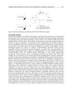
Some authors advocate incorporating a small “buoy flap” of skin with the vascularized fibula graft
to be used for monitoring of the circulation to the graft (27,124) (see Fig. 9). The vascular supply to
the “buoy flap” is via perforating cutaneous branches of the peroneal artery, and is therefore in
continuity with that of the fibula (27,55,124). By constantly observing the color of the skin island, it
is possible to determine immediately whether the anastomosis has become thrombosed. Because this
can be observed immediately, some form of surgical intervention could theoretically salvage the vas-
cularity of the fibula graft (124). This is an advantage over bone scanning, which gives information
at only one point in time. Others report that the use of such a monitoring flap is unreliable because the
quality of the perforating branches may be insufficient (22,126). Moreover, the circulation to the
monitoring flap may not fully correspond with that of the transferred fibula (34).
Fig. 8. Intraoperative photograph demonstrating the vasularized fibula graft in its tissue bed after the proxi-
mal and distal osteotomies have been completed. The clamp is on the distal aspect of the graft and the arrows
are pointing to the peroneal vascular pedicle.
This is trial version
www.adultpdf.com
Vascularized Fibula Grafts 329
Various other methods for postoperative monitoring of the circulation to the transferred fibula
have been advocated, including laser Doppler flowmetry (121), Doppler color-flow imaging (123),
implanted thermocouple probes (120), and measurement of hydrogen washout (122). These methods
allow for continuous monitoring of the flap, without the limitations associated with the “buoy flap.”
In addition, they do not require an additional surgical step, as does the incorporation of a “buoy flap”
into the transferred fibula. In our recent practice, we have not routinely employed the previously
discussed methods for postoperative monitoring of the graft, and rarely harvest a “buoy flap” for
postoperative vascular monitoring. Evidence of early callus formation, healing at the graft junctions,
and graft hypertrophy are used as indirect evidence of vessel patency.
COMPLICATIONS
Stress Fracture
Complications secondary to vascularized fibula transfer include stress fracture (10,28,94,98,101,
119,127–130), delayed and nonunion (4,89,101,127,131), thrombosis (15,121,124), infection (49,127,
132), and those related to the fibula donor site (4,75,78,93,111,117,118,133–136). Stress fracture of
Fig. 9. Diagram depicting a vascularized fibula graft isolated on its peroneal vascular pedicle with a “buoy
flap.” (From Yoshimura, M., Shimamura, K., Iwai, Y., Yamauchi, S., and Ueno, T. [1983] Free vascularized
fibular transplant. A new method for monitoring circulation of the grafted fibula. J. Bone Joint Surg. 65A(9),
1295–1301. Reprinted with permission.)
This is trial version
www.adultpdf.com
330 Gilbert and Wolfe
the graft after union, particularly in the lower extremity, is the most commonly reported complication in
the literature (10,127). De Boer and Wood studied 62 cases of vascularized transfer and reported a
25% stress fracture rate, occurring at an average of 8 mo postoperative (10). Overall, reported stress
fracture rates vary from 20% to 40% (10,128–130), the majority occurring within the first postopera-
tive year (10,98,101,117).
Stress fracture is significantly less common in transfers to the upper extremity, perhaps due to
lower applied loads (10,40,98,137). Vascularized fibula transfers in the upper extremity usually hyper-
trophy and incorporate rapidly (10,114). In de Boer and Wood’s study, fractures occurred only in the
grafts transferred to the lower extremity (10). Stress fractures are a result of excessive loading during
the hypertrophy phase, before adequate incorporation has occurred (10). Most occur within the middle
of the transferred fibula, rather than at the junction sites (28). Once fracture has occurred, provided
the graft is adequately vascularized, with proper immobilization and protection, exuberant callus and
hypertrophy usually results (10,28). Secondary bone grafting procedures are sometimes required (98).
To limit the incidence of stress fracture in the transferred fibula, the graft should be protected from
excessive mechanical loading until hypertrophy is well established (10,40,98). This usually occurs
by 1 yr, and can be followed by serial radiographs (4,10,32). Limited mechanical loading, however,
will enhance hypertrophy and remodeling (10). Stress fractures are particularly prevalent in vascu-
larized fibula transfer to reconstruct the femur, because of the disparity between the cross-sectional
area of the femoral and fibular shafts (94). These can potentially be avoided by dividing the fibula
into two struts as a “double-barrel” graft, preserving the vascular supply to both (22,94–97).
Delayed and Nonunion
Delayed or nonunion at one or both junctions of a vascularized fibula transfer is not uncommon.
Rates in the literature vary, but nonunion generally is reported to occur in 10–20% of cases, when
patients who had secondary grafting procedures are included (4,127,131). A review of 478 vascular-
ized fibula grafts performed for all indications documented a primary union rate of 68% and an overall
rate of 82% after supplemental bone grafting procedures (89). The Mayo Clinic reported a primary
union rate of 62% of 132 vascularized fibula transfers (4). After secondary grafting procedures, they
reported an overall union rate of 80%, at an average follow-up of 42 mo. Weiland reviewed 123 vas-
cularized fibula grafts and reported an ultimate union rate of 87%, with 10% of the patients requiring
supplemental bone grafts (131).
The incidence of nonunion differs depending on the underlying pathology of the patient. The results
for osteomyelitis are much less favorable than those for tumor, trauma, or nonunion reconstruction
(101). De Boer et al. reported an overall union rate of 93% in patients who underwent vascularized
fibula transfer for a diagnosis of tumor or trauma, compared to a 59% union rate for those whose under-
lying diagnosis was osteomyelitis (101). Nonunions are also more common in fibula grafts transferred
to the lower extremity, as compared to the upper extremity (4). Stable initial fixation, most commonly
with plates and/or screws, has been shown by some to correlate with higher rates of union, as com-
pared to other fixation methods, such as external fixation (4,101). The addition of nonvascularized
bone graft at the fibula–recipient junctions at the time of transfer has also been demonstrated to
increase primary union rates (101,138). Nonunions are treated with secondary bone grafting proce-
dures, which lead to eventual healing in most instances (101,132).
Thrombosis
Thrombosis occurs in approximately 10% of vascularized fibula grafts in the early postoperative
period, as diagnosed by continual laser Doppler flowmetry and confirmed by surgical exploration
(121). Whether or not surgical exploration of a thrombosed vessel of the pedicle is indicated remains
controversial (15,124). Experimentally, Siegert and Wood demonstrated that the viability of a throm-
bosed vascularized bone graft is less than that of a conventional nonvascularized graft (139).
This is trial version
www.adultpdf.com
Vascularized Fibula Grafts 331
Infection
The incidence of infection in vascularized fibula transfer ranges from approximately a 14% deep
infection rate to a 33% superficial infection rate (127). Deep infection appears to be more common
following reconstruction for the diagnosis of osteomyelitis or tumor (132). Experimentally, vascular-
ized bone grafts have been shown to become infected less often than do conventional nonvascularized
bone grafts (140). When fibula grafts do become infected, the infection is easier to eradicate in a suc-
cessfully vascularized graft, as compared to a nonviable transfer (49). Infection is usually more wide-
spread in nonviable grafts. When deep infection does occur, the response of a viable vascularized fibula
graft is similar to that of normal cortical bone. Treatment should consist of intravenous antibiotics with
debridement as necessary (49).
Fibula Donor-Site Morbidity
Weiland, Jupiter, and others have documented minimal or no morbidity at the fibula donor site (1,
22,141). Others, however, have found a number of associated complications (4,93,111,134–136). Most
commonly, these include residual paresthesias (134,136), occasional pain and cramps (135,136),
altered gait (93,135,136), weakness (93,136), reduced walking distance (134), and cold intolerance
(134). Gore et al. reported on fibula donor-site morbidity in 41 patients at an average of 27 mo post-
operative (135). They found that 42% had pain, 7% complained of muscle pain on exertion, 10% com-
plained of a tired, weak feeling associated with vigorous activity, and 2% had trouble with balance
wearing high-healed shoes. A review of 132 vascularized fibula grafts performed at the Mayo Clinic
demonstrated donor-site complications in 8% of the patients (4). These included flexor hallucis lon-
gus contracture, transient peroneal nerve palsy, compartment syndrome of the leg, and stress fracture
of the ipsilateral tibia. Youdas et al. evaluated the gait mechanics of 11 patients who had vascularized
fibula transfer to the upper extremity (93). They found muscle strength, especially foot inversion and
eversion, to be significantly impaired. There existed an inverse relationship between the length of the
resected fibula and the strength of the evertor muscles of the ankle.
The development of an ankle valgus deformity after vascularized fibula graft harvest in patients
with open physes is a complication which is well documented in the literature (75,78,117,118,133).
This has not been demonstrated to occur in the adult, provided that more than 6 cm of the distal fibula
is retained (22,136). In children, this deformity can be prevented by performing a distal tibio-fibular
synostosis proximal to the physis at the time of fibula harvest (45,117,118). Deformity has not been
demonstrated to occur proximally when the proximal fibular epiphysis is transferred in children (85,
136). The lateral collateral ligament which inserts into the fibular head should be reconstructed, how-
ever, to prevent instability of the knee joint (85).
CONCLUSION
Since the first report of a vascularized fibula transfer by Taylor et al. in 1975 (9), the indications
for this procedure have expanded widely. Today, it has become one of the established modalities for
the orthopedic surgeon in the reconstruction of extensive long bone defects following trauma, tumor
resection, and infection. Moreover, it is now widely employed in the treatment of osteonecrosis of the
femoral head, congenital tibial and forearm pseudarthrosis, congenital differences and pediatric trauma,
and to facilitate spine and joint arthrodesis. Although vascularized fibula transfer is a procedure
associated with a number of well-documented complications, these are far outweighed by its ultimate
clinical benefits. Future refinements in the use of the fibula as a free epiphyseal transfer and in the area
of postoperative monitoring are still needed.
REFERENCES
1. Moore, J. R., Weiland, A. J., and Daniel, R. K. (1983) Use of free vascularized bone grafts in the treatment of bone
tumors. Clin. Orthop. 175, 37–44.
This is trial version
www.adultpdf.com
332 Gilbert and Wolfe
2. Brunelli, G., Vigasio, B., Battiston, B., Di Rosa, F., and Brunelli, G. J. (1991) Free microvascular fibular versus con-
ventional bone grafts. Int. Surg. 76, 33–42.
3. Dell, P. C., Burckhardt, H., and Glowczewski, F. P. (1985) A roentgenographic, biomechanical, and histological eval-
uation of vascularized and nonvascularized segmental fibular canine autografts. J. Bone Joint Surg. 67A(1), 105–112.
4. Han, C. S., Wood, M. B., Bishop, A. T., and Cooney, W. P. III. (1992) Vascularized bone transfer. J. Bone Joint Surg.
74A, 1441–1449.
5. Osterman, A. L. and Bora, F. W. (1984) Free vascularized bone grafting for large-gap nonunion of long bones. Orthop.
Clin. N. Am. 15, 131–142.
6. Barth, H. (1895) Histologische Untersuchungen uber Knochen Transplantation. Beitr. Parthol. Anat Allg. Pathol. 17,
65–142.
7. Jacobson, J. H. II and Suarez, E. L. (1960) Microsurgery in anastomosis of small vessels. Surg. Forum 11, 243–245.
8. McKee, D. M. (1978) Microvascular bone transplantation. Clin. Plast. Surg. 5, 283–292.
9. Taylor, G. I., Miller, G. D., and Ham, F. J. (1975) The free vascularized bone graft. A clinical extension of microvascu-
lar techniques. Plast. Reconstr. Surg. 55, 533–544.
10. de Boer, H. H. and Wood, M. B. (1989) Bone changes in the vascularized fibular graft. J. Bone Joint Surg. 71B, 374–378.
11. Phemister, D. B. (1914) The fate of transplanted bone and regenerative power of its various constituents. Surg.
Gynecol. Obstet. 19, 303–333.
12. Abbott, L. C., Schottslaedt, E. R., Saunders, J. B. D M., and Bost, F. C. (1947) The evaluation of cortical and can-
cellous bone as grafting material: a clinical and experimental study. J. Bone Joint Surg. 29, 381–414.
13. Enneking, W. F., Burchardt, H., Puhl, J. J., and Piotrowski, G. (1975) Physical and biological aspects of repair in dog
cortical-bone transplants. J. Bone Joint Surg. 57A, 237–252.
14. Weiland, A. J., Kleinert, H. E., Kutz, J. E., and Daniel, R. K. (1979) Free vascularized bone grafts in surgery of the upper
extremity. J. Hand Surg. 4(2), 129–144.
15. Sowa, D. T. and Weiland, A. J. (1987) Clinical applications of vascularized bone autografts. Orthop. Clin. N. Am. 18,
257–273.
16. Arata, M. A., Wood, M. B., and Cooney, W. P. III. (1984) Revascularized segmental diaphyseal bone transfers in the
canine. An analysis of viability. J. Reconstr. Microsurg. 1, 11–19.
17. Berggren, A., Weiland, A. J., and Dorfman, H. (1982) The effect of prolonged ischemia time on osteocyte and
osteoblast survival in composite bone grafts revascularized by microvascular anastomoses. Plast. Reconstr. Surg. 69,
290–298.
18. Bos, K. E. (1979) Bone Scintigraphy of experimental composite bone grafts revascularized by microvascular anasto-
moses. Plast. Reconstr. Surg. 64, 353–360.
19. Doi, K., Tominaga, S., and Shibata, T. (1977) Bone grafts with microvascular anastomoses of vascular pedicles: an
experimental study in dogs. J. Bone Joint Surg. 59A, 809–815.
20. Weiland, A. J. (1981) Current concepts review: vascularized free bone transplants. J. Bone Joint Surg.
63A(1), 166–169.
21. Fujimaki, A. and Suda, H. (1994) Experimental stud and clinical observations on hypertrophy of vascularized bone
grafts. Microsurgery 15, 726–732.
22. Jupiter, J. B., Bour, C. J., and May, J. W. J. (1987) The reconstruction of defects in the femoral shaft with vascular-
ized transfers of fibular bone. J. Bone Joint Surg. 69A, 365–374.
23. Ostrup, L. T. and Fredrickson, J. M. (1974) Distant transfer of a free, living bone graft by microvascular anastomoses.
An experimental study. Plast. Reconstr. Surg. 54, 274–285.
24. Shaffer, J. W., Field, G. A., Goldberg, V. M., and Davy, D. T. (1985) Fate of vascularized and non-vascularized auto-
grafts. Clin. Orthop. 197, 32–43.
25. Davis, P. K., Mazur, J. M., and Coleman, G. N. (1982) A torsional strength comparison of vascularized and nonvas-
cularized bone grafts. J. Biomech. 15, 875–880.
26. Baudet, J., Panconi, B., Cai, P., Schoofs, M., Amarante, J., and Kaddoura, R. (1982) The composite fibula and soleus
transfer. Int. J. Microsurg. 4, 10–26.
27. Chen, Z. W. and Yan, W. (1983) The study and clinical applications of the osteocutaneous flap of fibula. Microsurgery
4, 11–16.
28. Harrison, D. H. (1986) The osteocutaneous free fibular graft. J. Bone Joint Surg. 68B, 804–807.
29. Jupiter, J. B. (1990) Complex non-union of the humeral diaphysis: treatment with a medial approach, an anterior plate,
and a vascularized fibular graft. J. Bone Joint Surg. 72A(5), 701–707.
30. Jupiter, J. B., Gerhard, H. J., Guerrero, J., Nunley, J. A., and Levin, L. S. (1997) Treatment of segmental defects
of the radius with use of the vascularized osteoseptocutaneous fibular autogenous graft. J. Bone Joint Surg. 79A(4),
542–550.
31. Koshima, I., Higaki, H., and Soeda, S. (1991) Combined vascularized fibula and peroneal composite-flap transfer for
severe heat-press injury of the forearm. Plast. Reconstr. Surg. 88, 338–341.
32. Malizos, K. N., Nunley, J. A., Goldner, R. D., Urbaniak, J. R., and Harrelson, J. M. (1993) Free vascularized fibula in
traumatic long bone defects and in limb salvaging following tumor resection: comparative study. Microsurgery 14,
368–374.
This is trial version
www.adultpdf.com
Vascularized Fibula Grafts 333
33. Newington, D. P. and Sykes, P. J. (1991) The versatility of the free fibula flap in the management of traumatic long
bone defects. Injury 22, 275–281.
34. Wei, F. C., Chen, H. C., Chuang, C. C., and Noordhoff, M. S. (1986) Fibular osteoseptocutaneous flap: anatomic
study and clinical application. Plast. Reconstr. Surg. 78(2), 191–199.
35. Wei, F. C., El-Gammal, T. A., Lin, C. H., and Ueng, W. N. (1997) Free fibular osteoseptocutaneous graft for recon-
struction of segmental femoral shaft defects. J. Trauma 43(5), 784–792.
36. Weiland, A. J. (1984) Vascularized bone transfers. Instruct. Course Lect. 33, 446–460.
37. Weiland, A. J., Moore, J. R., and Daniel, R. K. (1983) Vascularized bone autografts. Experience with 41 cases. Clin.
Orthop. 174, 87–95.
38. Aberg, M., Rydholm, A., Holmberg, J., and Wieslander, J. B. (1988) Reconstruction with a free vascularized fibular
graft for malignant bone tumor. Acta Orthop. Scand. 59, 430–437.
39. Bajec, J. and Gang, R. K. (1993) Bone reconstruction with a free vascularized fibular graft after giant cell tumour resec-
tion. J. Hand Surg. 18B(5), 565–567.
40. Hsu, R. W. W., Wood, M. B., Sim, F. H., and Chao, E. Y. S. (1997) Free vascularized fibular grafting for reconstruction
after tumour resection. J. Bone Joint Surg. 19B(1), 36–42.
41. Leung, P. C. and Chan, K. T. (1986) Giant cell tumor of the distal end of the radius treated by the resection and free
vascularized iliac crest graft. Clin. Orthop. 202, 232–236.
42. Ono, H., Yajima, H., Mizumoto, S., Miyauchi, Y., Mii, Y., and Tamai, S. (1997) Vascularized fibular graft for recon-
struction of the wrist after excision of giant cell tumor. Plast. Reconstr. Surg. 99(4), 1086–1093.
43. Pho, R. W. (1979) Free vascularized fibular transplant for replacement of the lower radius. J. Bone Joint Surg. 61B(3),
362–365.
44. Pho, R. W. (1981) Malignant giant-cell tumor of the distal end of the radius treated by a free vascularized fibular trans-
plant. J. Bone Joint Surg. 63A(6), 877–884.
45. Shea, K. G., Coleman, D. A., Scott, S. M., Coleman, S. S., and Christianson, M. (1997) Microvascularized free fibular
grafts for reconstruction of skeletal defects after tumor resection. J. Pediatr. Orthop. 17(4), 424–432.
46. Dell, P. C. and Sheppard, J. E. (1984) Vascularized bone grafts in the treatment of infected forearm nonunions. J. Hand
Surg. 9A, 653–658.
47. Doi, K., Kawakami, F., Hiura, Y., Oda, T., Sakai, K., and Kawai, S. (1995) One-stage treatment of infected bone
defects of the tibia with skin loss by free vascularized osteocutaneous grafts. Microsurgery 16, 704–712.
48. Lee, K. S., Chung, H. K., and Kim, K. H. (1991) Vascularized osteocutaneous fibular transfer to the tibia. Int. Orthop.
15, 199–203.
49. Low, C. K., Pho, R. W. H., Kour, A. K., Satku, K., and Kumar, V. P. (1996) Infection of vascularized fibular grafts. Clin.
Orthop. 323, 163–172.
50. Mattar, R., Azze, R. J., Ferreira, M. C., Starck, R., and Canedo, A. C. (1994) Vascularized fibular graft for manage-
ment of severe osteomyelitis of the upper extremity. Microsurgery 15, 22–27.
51. Nonnenmacher, J., Bahm, J., and Moui, Y. (1995) The free vascularised fibular transfer as a definitive treatment in
femoral septic non-unions. Microsurgery 16, 383–387.
52. Vitkus, K. and Vitkus, M. (1992) Reconstruction of large infected tibia defects.
Ann. Plast. Surg. 29(2), 97–108.
53. Wood, M. B. and Cooney, W. P. III. (1984) Vascularized bone segment transfers for management of chronic osteo-
myelitis. Orthop. Clin. N. Am. 15, 461–472.
54. Yajima, H., Tamai, S., Mizumoto, S., and Inada, Y. (1993) Vascularized fibular grafts in the treatment of osteomyelitis
and infected nonunion. Clin. Orthop. 293, 256–264.
55. Cho, B. C., Kim, S. Y., Lee, J. H., Ramasastry, S. S., Weinweig, N., and Baik, B. S. (1998) Treatment of osteonecro-
sis of the femoral head with free vascularized fibular transfer. Ann. Plastic Surg. 40(6), 586–593.
56. Malizos, K. N., Soucacos, P. N., and Beris, A. E. (1995) Osteonecrosis of the femoral head: hip salvaging with implan-
tation of a vascularized fibular graft. Clin. Orthop. 314, 67–75.
57. Malizos, K. N., Soucacos, P. N., Beris, A. E., Korobilias, A. B., and Xenakis, T. A. (1994) Osteonecrosis of the femoral
head in immunosuppressed patients: hip salvaging with implantation of a vascularized fibular graft. Microsurgery 15,
485–491.
58. Scully, S. P., Aaron, R. K., and Urbaniak, J. R. (1998) Survival analysis of hips treated with core decompression or
vascularized fibular grafting because of avascular necrosis. J. Bone Joint Surg. 80A(9), 1270–1275.
59. Sotereanos, D. G., Plakseychuk, A. Y., and Rubash, H. E. (1997) Free vascularized fibula grafting for the treatment
of osteonecrosis of the femoral head. Clin. Orthop. 344, 243–256.
60. Urbaniak, J. R., Coogan, P. G., Gunneson, E. B., and Nunley, J. A. (1995) Treatment of osteonecrosis of the femoral
head with free vascularized fibular grafting. A long-term follow-up study of one hundred and three hips. J. Bone Joint
Surg. 77A, 681–694.
61. Urbaniak, J. R. and Harvey, E. J. (1998) Revascularization of the femoral head in osteonecrosis. J. Am. Acad. Orthop.
Surg. 6(1), 44–54.
62. Yoo, M. C., Chung, D. W., and Hahn, C. S. (1992) Free vascularized fibula grafting for the treatment of osteonecrosis
of the femoral head. Clin. Orthop. 277, 128–138.
This is trial version
www.adultpdf.com
334 Gilbert and Wolfe
63. Doi, K., Kawai, S., Sumiura, S., and Sakai, K. (1988) Anterior cervical fusion using the free vascularized fibular
graft. Spine 13(11), 1239–1244.
64. Freidberg, S. R., Gumley, G. J., Pfeifer, B. A., and Hybels, R. L. (1989) Vascularized fibular graft to replace resected
cervical vertebral bodies: case report. J. Neurosurg. 71, 283–286.
65. Hallock, G. G. (1997) The role of pedicled or free fibular grafts in knee arthrodesis. Ann. Plast. Surg. 39(1), 60–63.
66. Hubbard, L. F., Herndon, J. H., and Buonanno, R. (1985) Free vascularized fibula transfer for stabilization of the thora-
columbar spine: a case report. Spine 10(10), 891–893.
67. Kaneda, K., Kurakami, C., and Minami, A. (1988) Free vascularized fibular strut graft in the treatment of kyphosis.
Spine 13(11), 1273–1277.
68. Minami, A., Kaneda, K., Satoh, S., Abumi, K., and Kutsumi, K. (1997) Free vascularized fibular strut graft for anterior
spinal fusion. J. Bone Joint Surg. 79B(1), 43–47.
69. Rasmussen, M. R., Bishop, A. T., and Wood, M. B. (1995) Arthrodesis of the knee with a vascularized fibular rotatory
graft. J. Bone Joint Surg. 77A(5), 751–759.
70. Usui, M., Ischii, S., Naito, T., et al. (1996) Arthrodesis of knee joint by vascularized fibular graft. Microsurgery 17, 2–8.
71. Bos, K. E., Besselaar, P. P., Van Der Eyken, J. W., Taminiau, A. H. M., and Verbout, A. J. (1993) Reconstruction of
congenital tibial pseudarthrosis by revascularized fibular transplants. Microsurgery 14, 558–562.
72. Dormans, J. P., Krajbich, J. I., Zuker, R., and Demuynk, M. (1990) Congenital pseudarthrosis of the tibia: treatment
with free vascularized fibular grafts. J. Pediatr. Orthop. 10(5), 623–628.
73. Gilbert, A. and Brockman, R. (1995) Congenital pseudarthrosis of the tibia: long-term followup of 29 cases treated
by microvascular bone transfer. Clin. Orthop. 314, 37–44.
74. Judet, J., Gilbert, Judet H., and Servan, M. (1978) Apport de la micro-chirurgie a la chirurgie osseuse. Chirurgie 104
(10), 921–924.
75. Paterson, D. (1989) Congenital pseudarthrosis of the tibia: an overview. Clin. Orthop. 247, 44–54.
76. Simonis, R. B., Shirali, H. R., and Mayou, B. (1991) Free vascularized fibular grafts for congenital pseudarthrosis of
the tibia. J. Bone Joint Surg. 73B(2), 211–215.
77. Uchida, Y., Kojima, T., and Sugioka, Y. (1991) Vascularized fibular graft for congenital pseudarthrosis of the tibia.
J. Bone Joint Surg. 73B(5), 846–850.
78. Weiland, A. J., Weiss, A. P. C., Moore, J. R., and Tolo, V. T. (1990) Vascularized fibular grafts in the treatment of
congenital pseudarthrosis of the tibia. J. Bone Joint Surg. 72A(5), 654–662.
79. Allieu, Y., Gomis, R., Yoshimura, M., Dimeglio, A., and Bonnel, F. (1981) Congenital pseudarthrosis of the forearm
—two cases treated by free vascularized fibular graft. J. Hand Surg. 6(5), 475–481.
80. Allieu, Y., zu Reckendorf, G. M., Chammas, M., and Gomis, R. (1999) Congenital pseudarthrosis of both forearm
bones: long-term results of two cases managed by free vascularized fibular graft. J. Hand Surg. 42A(3), 604–608.
81. Mathoulin, C., Gilbert, A., and Azze, R. G. (1993) Congenital pseudarthrosis of the forearm: treatment of six cases
with vascularized fibular graft and a review of the literature. Microsurgery 14, 252–259.
82. Witoonchart, K., Uerpairojkit, C., Leechavengvongs, S., and Thuvasethakul, P. (1999) Congenital pseudarthrosis of
the forearm treated by free vascularized fibular graft: a report of three cases and a review of the literature. J. Hand
Surg. 24A(5), 1045–1055.
83. Pho, R. W. H., Patterson, M. H., Kour, A. K., and Kumar, V. P. (1988) Free vascularized epiphyseal transplantation
in upper extremity reconstruction. J. Hand Surg. 13B(4), 440–447.
84. Shea, K. G., Coleman, S. S., and Coleman, D. A. (1997) Growth of the proximal fibular physis and remodeling of the
epiphysis after microvascular transfer. J. Bone Joint Surg. 79A(4), 583–596.
85. Tsai, T. M., Ludwig, L., and Tonkin, M. (1986) Vascularized fibular epiphyseal transfer. A clinical study. Clin. Orthop.
210, 228–234.
86. Taylor, G. I. (1977) Microvascular free bone transfer. Orthop. Clin. N. Am. 8, 425–446.
87. Mckee, N. H., Haw, P., and Vettesse, T. (1984) Anatomic study of the nutrient foramen in the shaft of the fibula. Clin.
Orthop. 184, 141–144.
88. van Twisk, R., Pavlov, P. W., and Sonneveld, J. (1988) Reconstruction of bone and soft tissue defects with free fibula
transfer. Ann. Plastic Surg. 21(6), 555–558.
89. Bishop, A. T. (1999) Vascularized bone grafting, in Green’s Operative Hand Surgery, 4th ed. (Green, D. P., Hotchkiss,
R. N., and Pederson, W. C., eds.), Churchill Livingstone, Philadelphia, pp. 1221–1250.
90. Bowen, C. V. and Tomaino, M. (1996) Vascularized bone transfers, in Surgery of the Hand and Upper Extremity
(Peimer, C. A., ed.), McGraw-Hill, New York, pp. 1941–1974.
91. Serafin, D. (1996) Atlas of Microsurgical Composite Tissue Transplantation. Saunders, Philadelphia, pp. 547–573.
92. Taylor, G. I. (1983) The current status of free vascularized bone grafts. Clin. Plast. Surg. 10, 185–209.
93. Youdas, J. W., Wood, M. B., Cahalan, T. D., and Chao, E. Y. S. (1988) A quantitative analysis of donor site morbid-
ity after vascularized fibula transfer. J. Orthop. Res. 6(5), 621–629.
94. Jones, N. F., Swartz, W. M., Mears, D. C., Jupiter, J. B., and Grossman, A. (1988) The “double barrel” free vascularized
fibular bone graft. Plast. Reconstr. Surg. 81(3), 378–385.
This is trial version
www.adultpdf.com
Vascularized Fibula Grafts 335
95. O’Brien, B. M., Gumley, G. J., Dooley, B. J., and Pribaz, J. J. (1988) Folded free vascularized fibula transfer. Plast.
Reconstr. Surg. 82(2), 311–318.
96. Tomita, Y., Murota, K., Takahashi, F., Moriyama, M., and Beppu, M. (1994) Postoperative results of vascularized
double fibula grafts for femoral pseudoarthrosis with large bony defect. Microsurgery 15, 316–321.
97. Yajima, H. and Tamai, S. (1994) Twin-barrelled vascularized fibular grafting to the pelvis and lower extremity. Clin.
Orthop. 303, 178–184.
98. Kasashima, T., Minami, A., and Kutsumi, K. (1998) Late fracture of vascularized fibular grafts. Microsurgery 18,
337–343.
99. Friedlaender, G. E., Tross, R. B., Doganis, A. C., Kirkwood, J. M., and Baron, R. (1984) Effects of chemotherapeutic
agents on bone. I. Short-term methotrexate and doxorubicin (adriamycin) treatment in a rat model. J. Bone Joint Surg.
66A(4), 602–607.
100. Chew, W. Y. C., Low, C. K., and Tan, S. K. (1995) Long-term results of free vascularized fibular graft. A clinical and
radiographic evaluation. Clin. Orthop. 311, 258–261.
101. de Boer, H. H., Wood, M. B., and Hermans, J. (1990) Reconstruction of large skeletal defects by vascularized fibula
transfer. Factors that influenced the outcome of union in 62 cases. Int. Orthop. 14, 121–128.
102. Streitz, W., Brown, J. C., and Bonnett, C. A. (1977) Anterior fibular strut grafting in the treatment of kyphosis. Clin.
Orthop. 128, 140–148.
103. Bradford, D. S. (1980) Anterior vascular pedicle bone grafting for the treatment of kyphosis. Spine 5, 318–323.
104. Morrissy, R. T., Riseborough, E. J., and Hall, J. E. (1981) Congenital pseudarthrosis of the tibia. J. Bone Joint Surg.
63B(3), 367–375.
105. Zumiotti, A. and Ferreira, M. C. (1994) Treatment of congenital pseudarthrosis of the tibia by microsurgical fibula trans-
fer. Microsurgery 15, 37–43.
106. Hagan, K. F. and Buncke, H. J. (1982) Treatment of congenital pseudarthrosis of the tibia with free vascularized bone
graft. Clin. Orthop. 166, 34–44.
107. Kort, J. S., Schink, M. M., Mitchel, S. N., and Bassett, C. A. L. (1962) Congenital pseudarthrosis of the tibia: treat-
ment with pulsing electromagnetic fields. Clin. Orthop. 165, 124–137.
108. Disa, J. J. and Cordeiro, P. G. (1998) The current role of preoperative arteriography in free fibula flaps. Plast. Recon-
str. Surg. 102(4), 1083–1088.
109. Lutz, B. S., Ng, S. H., Cabailo, R., Lin, C. H., and Wei, F. C. (1998) Value of routine angiography before traumatic
lower-limb reconstruction with microvascular free tissue transplantation. J. Trauma 44(4), 682–686.
110. Lutz, B. S., Wei, F. C., Ng, S. H., Chen, I. H., and Chen, S. H. T. (1999) Routine donor leg angiography before
vascularized free fibula transplantation is not necessary: a prospective study in 120 clinical cases. Plast. Reconstr.
Surg. 103(1), 121–127.
111. Vail, T. P. and Urbaniak, J. R. (1996) Donor-site morbidity with use of vascularized autogenous fibular grafts. J. Bone
Joint Surg. 78A(2),
204–211.
112. Carroll, W. R. and Esclamado, R. (1996) Preoperative vascular imaging for the fibular osteocutaneous flap. Arch.
Otolaryngol. Head Neck Surg. 122, 708–712.
113. Manaster, B. J., Coleman, D. A., and Bell, D. A. (1990) Magnetic resonance imaging of vascular anatomy before vas-
cularized fibular grafting. J. Bone Joint Surg. 72A(3), 409–414.
114. Manaster, B. J., Coleman, D. A., and Bell, D. A. (1990) Pre- and postoperative imaging of vascularized fibular grafts.
Radiology 176(1), 161–166.
115. Young, D. M., Trabulsky, P. P., and Anthony, J. P. (1994) The need for preoperative leg angiography in fibula free
flaps. J. Reconstr. Microsurg. 10, 283–287.
116. Hallock, G. G. (1994) Evaluation of fasciocutaneous perforators using color duplex imaging. Plast. Reconstr. Surg.
94, 644–651.
117. Minami, A., Kaneda, K., Itoga, H., and Usui, M. (1989) Free vascularized fibular grafts. J. Reconstr. Microsurg. 5,
37–43.
118. Omokawa, S., Tamai, S., Takakura, Y., Yajima, H., and Kawanishi, K. (1996) A long-term study of the donor-site
ankle after vascularized fibula grafts in children. Microsurgery 17, 162–166.
119. Itoh, K., Minami, A., Sakuma, T., and Furudate, M. (1989) The use of three-phase bone imaging in vascularized fibu-
lar and iliac bone grafts. Clin. Nuclear Med. 14(7), 494–500.
120. May, J. W., Lukash, F. N., Gallico, G. G. 3rd, and Stirrat, C. R. (1983) Removable thermocouple probe microvascu-
lar patency monitor: an experimental and clinical study. Plast. Reconstr. Surg. 72, 366–379.
121. Schuurman, A. H., Bos, K. E., and Van Nus, Y. H. (1987) Laser doppler bone probe in vascularized fibula transfers:
a preliminary report. Microsurgery 8, 186–189.
122. Shima, I., Yamauchi, S., Matsumoto, T., et al. (1985) A new method for monitoring circulation of grafted bone by use
of electrochemically generated hydrogen. Clin. Orthop. 198, 244–249.
123. Stevenson, T. R., Rubin, J. M., and Herzenberg, J. E. (1988) Vascular patency of fibular free graft: assessment by
doppler color-flow imager. A case report. J. Reconstr. Microsurg. 4, 409–413.
This is trial version
www.adultpdf.com
336 Gilbert and Wolfe
124. Yoshimura, M., Shimamura, K., Iwai, Y., Yamauchi, S., and Ueno, T. (1983) Free vascularized fibular transplant.
A new method for monitoring circulation of the grafted fibula. J. Bone Joint Surg. 65A(9), 1295–1301.
125. Berggren, A., Weiland, A. J., and Ostrup, L. T. (1982) Bone scintigraphy in evaluating the viability of composite
bone grafts revascularized by microvascular anastomoses, conventional autogenous bone grafts, and free nonrevas-
cularized periosteal grafts. J. Bone Joint Surg. 64A, 799–809.
126. Yajima, H., Tamai, S., Mizumoto, S., and Ono, H. (1993) Vascularized fibular grafts for reconstruction of the femur.
J. Bone Joint Surg. 75B(1), 123–128.
127. Coghlan, B. A. and Townsend, P. L. G. (1993) The morbidity of the free vascularised fibula flap. Br. J. Plast. Surg.
46, 466–469.
128. Ihara, K., Doi, K., Sakai, K., Kuwata, N., and Kawai, S. (1992) Fracture of free vascularized fibular grafts. J. Jpn. Soc.
Reconstr. Microsurg. 5, 86–95.
129. Minami, A., Kimura, T., Matsumoto, O., and Kutsumi, K. (1993) Fracture through united vascularized bone grafts. J.
Reconstr. Microsurg. 9, 227–232.
130. Tamai, S. (1991) Treatment of traumatic extensive bony defect in the lower leg: with vascularized fibula graft. J. Jpn.
Soc. Reconstr. Microsurg. 4, 152–155.
131. Weiland, A. J. (1991) Clinical applications of vascularized bone grafts, in Bone and Cartilage Allografts (Friedlaender,
G. E. and Goldberg, V. M., eds.), American Academy of Orthopaedic Surgeons, Park Ridge, IL, pp. 239–245.
132. Moran, C. G. and Wood, M. B. (1993) Vascularized bone autografts. Orthop. Rev. 22, 187–197.
133. Coleman, S. S. and Coleman, D. A. (1994) Congenital pseudoarthrosis of the tibia: treatment by transfer of the ipsilat-
eral fibula with vascular pedicle. J. Pediatr. Orthop. 14, 156–160.
134. Goodacre, T. E. E., Walker, C. J., Jawad, A. S., Jackson, A. M., and Brough, M. D. (1990) Donor site morbidity fol-
lowing osteocutaneous free fibula transfer. Br. J. Plast. Surg. 43, 410–412.
135. Gore, D. R., Gardner, G. M., Sepic, S. B., Mollinger, L. A., and Murray, M. P. (1987) Function following partial fibul-
ectomy. Clin. Orthop. 220, 206–210.
136. Lee, E. H., Goh, J. C. H., Helm, R., and Pho, R. W. H. (1990) Donor site morbidity following resection of the fibula.
J. Bone Joint Surg. 72B(1), 129–131.
137. Olekas, J. and Guobys, A. (1991) Vascularised bone transfer for defects and pseudarthroses of forearm bones. J. Hand
Surg. 16B(4), 406–408.
138. Pirela-Cruz, M. A. and DeCoster, T. A. (1994) Vascularized bone grafts. Orthopedics 17(5), 407–412.
139. Siegert, J. J. and Wood, M. B. (1987) Thrombosed vascularized bone graft: viability compared with a conventional
bone graft. J. Reconstr. Microsurg.
3, 99–103.
140. Haw, C. S., O’Brien, B. C., and Kurata, T. (1978) The microsurgical revascularization of resected segments of tibia
in the dog. J. Bone Joint Surg. 60B, 266–269.
141. Tang, C. L., Mahoney, J. L., McKee, M. D., et al. (1998) Donor site morbidity following vascularized fibular graft-
ing. Microsurgery 18, 383–386.
This is trial version
www.adultpdf.com
Craniofacial Repair 337
337
From: Bone Regeneration and Repair: Biology and Clinical Applications
Edited by: J. R. Lieberman and G. E. Friedlaender © Humana Press Inc., Totowa, NJ
17
Craniofacial Repair
Bruce A. Doll, DDS, PhD, Charles Sfeir, DDS, PhD, Kodi Azari, MD,
Sarah Holland,
MD, and Jeffrey O. Hollinger, DDS, PhD
INTRODUCTION
Annually, skeletal injury and specifically craniofacial injury total approx 12.2 million people in
the United States (1). Advances in craniofacial therapy, founded on developing knowledge of the
molecular signals and intercellular communication, has greatly improved the restoration of form and
function. Fracture healing is a complex physiological process. Cellular and biochemical processes
that occur during fracture healing parallel those that take place in the growth plate during develop-
ment, except in fracture healing these processes occur on a temporal scale (2–4). Similarities in the
processes occurring at the growth plate and at the fracture site permit some knowledge from growth-
plate analysis to comprehend events in fracture healing. Fracture healing involves a series of distinct
cellular responses. Specific paracrine and autocrine intercellular signaling pathways control cellular
and osseous tissue mineralization (Fig. 1). However, extrapolation of knowledge of growth-plate molec-
ular dynamics is insufficient to achieve consistently optimal bone regeneration during primary and
secondary fracture healing.
Fracture healing has been divided into primary fracture healing and secondary fracture healing.
Attempts by the cortex to reestablish itself once it has become interrupted characterize primary frac-
ture healing (5,6). Responses in the periosteum and external soft tissues lead to callus formation dur-
ing secondary healing. Bone on one side of the cortex unites with bone on the other side of the cortex
to reestablish mechanical continuity. Anatomical restoration of the fracture is favored when the frag-
ments are coapted and stable (7). Under these conditions, bone-resorbing cells on one side of the
fracture undergo a tunneling resorptive response whereby they reestablish new Haversian systems by
providing pathways for the penetration by blood vessels. These new blood vessels are accompanied
by endothelial cells and perivascular mesenchymal cells, which become the osteoprogenitor cells for
osteoblasts.
The regeneration of the bone form and function appears to have limits. Some fractures heal slowly
or not at all. Destruction of a critical mass of osseous topography, i.e., a critical-sized defect (CSD),
does not regenerate completely. A complex series of molecular cues temporally and spatially influ-
ence healing. A critical-sized defect has been defined as an intraosseous deficiency that will not heal
with more than 10% new bone formation within the life expectancy of the patient (where a patient may
be human or nonhuman) (8). A critical-sized defect heals with scar formation—a fibrous unification
of osseous cortical plates.
Overcoming the predisposition for skeletal nonunion requires supplemental treatments. Surgeons
may circumvent scar formation by numerous approaches, some based on empirical evidence (9–11).
Accounts richly detail treatments that surgeons have used to augment fracture healing and continuity
defect regeneration (reviewed in ref. 12). Present treatments include bone grafts, alloplasts, electrical
This is trial version
www.adultpdf.com
338 Doll et al.
Fig. 1. Four phases of fracture healing are noted. A controlled temporal and spatial cascade controls the behavior of cellulars elements. Each phase is char-
acterized by bone formation and remodeling, exhibiting the coupling of osteoblastic and osteoclastic activities. (Color illustration in insert following p. 212.)
338
This is trial version
www.adultpdf.com
Craniofacial Repair 339
stimulation, distraction osteogenesis, guided bone regeneration, and growth factors, whereas targeted-
cells and gene delivery represent a relatively new approach for enhanced bone growth (reviewed in
refs. 13–17).
Comprehensive reviews summarizing historical origins of enhanced bone healing are available and
are cited for completeness. However, the present review emphasizes selected bone regeneration options
that may be new to many surgeons, offering potential for establishing new standards of care in the
near future. The review emphasizes contemporary therapies in the context of an ongoing elaboration
of bone biology and healing. Present therapies must focus on the controlled delivery of a single agent
or rely on the presence of numerous factors purported for bone auto- and allografts. Effectiveness of
grafting is attributed to the host of factors released from the graft and the host’s support of the graft.
The graft lacks temporal, spatial, and stoichiometric precision for dispersion of the factors encouraging
bone growth. Therefore, fine predictability for graft success is not currently possible. Successful ther-
apies anticipate the necessity for delivery vehicles (18). The review underscores the important affili-
ation at several levels—scaffold and factor, chemist, engineer, biologist, and clinician—to achieve a
predictable, regenerative therapy.
CELL AND MOLECULAR BIOLOGY OF FRACTURE HEALING
Treatment of most fractures accepts a degree of motion (19,20). The majority of fractures heal by
secondary facture healing involving a combination of intramembranous and endochondral ossification.
Both processes contribute to repair in an orchestrated sequence of four or five phases of healing. Hema-
toma and inflammation precede angiogenesis (18) and chondrogenesis. Cartilage is removed and osteo-
progenitor cells induce bone formation and remodeling in concert with osteoclastogenesis (21).
Multiple events occur during bone injury. Fracture is an injury that incites an inflammatory response,
activation of complement ensues, and vascular damage leads to fluid extravasation (Fig. 1). At the
fracture, there is a decrease in pH to 4–5 (the acidotic state), monocyte and macrophage recruitment,
platelet degranulation, and disruption of bone marrow architecture (22). Proteolytic degradation of
extracellular matrix (ECM) produces chemotactic remnants attracting monocytes and macrophages
to the wound. Chemokines at the fracture site establish selective migration gradients for polymorpho-
nuclear leukocytes (PMNs), and growth factors released from the alpha granules of degranulating plate-
lets attract additional PMNs, as well as lymphocytes, monocytes, and macrophages. Activation of
macrophages elicits secretion of fibroblast growth factor (FGF) and vascular endothelial growth fac-
tor (VEGF), stimulating endothelial cells to express plasminogen activator and procollagenase (22).
Hematoma forms from extravasated blood and establishes a hemostatic plug. The hematoma may
be a source of signaling molecules that have the capacity to initiate the cascades of cellular events
critical to fracture healing. Blood-volume depletion is minimized. Aggregated platelets provide hemo-
stasis control and mediator-signaling through isoforms of platelet-derived growth factor (PDGF), trans-
forming growth factor-β (TGF-β), insulin-like growth factors (IGFs), and fibroblast growth factors (FGFs)
(23). Inflammatory cells that secrete cytokines such as interleukin-1 (IL-1) and IL-6 may be important
in regulating the early events in the fracture-healing process.
Macrophages remove cellular and tissue remnants. Macrophages can develop into polykaryon, multi-
nucleated giant cells to manage a protracted bacterial presence. Macrophages synthesize cytokines,
interleukins (e.g., IL-1, IL-5, IL-6), tumor necrosis factor (TNF), and macrophage colony-stimulating
factor, in addition to PDGF and TGF-β isoforms that stimulate cell activity, recruit cells, and provoke
mitogenesis and chemotaxis. Il-5 can induce ectopic ossification. During the first 24–36 h, the envi-
ronment is characterized by acidotic, hypoxic conditions favorable to PMNs and macrophage activi-
ties. PMNs remove microbes and cellular debris (24).
Approximately 3–5 d after fracture, a blastema develops (25). The blastema is similar to an embryo-
logical environment: new blood vessels, collagen isotypes, pluripotential cells, supportive ECM, and asso-
ciated signaling molecules such as growth factors, chemokines, cytokines, and interleukins. Within
This is trial version
www.adultpdf.com
340 Doll et al.
the blastema, preferential, selective binding of growth factors to collagens (e.g., bone morphogenetic
protein and type IV collagen) may localize, protect, and position growth factors to optimize cell inter-
actions. Inflamatory cells secrete cytokines (Il-1, IL-6) and may contribute to regulation of early frac-
ture healing. The blastema-rich collagen facilitates molecular interactions with receptive cells and
offers a provisional solid-state matrix for differential cell attachment and promotion of cell transduc-
tive mechanisms. Undifferentiated cells traversing neovasculature and osteoprogenitor cells local-
ized to periosteum and endosteum, guided to the fracture site by chemotactic signals (e.g., TGF-β, bone
morphogenic proteins [BMPs]), anchor to the collagen-ECM and differentiate into chondrocytes and
osteoblasts. The orchestration of cell anchorage, mechanotransduction, and cell-factor interaction pro-
motes cell differentiation to specific phenotypes to favor fracture-wound healing (26). The periosteum
undergoes an intramembranous bone formation response, and histological evidence shows formation
of woven bone opposed to the cortex within a few millimeters from the site of the fracture during the
first 7–10 d. Concurrently, chondrogenesis commences within the callus overlying the fracture site.
Cell differentiation in the adjacent periosteum and external soft tissues, accumulation of their expres-
sion products, and maturation of ECM leads to callus formation. The process extends over several
weeks. Callus formation is survival-linked. Fracture chondrogenesis and callus provide rapid stabili-
zation of unstable skeletal parts. Callus components include vascular elements, a community of cells
(such as, chondrocytes, chondroclasts, fibroblasts, endothelial cells, smooth muscle cells, preosteo-
blasts, and pluripotential cells), cartilage, and bone. Cartilage is a normal event during fracture heal-
ing in the endochondrally derived appendicular skeleton. However, in the intramembranous flat bones
of the craniofacial complex, cartilage during fracture healing is indicative of an unstable fracture. A
probable etiology for cartilage in the wound is localized motion of unstable bone that provokes cell-
shape alterations (26–31). Possibly, the pluripotent cellular population susceptible to mechanotrans-
duction progresses to a cartilage phenotype with minimal vascularity.
Hypertrophic chondrocytes become embedded in a calcified matrix. Tissue and cells are removed
as woven bone develops. The sequence of tissue and cell removal is consistent. The removal of chon-
drocytes during endochondral fracture healing probably involves an ordered process of programmed
cell death (apoptosis) (32). Elongated proliferative chondrocytes undergo mitosis and divide approxi-
mately 9 d after fracture. Shortly, cell proliferation decreases and hypertrophic chondrocytes become
the dominant cell type in the callus. Membrane structures bud from the hypertrophic chondrocytes to
form vesicularized bodies, known as matrix vesicles. The matrix vesicles migrate to the extracellular
matrix and participate in the regulation of calcification (21). The mitochondria in these cells store and
release calcium for transport by matrix vesicles (33). Einhorn and coworkers demonstrated that the
matrix vesicles contain enzymes important in proteolytic degradation of the matrix, a necessary step
in the preparation of the callus for calcification (34). In addition, matrix vesicles possess phosphatases
needed to degrade matrix phosphodiesters to release phosphate ions for precipitation with calcium. A
peak in all types of neutral proteases occurs at approx 14 d after fracture, with the peak in alkaline phos-
phatase occurring at approx 17 d (34). Importantly, a temporal and spatial distribution of enzymes is
supportive of the concept that large proteins and proteoglycans in the extracellular matrix of the callus
may inhibit calcification until they are degraded sufficiently.
Cellular migration into the wound site is dependent on a scaffold of molecules that mediate adhesion
and migration in fracture repair. Fibroblasts, chondrocytes, and osteoblasts produce fibronectin in the
callus. Fibronectin was detected in the fracture hematoma during the first 3 d after fracture. Fibronectin
was distributed in the fibrous portions of the matrices and, to a lesser extent, in cartilage matrix. Sub-
periosteal woven bone did not contain fibronectin. In situ hybridization showed a moderate signal in
poorly differentiated mesenchymal cells and immature chondrocytes a week after fracture. There was
no evidence of this signal in the periosteum or in osteoblasts and osteocytes of periosteal woven
bone. Northern hybridization showed low levels of fibronectin mRNA in intact bone but marked eleva-
tions in expression in the soft callus within 3 d after fracture. These levels increased with time, reaching
This is trial version
www.adultpdf.com