Nghiên cứu đặc điểm lâm sàng, điện sinh lý thần kinh và điều trị hội chứng ống cổ tay vô căn ở người trưởng thành tt tiếng anh
Bạn đang xem bản rút gọn của tài liệu. Xem và tải ngay bản đầy đủ của tài liệu tại đây (286.16 KB, 29 trang )
1
INTRODUCTION
1. The importance of thesis
Carpal tunnel syndrome (CTS) is a symptomatic compression
neuropathy of the median nerve in carpal tunnel at the wrist and is the
most common entrapment neuropathy. The prevalence of CTS in the
United States is approximately 5%. Early diagnosis and treatment
results in complete cure, but delay can result in irreversible median
nerve damage with persistent symptoms and permanent disability. Up
to now, in Viet Nam there has been no study about both clinical,
electrophysiological characteristics and treatment of CTS. So we
conducted the thesis “Study the clinical, electrophysiological
characteristics and treatment of idiopathic carpal tunnel syndrome
in adult patients” with three objectives:
1. Study the clinical, electrophysiological characteristics of median
nerve of idiopathic CTS in adult patients.
2. Study the relationships between clinical and electrophysiological
characteristics of median nerve of idiopathic CTS in adult
patients.
3. Evaluate the efficacy of the treatment methods of idiopathic CTS
in adult patients.
2. Thesis structure
The thesis consists of 134 pages, 18 tables, 21 charts including:
Introduction (2 pages), overview (37 pages), objectives and methods
of the study (22 pages), results (27 pages), discussions (43 pages),
conclusions (2 pages), recommendations (1 page), 156 references
(Vietnamese and English).
3. New findings of the thesis
2
- Identifying the relationships between Boston scale scores and
electrophysiological parameters of median nerve in idiopathic CTS.
- This is first study in Vietnam which compared the efficacy of local
steroid injection with open carpal tunnel release in the treatment of
moderate idiopathic CTS.
CHAPTER 1: OVERVIEW
1.1. Anatomy of the median nerve and carpal tunnel
The carpal tunnel is a narrow structure in the wrist. The roof of the
canal is formed by transverse carpal ligament. The bottom and the
sides of the carpal tunnel are formed by the carpal bones. The median
nerve passes through the carpal tunnel with nine flexor tendons (four
flexor digitorum superficialis, four flexor digitorum profundus
tendons and the flexor pollicis longus).
In the palm, the median nerve is divided into motor and sensory
divisions.
+ Sensory fibers supply the thumb, index finger, middle finger and
radial half of the ring finger.
+ Motor division supplies the first and second lumbricals, opponens
pollicis, abductor pollicis brevis.
1.2. Pathophysiology
- Increased pressure in the carpal tunnel
- Median nerve injury
- Median nerve tethering
- Involvement of small fibers of median nerve
- Breakdown in the blood - nerve barrier
3
- Ischemic injury of the median nerve
- Inflammation of synovial tissue in carpal tunnel.
The pathophysiology of CTS is multifactorial. Increased pressure in
the carpal tunnel plays a key role in the development of clinical CTS.
1.3. Clinical features
1.3.1. Clinical symptoms
- Sensory symptoms: pain, numbness and tingling, sensory loss in the
median nerve distribution of the hand (thumb, index, middle fingers
and radial half of the ring finger). Sensory symptoms are often worse
at night and driving.
- Motor symptoms: Weakness of abductor pollicis brevis and
opponens pollicis and atrophy of the thenar muscles may occur in the
late stage of the disease
1.3.2. Clinical tests
- Tinel’s test: Sensitivity 50 - 60% and specificity 67-87%
- Phalen’s test: Sensitivity 68% and specificity 73%
- Carpal compression test : Sensitivity 64% and specificity 83%
1.3.3. Clinical grading of severity of CTS: The classification of
severity of symptoms and functional status in CTS patients based on
BQ scores: normal, mild, moderate, severe and very severe.
1.4. Diagnosis
1.4.1. The diagnostic criteria: CTS diagnostic criteria of the
American Academy of Neurology include clinical symptoms of CTS
4
and evidence of the median nerve injuries on the nerve conduction
studies while the other nerves (radial, ulnar) are normal.
1.4.2. Differential diagnosis
- Pronator syndrome
- Cervical radiculopathy
- Cervical spinal cord diseases
- Brachial plexopathy
- Polyneuropathy
5
1.5. Nerve conduction study
1.5.1. The electrodiagnostic evaluation for CTS
- Motor nerve conduction studies
- Sensory nerve conduction studies
- Needle electromyography
1.5.2. Electrophysiological grading of the severity:
The electrophysiological severity of CTS was assessed according to
Padua: normal, very mild, mild, moderate, severe and very severe.
1.6. Treatment
1.6.1. Non – surgical treatment
- Ergonomics
- Wrist splints
- Local steroid injection
- Medication
- Physical therapy
1.6.2. Surgical treatment
- Open carpal tunnel release
- Endoscopic carpal tunnel release
CHAPTER 2: PATIENTS AND METHODS OF THE STUDY
2.1. Patients: Our study included 132 patients with 197 hands
were diagnosed idiopathic CTS.
2.1.1. Inclusion criteria
- Adult (over 18 years old)
- Was diagnosed idiopathic CTS.
6
2.1.2. The diagnostic criteria of CTS: We used the CTS diagnostic
criteria of the American Academy of Neurology (AAN).
- Pain, numbness, tingling, sensory loss in the median nerve
distribution of the hand
- Weakness or atrophy in the thenar muscles.
- Clinical tests are positive.
- Evidences of the median nerve injuries on the nerve conduction
studies while the other nerves are normal.
2.1.3. Exclusion criteria
- Secondary CTS: tumors, wrist trauma, distal radius fracture,
infectious, rheumatoid arthritis, gout, diabetes mellitus, acromegaly,
hypothyroidism, chronic renal failure hemodialysis and pregnancy...
- Coexisting disorders or conditions that may mimic CTS such as
cervical radiculopathy, cervical spinal cord injury,
brachial
plexopathy, pronator syndrome and polyneuropathy...
- Patients have history of treatment CTS (steroid injection or
surgical decompression).
- Patients have contraindications for steroid injection and surgical
decopmression.
- Patients refuse to participate in the study.
2.1.4. Time and place of the study
- Place: Outpatient department for required services of Bach Mai
hospital.
+ Steroid injection: Outpatient for require department of BachMai
hospital.
+ Surgical treatment: Department of Neurosurgery of BachMai
hospital and Neurosurgery Center of VietDuc hospital
7
- Time of the study: from 2012 to 2018.
8
2.2. Methods of the study
2.2.1. Method: follow - up study
2.2.2. Sample size:
n = Z2(α,β)
Minimal size is 60.
2.2.3. Clinical examination:All the patients were examined before
and at the first, second and third months after the treatment. Outcome
was assessed by using the Boston questionnaire (BQ) for symptom
severity and functional scores
2.2.4. Nerve conduction study (NCS): NCS was performed in
Electrophysiological Laboratory of National Geriatric Hospital. The
electrophysiological severity of CTS was assessed according to
Padua: normal, very mild, mild, moderate, severe and very severe.
The electrophysiological parameters of median nerve were:
+ Distal motor and sensory latencies: DML and DSL
+ Motor and sensory amplitudes: MMAP and SAMP
+ Motor and sensory conduction velocities: MCV and SCV
+ Median-ulnar motor, sensory latencies difference:DMLm-u,DSLm-u
2.2.5. Treatment
- Local steroid injection
+ Indication: Very mild, mild and moderate CTS
+ Medication and technique: Used technique of Jacob with single
injection of 20mg methyprednisolon acetate.
- Surgical treatment
+ Indication: Moderate, servere and very severe CTS.
+ Surgical method: Open carpal tunnel release.
2.3. Study diagram
9
Suspected CTS patients
Clinical examination
Nerve conduction study (NCS)
Laboratory procedures
Exclude
Not CTS
Steroid injection
n = 154 (hands)
Idiopathic CTS
n = 197 (hands)
Exclude
Secondary CTS
Surgical decompression
n = 43 (hands)
Clinical examination
NCS
After
Clinical examination
1 month NCS
Clinical examination
NCS
After
2 months Clinical examination
NCS
Clinical examination
NCS
Clinical examination
After
3 months NCS
Data analysis
Data analysed
10
2.4. Statistical Analysis
Data were analyzed using the Stata 14 statistical software.
CHAPTER 3: RESULTS
3.1. Patient characteristics
Female patients were 125 (94.7%), male patients: 7(5.3%).
Female/male ratio: 17.9/1. The mean age was 46.84 ± 9.31 (26-66).
The most common age range was 41-60 (66.67%). Farmers were
20.46%, housewives: 18.18%, sellers: 17.42%, workers and
handicraftsmans: 15.91%, teachers and office workers: 8.33% and
7.58%.
3.2. Clinical features
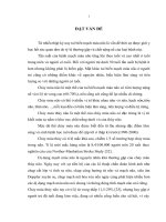
3.2.1. Clinical symptoms
90
80
70
60
50
40
30
20
10
0
88.32
67.51
25.89
31.98
29.95
Chart 3.7. Clinical symptoms
3.2.2. Clinical symptom characteristics
36.55
15.23
11
Sensory disorders in the median nerve distribution of the hand:
97.97%. Pain and paresthesia radiated into forearm, arm and
shoulder: 27.92%. Sensory symptomps are worse at night: 85.79%,
during driving: 88.32%.
3.2.3. Clinical tests: Phalen’s test: 85.77%, Tinel’s test: 77.66% and
carpal compression test: 67.51%.
3.3. The electrophysiological characteristics
Table 3.3. Percentage of abnormal electrophysiological parameters
Parameters
Number of hands (n=197)
%
Prolonged DMLm
120
60.91
Low MAMPm
39
20.31
Slow MCVm
26
13.20
Prolonged DSLm
107
54.31
Low SAMPm
103
52.28
Slow SCVm
180
91.37
Abnormal DMLm-u
171
86.80
Abnormal DSLm-u
182
92.39
3.4. Relationships between clinical and electrophysiological
characteristics
3.4.1. Between clinical symptoms and electrophysiological severity
-The burning sensation and pain were related with the
electrophysiological severity (p<0.05).
- The sensory loss, weakness and thenar atrophy were related closely with
the electrophysiological severity (p<0.001).
12
- There were no relationships between numbness, tingling and the
electrophysiological severity (p>0.05).
3.4.2. Between Boston scores and electrophysiological parameters
- There were the positive correlations between Boston scores, Boston
scales severity and electrophysiological severity (r=0.48;0.37;0.43;
0.36. p<0.05), between distal motor and sensory latencies, medianulnar motor and sensory latencies difference and Boston scales
severity (r= 0.37; 0.36; 0.40; 0.37; 0.30; 0.28; 0.31; 0.27; p<0.05).
- The negative correlations were observed between sensory conduction
velocity and Boston scales severity (r= -0.41; -0.29; p<0.05), between
motor amplitude and Boston functional severity (r= -0.32; p<0.05).
- There were no relationships between sensory amplitude, motor
conduction velocity and Boston scales severity, between motor
amplitude and Boston symptom severity (p>0.05).
3.4.3. Between symptom duration and electrophysiological severity
- There was the positive correlation between symptom duration and
the electrophysiological severity (r=0.23. p<0.05).
3.5. The efficacy of local steroid injection
3.5.1. The clinical assessment
3.5.1.1. The mean Boston scores
Table 3.10. The mean Boston scores in injection group
Boston score
Before injection
Symptom
Functional
score
score
1.83±0.34
1.32±0.44
n
15
4
1 month after injection
1.10±0.15
1.02±0.09
15
13
4
2 months after injection
1.08±0.13
1.00±0.00
92
3 months after injection
1.09±0.15
1.01±0.02
78
<0.001
<0.001
p3.2.1-0
p3-2
>0.05
<0.05
3.5.1.2. Boston scales severity
- Boston symptom severity: After 1 month, 45.45% hands had no
symptoms and after 3 months, 57.69% hands were completely
recovered. At the first month after injection, there were no moderate
cases and the number of mild cases was decreased (p<0.001).
- Boston functional severity: After 1 month, there was no moderate
case, the number of mild cases was decreased. The number of normal
cases was increased from 57.14% to 100% at the second month and
was decreased to 96.15% at the third month (p<0.001).
3.5.2. The electrophysiological assessment
3.5.2.1. Electrophysiological parameters of median nerve
Table 3.11. Electrophysiological parameters in injection group
Parameter
Before
1 month after 2 months after 3 months after
s
injection
injection
injection
injection
DMLm
4.90±1.48
4.57±1.26
4.32±1.03
4.34±1.12
p1-0<0.01
p2-0<0.01
p3-0<0.01
7.14±2.95
7.25±3.11
7.57±3.05
p1-0>0.05
p2-0>0.05
p3-0<0.05
55.86±6.53
55.37±7.13
55.33±5.49
p1-0>0.05
p2-0>0.05
p3-0>0.05
3.34±1.88
3.08±0.68
3.08±0.65
p1-0>0.05
p2-0<0.001
p3-0<0.001
(ms)
MAMPm
6.61±2.94
(mV)
MCVm
56.41±7.71
(m/s)
DSLm
(ms)
3.53±1.01
14
SAMPm
(µV)
SCVm
(m/s)
24.20±14.8
27.76±16.7 30.93±18.93 28.86±15.32
0
3
p2-0<0.05
p3-0<0.05
p1-0<0.05
39.82 ±9.12
43.59±8.41
44.70±7.74
44.15±7.81
p1-0<0.001
p2-0<0.001
p3-0<0.001
3.5.2.2. Electrophysiological grading of the severity: After 3 months,
the number of moderate group was decreased from 58.44% to
34.42%. After 1 month, 17.53% cases became normal in the nerve
conduction study and increased to 20.51% after 3 months. The mild
cases were increased after 3 months (p<0.001).
3.6. The efficacy of surgical decompression
3.6.1. The clinical assessment
3.6.1.1. The mean Boston scores
Table 3.12. The mean Boston scores in surgical group
Mean Boston score Symptom score
Functional
n
score
Before surgical
2.50±0.46
2.15±0.41
43
After 1 month
1.34±0.25
1.30±0.40
43
After 2 months
1.23±0.19
1.20±0.35
23
After 3 months
1.19±0.22
1.13±0.28
31
P3.2.1-0
<0.001
<0.001
3.6.1.2. Boston scales severity
- Boston symptom severity: After 1 month, there were no severe or
moderate cases. The percentage of normal group was 13.95% after 1
month and 32.26% after 3 months (p<0.001).
- Boston functional severity: The moderate cases were decreased
from 65.12% to 4.65% after 1 month and there were no moderate
cases after 2 months. After 1 month, there was 53.49% cases
completely recovered and increased to 70.97% after 3 months, the
number of mild cases was decreased after 3 months (p<0.001).
15
3.6.2. The electrophysiological assessment
3.6.2.1. Electrophysiological parameters of median nerve
Table 3.13. Electrophysiological parameters in surgical group
Parameters Before
After
After
After
surgical
1 month
2 months
3 months
DMLm
5.62±1.60
4.85±2.05
4.37±1.18
4.10±1.23
(ms)
p1-0<0.05
p2-0<0.01
p3-0<0.001
MAMPm 5.54±3.51
5.67±3.41
5.94±3.77
7.30±4.00
(mV)
p1-0>0.05
p2-0>0.05
p3-0<0.05
54.33±7.70 53.14±11.2 52.96±5.47 51.93±8.40
MCVm
7
p2-0>0.05
p3-0>0.05
(m/s)
p1-0>0.05
DSLm
4.85±3.02
3.53±1.25
3.36±1.37
3.10±0.81
(ms)
p1-0<0.01
p2-0<0.05
p3-0<0.01
20.05±13.28 20.81±14.5 22.04±15.6 25.50±20.9
SAMPm
5
7
7
(µV)
p1-0>0.05
p2-0>0.05
p3-0>0.05
33.16±10.41 39.83±8.98 42.88±11.1 44.67±9.37
SCVm
p1-0<0.01
8
p3-0<0.001
(m/s)
p2-0<0.001
3.6.2.2. Electrophysiological grading of the severity
- The percentage of very severe and severe cases were decreased
from 6.98% and 4.65% to 2.32% and 2.32% after 1 month. After 2
months there was no severe and very severe case.
- The percentage of moderate cases were decreased from 88.37% to
41.93% after 3 months. After 1 month, there was 2.32% cases
became normal in the nerve conduction study and increased to
25.81% after 3 months (p<0.001).
3.7. Comparison of local steroid injection and surgical
decompression in the treatment of moderate idiopathic CTS
3.7.1. Comparison of the clinical improvement
16
Table 3.15. Comparison of the improvement of Boston scores
The improvement of Boston
symptom score
The improvement of Boston
functional score
Steroid
Injection
Steroid
Injection
(n=90)
After 1
month
Surgical
(n=38)
p
Surgical
(n=38)
(n=90)
p
0.77±0.04
<0.05
1.08±0.08
0.35±0.05
0.82±0.09
<0.05
After 2
months 0.79±0.05
<0.05
1.19±0.09
0.37±0.06
0.92±0.10
<0.05
After 3
months 0.80±0.05
<0.05
1.24±0.08
0.36±0.07
0.98±0.09
<0.05
>0.05
<0.05
P3-2
>0.05
<0.05
3.7.2. Comparison of electrophysiological recovery
17
Table 3.17. Comparison of the recovery of electrophysiological parameters
Parameter
Injection
Surgical
Time
p
s
Group
Group
After 1 month
DMLm
(ms)
- 0.55±0.20
- 0.77±0.42
- 1.02±0.21
- 1.25±0.41
- 0.91±0.23
- 1.52±0.37
p3-2<0.05
p3-2<0.05
0.54±0.40
0.13±0.79
0.97±0.49
0.39±0.99
0.77±0.49
1.76±0.94
p3-2<0.05
p3-2<0.001
- 1.29±1.19
- 1.19±2.21
- 1.43±1.49
- 1.39±1.94
- 1.83±1.43
- 2.40±2.03
p3-2>0.05
p3-2>0.05
After 1 month
- 0.42±0.15
- 1.32±0.53
After 2
- 0.60±0.17
- 1.49±0.71
After 2
months
After 3
months
After 1 month
After 2
MAMPm
months
(mV)
After 3
months
After 1 month
After 2
MCVm
months
(m/s)
After 3
months
DSLm
(ms)
months
<0.0
5
<0.0
5
<0.0
5
<0.0
5
<0.0
5
<0.0
5
>0.0
5
>0.0
5
<0.0
5
<0.0
5
<0.0
5
18
After 3
months
After 1 month
SAMPm
(µV)
After 2
months
After 3
months
After 1 month
SCVm
(m/s)
After 2
months
After 3
months
- 0.65±0.18
- 1.75±0.61
p3-2>0.05
p3-2>0.05
1.96±2.02
0.76±3.20
8.80±2.82
1.99±3.90
5.21±2.37
5.45±4.28
p3-2<0.001
p3-2<0.01
4.16±1.03
6.68±2.23
6.20±1.25
9.72±2.95
6.23±1.30
11.51±2.55
p3-2>0.05
p3-2<0.05
<0.0
5
<0.0
5
<0.0
5
>0.0
5
<0.0
5
<0.0
5
<0.0
5
3.8. Complications
No major complications were reported in both groups. In the
injection group, 35 patients (22.73%) had mild pain at the injection
site. In the surgical group, 4 patients (9.30%) had wound pain.
CHAPTER 4: DISCUSSION
4.1. Patient characteristics
4.1.1. Gender
19
The females were dominant with 125 patients (94.70%), males
were 7 patients (5.30%). Our results were similar to the results of
other authors.
4.1.2. Age
The mean age was 46.84 ± 9.31.The youngest was 26 and the
oldest was 66. The most common age range was 41- 60 (66.67%).
Almost authors agreed that CTS often occur in the midle-age people.
4.1.3. Occupation
In our study, farmers were 20.46%, housewives 18.18%, sellers
17.42%, workers and handicraftsmen 15.91%, teachers and office
workers 7.58%. In other studies, the rate of CTS was higher in
occupational groups which have to work with vibration, high-force
and repetitive movements of the wrist.
4.2. Clinical features
4.2.1. Clinical symptoms
The sensory symptoms were most common symptoms, numbness
88.32%, tingling 67.51% and occurred in early stage. Pain was
31.98%, burning 25.89%, sensory loss 29.95%.
The motor symptoms were less common, weakness 36.55%, thenar
atrophy 15.23% and often occurred in severe cases ( chart 3.7).
Our results were similar to the results of Le Thi Lieu, Nguyen Le
Trung Hieu, Nora and Steven. The sensory disorders are more
common than motor disorders in CTS because the sensory fibers are
more sensitive to compression than motor fibers.
20
4.2.2. Clinical symptoms characteristics
Sensory disorders in the median nerve distribution of the hand:
97.97%. Pain and paresthesia radiated into forearm, arm and
shoulder: 27.92%. Sensory symptomps are worse at night: 85.79%,
during driving: 88.32%. These symptoms were often intermittent:
81.22%. Our results were similar to the results of other authors.
4.2.3. Clinical tests
Phalen’s maneuver was positive in 85.77% cases, Tinel’s test was
77.66% and carpal compression test was 67.51%. Other authors had the
same conclusion that these tests are clinical tests with high sensitivity in
the diagnosis of CTS.
4.3. The electrophysiological characteristics
4.3.1. Motor conduction studies of median nerve
Prolonged distal motor latency of median nerve was 60.91%, low
motor amplitude 20.31%, slow motor conduction velocity 13.2%
(table 3.3). According to Nguyen Thanh Binh, prolonged distal motor
latency of median nerve was 68.2%, low motor amplitude 28.8%,
slow motor conduction velocity 31.8%. In the study of Kimura,
sensitivity of prolonged distal motor latency was 61%.
The motor fibers are often less involved than sensory fibers in CTS. In
addition, when there is damage to motor fibers, there are compensatory
mechanisms to preserve function. It can explain why the motor
conduction studies are less sensitive for CTS than the sensory
conduction studies.
4.3.2. Sensory conduction studies of median nerve
21
Sensory conduction studies are more sensitive than motor
conduction studies. In our study, slow sensory conduction velocity of
media nerve was 91.37%, prolonged distal sensory latency: 54.31%
and low sensory amplitude was 52.28% (table 3.3).
4.3.3. Median-ulnar motor and sensory latency difference
Abnormal median-ulnar sensory latency difference was the most
sensitive parameter (92.39%). Abnormal median-ulnar motor latency
difference was 86.8% (table 3.3).
The study of Vo Hien Hanh and Nguyen Huu Cong showed that,
abnormal median-ulnar motor and sensory latency differences were
95.5% and 98.9%. Other authors also concluded that, these
parameters had high sensitivity (85-88%) and specificity (100%) in
the diagnosis of CTS.
4.4. The relationship between clinical and electrophysiological
characteristics
4.4.1.Between clinical symptoms and electrophysiological severity
The burning sensation and pain were related with the
electrophysiological severity (p<0.05).
The sensory loss, weakness and thenar atrophy were related
closely with the electrophysiological severity (p<0.001).
There was no relationship between numbness, tingling and the
electrophysiological severity (p>0.05).
Our results were similar to the results of Bui Thi Ngoc, Nguyen Le
Trung Hieu, Padua. Numbness, tingling are associated with large nerve
fibre dysfunction, which often occurs early. Burning, pain are associated
with small nerve fibre dysfunction, occur in later stage of the disease. The
22
motor fibers may become involved in more advanced stage and the
motor symptoms often occur in severe cases.
4.4.2. Between Boston scores and electrophysiological parameters
There were positive correlations between Boston scores, Boston
scales severity and electrophysiological severity (r=0.48;0.37;0.43;
0.36; p<0.05). Our results were similar to the results of Le Thi Lieu,
Giannini and Karadag.
There were positive correlations between distal motor and sensory
latencies, median-ulnar motor and sensory latencies difference and
Boston scales severity (r= 0.37; 0.36; 0.40; 0.37; 0.30; 0.28; 0.31;
0.27; p<0.05).
The negative correlations were observed between sensory conduction
velocity and Boston scales severity (r= -0.41; -0.29; p<0.05), between
motor amplitude and Boston functional severity (r= -0.32; p<0.05).
There were no relationships between sensory amplitude, motor
conduction velocity and Boston scales severity, between motor
amplitude and Boston symptom severity (p>0.05).
The study of You.H showed that, there were positive correlations
between Boston symptom score and sensory amplitude, distal
sensory latency, sensory conduction velocity, distal motor latency
(r=-0.41; 0.53; 0.49; 0.46; p<0.01). There was no relationship
between Boston symptom score and motor amplitude.
4.4.3. Between symptom duration and electrophysiological severity
There was positive correlation between symptom duration and the
electrophysiological severity (r=0.23; p<0.05). Our results were
similar to the results of Longstaff and Padua. The authors concluded
23
that, there was a poor correlation between symptom duration and the
electrophysiological severity.
4.5. Efficacy of local steroid injection
4.5.1. The clinical assessment
Both Boston scores and Boston severity were improved
significantly after steroid injection (p<0.001) (table 3.10). These
results were similar to the results of Nguyen Van Huong, Atroshi and
Amstrong. In the study of Agarwal, after 3 months, 34.42% hands
had no clinical symptoms and 52.17% were recovered completely
hand functions.
4.5.2. The electrophysiological assessment
After injection, there was remarkable recovery of distal motor and
sensory latencies, motor and sensory amplitudes, sensory conduction
velocity (p<0.05). Our results were similar to studies of Nguyen Van
Lieu, Cartwright, Gupta and Celik.
The electrophysiological severity was also improved. A number of
moderate group was recovered to mild and normal groups (p<0.001).
The study of Agarwal showed that, there was 64% cases became
normal in the nerve conduction study after 3 months. Steroids reduce
synovial swelling, vascular congestion and the pressure in the carpal
tunnel. It can result the improvement of clinical symptoms and nerve
conduction. The sensory conduction often recovered earlier than
motor conduction.
4.5.3. Complications of steroid injection
No major complications (median nerve, tendon or muscle injuries,
hematoma or infectious) were reported. There were 35 patients
24
(22.73%) had mild pain at the injection site, which disappeared after
2 or 3 days without treatment. Other studies showed the results
similar to our result.
25
4.6. Efficacy of surgical decompression
4.6.1. The clinical assessment
After operation, there were significant improvement of Boston
symptom and functional scores (p<0.001) (table 3.12). The studies of
Tran Trung Dung, Mondelli and Heybeli showed that surgical
decompression result the improvement of clinical symptoms in the
first months.
Clinical grading of severity improved significantly after surgical
decompression (p<0.001), this result was similar to the results of
Pham Van Toan and Iida.
4.6.2. The electrophysiological assessment
Distal motor latency, distal sensory latency, motor conduction
velocity and motor amplitude improved significantly after surgical
decompression (p<0.05) (table 3.13). At baseline, there were 3 very
severe cases and 2 severe cases, but after 1 month there were only 2
severe cases and after 2 months there were no severe cases. Our
results were similar to the results of Tran Trung Dung, Celik and
Hui. AC.
- Almost patients had improved electrophysiological severity after
surgical decompression. Specially, there were 2.32% cases becoming
normal in the nerve conduction study after 1 month and increased to
25.81% after 3 months (p<0.001). The study of Ucan showed that,
there was 36.36% cases became normal in the nerve conduction study
after 3 months and increased to 45.45% after 6 months.
Surgical treatment reduced the pressure in carpal tunnel, the
compression on the median nerve and relieved local ischemia. It
induced an improvement of the clinical symptoms and median nerve