Ebook Nutrition support for the critically ill: Part 2
Bạn đang xem bản rút gọn của tài liệu. Xem và tải ngay bản đầy đủ của tài liệu tại đây (4.49 MB, 181 trang )
Chapter 7
Access and Complications of Parenteral Nutrition
Dustin R. Neel
Keywords Parenteral nutrition • Venous anatomy • Peripheral venous catheter • Midline catheter •
Central venous catheter • Peripherally inserted central catheter (PICC) • Tunneled central venous
catheter • Implantable central venous port • Thrombophlebitis • Catheter-related infections • Blood
stream infections • Pneumothorax • Air embolism • Catheter malposition • Pinch-off syndrome •
Catheter occlusion • Catheter thrombosis • Hyperglycemia • Hypoglycemia • Hyperlipidemia •
Essential fatty acid deficiency • Hepatic steatosis • Nephromegaly • Metabolic bone disease •
Refeeding syndrome
Key Points
• Peripheral venous access is indicated for short-term parenteral nutrition in those with adequate
veins and those whom can tolerate high volumes of low osmolality solutions but cannot tolerate
short-term starvation. Peripheral parenteral nutrition is rarely necessary. The major complication
of peripheral venous access is thrombophlebitis.
• Non-tunneled central venous catheters are placed via the Seldinger technique. The majority of
complications including pneumothorax, air embolism, and bleeding, occur during initial placement. These catheters may be used for short-term parenteral nutrition therapy.
• Peripherally inserted central catheters (PICCs) are indicated for intermediate-term access.
Compared to central venous catheters, they have a lower infection risk but a higher incidence of
thrombophlebitis, but dislodgement and difficulty with daily activities remain the major
disadvantages.
• Tunneled central venous catheters are the preferred route of administration of parenteral nutrition
in those patients that require it for an extended period of time. Occlusion and thrombosis results
from a fibrin sheath formation which is a long-term complication of all access lines.
• Infection is the number one complication in central venous catheters, with a wide range of presentations. Sepsis is associated with significant morbidity and mortality. The most commonly isolated
D.R. Neel, MD (*)
Surgical Intensive Care Unit, Department of Surgery,
Truman Medical Center, Kansas City, MO, USA
e-mail:
D.S. Seres, C.W. Van Way, III (eds.), Nutrition Support for the Critically Ill, Nutrition and Health,
DOI 10.1007/978-3-319-21831-1_7, © Springer International Publishing Switzerland 2016
99
100
D.R. Neel
organism is Staphylococcus epidermidis. Tunneled, cuffed central venous lines placed in the subclavian vein carry the lowest infection risk.
• Hyperglycemia, hyperlipidemia, and refeeding syndrome are complications of parenteral nutrition
associated with its internal composition.
Introduction
Nutrition support has been a crucial component of medical practice for decades. While voluntary oral
nutrition is the best route of nourishment delivery for most patients, and enteral nutrition is considered
the best alternative, some patients ultimately require parenteral nutrition (PN) [1]. Since the first laboratory demonstration of its efficacy by Dudrick and Wilmore in 1968, PN has been used successfully
to support patients with intestinal failure [1–3].
The first patient received PN at home in 1968 [4, 5]. Home PN is used largely for short-bowel
syndrome, high-output enterocutaneous fistulae, or severe chronic gastrointestinal dysfunction, and is
used to treat both electrolyte abnormalities and to provide all required nutrients [6]. Over 40,000
patients require PN at home annually, and there are many others who require PN temporarily during
a medical or surgical crisis [1, 4, 7, 8]. Despite 45 years of successful clinical use, PN has been called
poison and condemned as being inferior to enteral nutrition [9–12]. However, when making comparisons of PN to enteral nutrition it is important to remember that PN should not be considered a replacement for enteral nutrition; rather, it is intended to treat patients who cannot sustain oral or enteral
feeding [5, 12]. In fact, all humans start their life on parenteral nutrition and adapt to enteral nutrition
after exiting the womb and entering the world [10]. In the words of Sir David Cuthbertson, a prominent biochemist and nutritionist, “Lest we forget, I would remind you that we all owe our fetal life till
parturition to the passage of the nutrients we require from the blood vessels of our mothers into blood
vessels as they transverse the chorionic villi in close relation” [13, 14].
Parenteral Access and Complications
In 1656, Sir Christopher Wren first experimented with PN. He administered wine, ale and morphine
to a dog via a goose quill attached to a pig’s bladder [5, 11]. Three hundred years later, Dudrick and
Wilmore used vinyl catheters in six beagle puppies to show that PN could support growth and development [10]. Today, PN is widely used, and we have a bewildering variety of catheters available [15].
Over 50% of hospitalized patients have either a peripheral or central catheter, and over five million
central venous catheters are inserted yearly [16, 17]. But despite all of the advances, vascular access
still remains one of the most important and challenging components of PN.
Vascular Anatomy and Physiology
Venous return is the most important characteristic in choosing which access to use in the delivery of
PN to the patient. An understanding of the basic anatomy and physiology of the vasculature is helpful
in determining best access locations and safe practices.
Veins have three layers: the tunica intima, tunica media, and tunica adventitia. The innermost layer
is the tunica intima which is in direct contact with the venous flow via a nonthrombogenic smooth
low-friction surface. The middle layer is the tunica media which contains connective tissue with
7 Access and Complications of Parenteral Nutrition
101
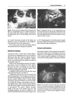
Internal Jugular Vein
External Jugular Vein
Subclavian Vein
Brachiocephalic
(innominant) Vein
Superior Vena Cava
Cephalic Vein
Basilic Vein
Inferior Vena Cava
Common Iliac Vein
Internal Iliac Vein
External Iliac Vein
Femoral Vein
(deep vein)
Greater Saphenous Vein
(superficial vein)
Fig. 7.1 Upper and lower extremity venous anatomy [21]. Reprinted by permission of SAGE Publications. Vanek VW,
Nutrition in Clinical Practice, 17(2), pp. 85–98, copyright © 2002 by SAGE Publications
elastic fibers. This allows the veins to stretch in order to tolerate changes in pressure. The outermost
layer is the tunica adventitia which contains the nutrient-supplying blood vessels to the walls of
the larger veins. These are known as the vasa vasorum [18–20]. The major veins are diagrammed in
Fig. 7.1.
The superficial veinsof the upper extremity include the cephalic, the basilic, and the median antebrachial veins. The basilic vein becomes the axillary vein at the lateral chest wall (teres minor). The
cephalic vein drains directly into the axillary vein. The average diameters of the basilic, cephalic, and
axillary are 8, 6, and 16 mm, respectively [18, 19, 21]. The axillary vein becomes the subclavian vein
as it crosses the first rib. The neck has two major veins: the internal jugular and the external jugular.
The external jugular vein drains the face and scalp, and it ultimately empties into the subclavian vein.
The average diameter of the subclavian vein is approximately 19 mm. The internal jugular vein drains
the head and brain and combines with the subclavian vein to create the brachiocephalic, also referred
to as the innominate vein. The left brachiocephalic crosses the chest to join the vertically oriented
right brachiocephalic to create the superior vena cava (SVC). The SVC measures approximately
20–30 mm, and is approximately 7 cm in length. The last centimeter of SVC is inside the pericardium,
where it joins the right atrium [18–22].
The veins in the lower extremity include both a superficial and a deep venous system. The deep
venous system has a rich collateral network and ultimately drains into the popliteal vein. The common
femoral vein is the continuation of the popliteal vein above the adductor (Hunter’s) canal. The superficial system drains into the greater saphenous vein and ultimately into the common femoral vein. The
profunda femoral vein also drains into the common femoral vein. The common femoral vein courses
superiorly and becomes the external iliac vein at the inferior border of the inguinal ligament. The
internal iliac vein joins the external iliac vein to become the common iliac vein. The right and the left
common iliac veins join to become the inferior vena cava (IVC) at approximately L5. The IVC then
drains into the right atrium [19, 21, 22].
102
D.R. Neel
Venous return to the heart is aided by many physiologic principles. Muscle contraction aids the
return of blood to central circulation via compression of the superficial veins of the lower extremities.
Paired valves within these veins prevent retrograde blood flow and the muscle contractions propel
blood towards the heart. Blood flow within central veins is not dependent on valves; instead the negative intrathoracic pressure of inspiration accelerates blood into the central circulation [18]. The SVC
and the IVC have large diameters to accommodate high blood flow. This makes the central veins the
preferred vessels for PN as it rapidly dilutes the hyperosmotic solution. The flow through the SVC is
estimated at 2000 mL per minute versus 150–250 mL/min in forearm veins [18, 21].
Peripheral Vein Access Versus Central Access
The position of the distal catheter tip, not the location of the entry site, determines whether or not the
vascular access is peripheral or central. Central access catheters have distal tips that terminate in the
SVC or the IVC [21], although it should be noted that the preferred terminus for catheters used for
central parenteral nutrition is at the vena caval entry into the atrium.
Peripheral Venous Access
Peripheral vein access should only be used for a short-term therapy, and because of the increasing
ease and safety of PICCs peripheral parenteral nutrition (PPN) is rarely necessary.
Peripheral venous access is simply placing an intravenous cannula into a peripheral vein. It remains
the safest, easiest and fastest ways to gain vascular access, in general, but is fraught with difficulties
when used for PPN. Examples of peripheral venous access include: needles, short peripheral catheters, and midline catheters. While midline catheters resemble PICCs, they are not central lines. They
are placed peripherally and terminate in larger veins usually in the upper arm. The main limitation of
peripheral access for patients requiring PN remains the high tonicity of the PN, which is often 1200
mOsm/L or more in centrally infused formulas [23]. Peripheral intravenous cannulas should not be
utilized for solutions greater than 900 mOsm/L as “burning” of the vein will occur [2, 21, 24, 25]. This
is why peripheral parenteral nutrition requires such a larger volume and why standard PN cannot be
infused peripherally, including via midline catheter.
PPN solutions can only be given through peripheral catheters for short periods; usually a few days.
This type of access is not approved for patients with inadequate veins, those requiring longer than 5
days of therapy, and those who cannot handle large volumes of fluid, as in patients with congestive
heart failure. PPN solutions should contain no more than a final concentration of 3 % amino acids and
no more than 10% dextrose [21]. The primary advantages of peripheral venous access are fewer infections, and easy access if adequate veins are present [21, 23, 24].
The primary complication of peripheral venous access is thrombophlebitis of the peripheral vein.
Infusion thrombophlebitis is the inflammation of a cannulated vein resulting in pain and discomfort
and occurs in a large percentage of patients with peripheral access [21, 22, 24, 26]. The inflammation
results in venous thrombosis and possible occlusion, and leads to skin changes and edema, erythema,
pain, and often a palpable venous cord. The main risk factors for peripheral thrombophlebitis are the
type and concentration of infusate, the location of the catheter, and the duration. Infusates including
dextrose, amino acids, lipids, and irritant drugs including antibiotics, chemotherapeutic drugs, acidic
solutions, and vasoactive agents also increase the risk of thrombophlebitis [21, 26]. Blood, medications, electrolytes, and other infusates not included in the PN should be given via separated peripheral access sites [24].
7 Access and Complications of Parenteral Nutrition
103
There is a marked increase in the incidence of thrombophlebitis after 48 h of infusion, which has
led to recommendations to change the site of the every 24–72 hours to decrease this risk [2, 23, 24,
26–28]. The lowest rate of thrombophlebitis occurs at solution osmolarity below 450 mOsm/L [21,
24]. For reference, the osmolality of normal saline is approximately 285 mOsm/l. Recent infusion
guidelines allow peripheral access to remain for 72 hours as long as the sites are free from visible
complications [27]. Access should be changed sooner if the patient develops pain, erythema, or other
signs of vascular site compromise, or a break in sterile technique occurs. If thrombophlebitis develops, rapid removal of the cannula should occur, and replacement should be distant from the original
site, preferably in an alternative limb [28]. Various techniques have been attempted to decrease the
risk of thrombophlebitis associated with PN, including topical anti-inflammatory agents, buffering
solutions, and heparin, but none has resulted in significant reductions [21, 23, 24]. Other complications include cellulitis and sepsis, discussed later in the chapter.
Midline Cathethers
Midline catheters are also considered peripheral access, and are not recommended for infusion of
standard PN or any other caustic or highly concentrated solution. It is preferable to place a PICC for
central PN since the insertion techniques are similar, and the PICC has fewer downsides. Midline
catheters are usually approximately 8 in. long, and are inserted into the basilic vein with the distal tip
in the proximal basilic or axillary vein, but not into the subclavian vein. Due to the size of the vein,
there is a decreased risk of thrombophlebitis compared with standard peripheral lines when infusing
low osmolality solutions, but venous stenosis is a potential longer-term sequela. Midline catheters
function for a median of 7 days, but may be used in general for up to several weeks. Advantages of
midline catheters are the ease of placement by a specially trained nurse, longer dwell time, and minimal post-placement care. In addition, midline catheters have lower rates of thrombosis in the deep
brachial veins compared to PICCs. Disadvantages include the need to change the catheter every 14
days, increased cost compared with peripheral cannulas, and the lack of central access and the attendant issues related to PPN [21, 24].
Central Venous Access
As mentioned above, the determination of central versus peripheral is the location of the distal tip, not
the access location. Central venous catheters (CVC) have distal tips located in the central circulation,
specifically the SVC, the IVC, or the right atrium [2, 22]. A PICC, as noted above, is inserted in a
peripheral vein, usually the cephalic or basilic, and terminates in the SVC. Even though the tip of
PICCs is central, because the insertion technique and useful lives of PICCs and temporary central
catheters are significantly different, they are addressed in separate sections. For the sake of clarity, the
term CVC refers to temporary, non-tunneled central catheters other than PICCs, and are distinguished
from tunneled central venous catheters, discussed below. Common places for CVC puncture sites
include the subclavian, internal jugular, and femoral veins. CVCs have multiple uses including the
administration of solutions, including PN, that may cause phlebitis or sclerosis if infused peripherally.
These uses include PN, laboratory draws, as well as central venous pressure monitoring. Multiple
types of central catheters exist, each with their pros and cons [15].
Temporary non-tunneled CVCs are placed via the Seldinger technique. This involves the use of a
needle to pierce the vein, followed by the cannulation of the vein with a wire. One or more dilators
104
D.R. Neel
are used to dilate the tract, and the catheter is subsequently placed over the wire. With the exception
of PICCs, non-tunneled CVCs are most commonly placed in the internal jugular or subclavian, and
advanced to the SVC. Femoral access to the IVC may be performed in an emergency, but is not recommended for routine use, particularly for PN, because the risk of infection and venous thrombosis
are both higher [29].
CVCs have a high success rate of placement, providing immediate access for those needing central
access. Advantages include the availability of multiple lumens within the catheter for patients requiring multiple infusions, the ability to monitor central venous pressure, and the ability to draw frequent
labs without venipuncture. The complication rate associated with CVCs is approximately 10%, with
over half associated with the initial placement. Early complications include pneumothorax, great vessel injury, hemothorax, bleeding, air embolism, arrhythmia, cardiac tamponade, nerve injury, and
misplacement of the catheter [18, 19, 21, 22, 24, 30–32]. Pneumothorax is less common in internal
jugular access compared with subclavian access, and is a non-issue in femoral access. The increased
usage of ultrasound for placement has reduced but not eliminated complications [18, 27, 30, 33].
Because the risk of infection and thrombosis is higher in femoral access, the Center for Disease
Control (CDC) and most other authorities recommend using the subclavian or jugular access [2, 29,
30, 34].
Immediate Complications of Central Venous Access
As with any invasive procedure, central line insertion is associated with complications. Those specific
to PICC line insertions will be addressed in a separate section below. Pneumothorax occurs when the
pleura is nicked or punctured by the needle, introducer, or dilator. The incidence ranges widely, and is
probably most dependent on the experience of the operator. These are very rare with PICCs. The size
of the pneumothorax determines management [21, 31, 33]. If it is less than 10–15%, and the patient
is asymptomatic, it may be monitored simply with repeated chest radiographs. However, if the pneumothorax is larger, the patient is symptomatic, or the patient is ventilated with positive pressure, a
tube thoracostomy may be needed to re-expand the lung. Bleeding may result from the venous puncture or from accidental laceration of the vein or artery, especially if coagulation is impaired. At the
extremes bleeding may result in a simple hematoma, responsive to gentle pressure, or may create a
life-threatening exsanguination. Bleeding into the pleural space may result in a hemothorax.
Unrecognized misplacement of a CVC into the pleural space and infusion of fluids will result in
hydrothorax. The position of the tip of every CVC must be confirmed by X-ray or other proven methods, so this should be an extremely rare event. A chylothorax is also possible if the thoracic duct is
lacerated during CVC placement, most commonly occurring via the placement into the left subclavian
vein. While minor pleural complications may be simply observed with serial radiographs, more serious complications may require tube thoracostomy, video-assisted thoracotomy, or even a thoracotomy
to repair the complication [21, 33].
Injury to nearby arteries, particularly the internal carotid artery, the subclavian artery, and femoral
artery may occur. Direct pressure is effective for puncture injuries of internal jugular or femoral
arteries, but the subclavian artery cannot be easily be compressed. Any of these may on occasion,
require intervention with an intravascular stent or even an open surgical repair. Arterial bleeding can
cause airway compression, or even arteriovenous fistula, retrograde aortic dissection, or cerebrovascular events in extreme cases [31, 33]. Nerve injury of the phrenic, brachial plexus, vagus, recurrent
laryngeal, and cervical sympathetic chain may cause pain, numbness, paralysis, or autonomic dysfunction [21, 33].
Air embolism is a life-threatening complication from any central catheter insertion. Care must be
taken to prevent the catheter hub from being open during patient inspiration. Negative intrathoracic
7 Access and Complications of Parenteral Nutrition
105
pressure can suck air in through the catheter. Except for confirming blood flow from the catheter, the
hub should always be occluded. When air is pulled through the catheter, a froth of air bubbles and
blood develops within the right atrium. If nothing is done, the air bubbles can pass into the right ventricle, and these may block perfusion. The patient should be placed on his or her left side immediately,
leaving the catheter in place. The expectation is that air will rise to the right atrium and cava, thus
allowing aspiration via the recently placed catheter. Further, as long as the air remains in the atrium,
it will slowly be absorbed. Elevating the legs (decubitus Trendelenburg position) may also aid in
keeping the air bubbles from passing into the heart [32, 33, 35].
Cardiac arrhythmias often result from the guidewire “tickling the heart.” The wire is passed through
the central veins into the right atrium and right ventricle. The wire can irritate the ventricular endocardium, resulting in premature ventricular beats or even runs of ventricular tachycardia. The endocardium around the tricuspid valve is especially sensitive. Generally, the ectopic rhythm is corrected by
simply pulling the wire out of the heart. Perforation of the atrium or ventricle by a guide wire or dilator
may be catastrophic, but is very rare. This results in blood accumulating in the pericardium, cardiac
tamponade, cardiogenic obstructive shock, and ultimately cardiac arrest. Temporary life-saving treatment for cardiac tamponade is pericardiocentesis, but median sternotomy or thoracotomy may ultimately be required to repair this complication [19, 32, 33].
Malposition of CVCs occurs in 4–10% of central access insertions [21, 33]. To avoid the intrapericardial portion of the vena cava, the best location is 1–2 cm above the junction of the SVC and the
right atrium. But many authorities feel that placing it at the junction or in the atrium for 1–2 cm
decreases the risk of later occlusion by keeping the catheter tip in motion [21, 36]. The ideal location
of the distal tip is still a matter for disagreement. Common incorrect positions of the distal tip include:
the contralateral subclavian vein, the ipsilateral internal jugular vein, the right atrium, the right
ventricle, and IVC. As stated, a chest radiograph is required for confirmation of placement prior to use
to both detect and avoid this complication [2, 19, 21, 22, 32, 33].
Late Complications of Central Venous Access Catheters
Late complications occur beyond those events related to initial placement and are directly related to
the length of time the catheter is in place. Catheter dislodgement can be both a devastating and costly
complication. Multiple techniques have been developed to secure the catheter in place, including:
suturing, commercial devices that adhere to the skin, and a combination of the two. Catheters still
become dislodged despite these methods. This results in the need for replacement, exposing the
patient to the risks mentioned above that are associated with initial placement. In addition, secondary
delayed catheter migration and malposition have been reported [22].
Catheter occlusion and thrombosis are additional late complications that restrict the use of the
central catheters. Occlusion is the second most common complication behind infection, and the
incidence increases as catheter life span increases [8, 35]. Incidence varies from 7–40% per catheter-year [37]. Catheter thrombosis should be suspected if it is difficult to draw blood from the catheter or resistance is experienced during infusion. Occlusion is usually caused by the formation of a
fibrin sheath around the catheter tip. The central catheter injures and disrupts the venous intima,
resulting in the formation of a fibrous sheath around the catheter. The result is blockage or a plug at
the catheter tip [4, 8, 15, 21, 36, 38–40]. Venous thrombosis may develop as well. Patients at highest risk for thrombosis include those with hypercoagulable states, such as malignancy, renal failure,
and sepsis [4, 8, 40].
Thrombosis associated with central catheters occurs due to Virchow’s triad: intimal damage due to
the catheter tip, altered flow, and stasis [33, 36, 41]. Thrombosis of the central veins is related to the
elevated osmolality, change in pH and viscosity. Because of the rich collateral venous network
106
D.R. Neel
associated with the thorax, central vein thrombosis rarely results in skin changes [21, 40]. Central vein
stenosis and thrombosis occurs at a rate of 0.25 episodes per 1000 catheter access days [21]. The
subclavian vein and upper extremity veins can develop catheter-related venous thrombosis. These can
propagate and embolize [8, 35, 40], but pulmonary embolism (PE) rarely occurs in the presence of
upper extremity and chest thrombosis [4]. Another rare complication (incidence 0.03%) of venous
thrombosis is superior and inferior cava syndromes [4, 8, 42]. Intracardial thrombosis has also been
reported in those catheters with the tip in the right atrium [4]. The actual catheter-related venous
thrombosis rate is not entirely known because many patients may be asymptomatic [40, 43].
Thromboses and hematomas may become infected and result in septicemia [24, 36, 38, 40]. In fact,
thrombosis and infection are frequently found together [24, 40]. Infection will be discussed in further
detail with the long-term tunneled central venous catheters.
Peripherally Inserted Central Catheter (PICC)
As the name implies, PICCs are generally inserted into the superficial veins, usually the cephalic or
basilic veins of the arm, and advanced into the central veins. In 1957, Ross used peripherally inserted
central venous catheters to infuse hyperosmotic solutions [44]. In 1975, Hoshal described the first
long-term use of a PICC for intravenous nutrition [45, 46]. PICCs are longer than other CVCs so they
can be inserted in the antecubital fossa, or preferably under ultrasound guidance into the basilic vein
between the biceps and triceps medially, and subsequently advanced through the axillary vein into the
SVC [18, 21, 22, 44, 45]. Negotiating the acute angle of the cephalic-axillary vein confluence makes
the cephalic vein less appealing than the basilic vein.
PICCs are indicated for intermediate and long-term access, usually for an anticipated duration of 6
days or longer [15, 20, 30, 47]. They are used to provide PN, intravenous antibiotics, and intravenous
medications [30, 44, 45]. PICCs function for an average of 10–73 days, but have been kept in place
as long as 307–421 days [30, 44, 47]. Contraindications to PICC placement include thrombophlebitis
of the antecubital veins, active inflammation, cellulitis or burns, thrombosis, arteriovenous fistula,
history of axillary dissection or active lymphedema. As the law of Laplace states, liquid flow velocity
is inversely related to diameter and length of the tube. Due to their length and small lumens, most
PICCs are not recommended for high volume, rapid boluses or pressurized injections [20, 44, 46].
There are, however, newer versions of PICC catheters designed to both withstand rapid and higher
pressure infusions. These allow for both pressure monitoring and bolus infusions of substances such
as intravenous dyes for procedures such as CT scans.
Complications of PICC insertion include malposition, catheter occlusion, infection, thrombosis
and thrombophlebitis [15, 20, 22, 44–46]. As with other central catheters, the ideal location for the
distal tip is still in question; either above, at, or below the cavo-atrial junction, as described above.
Those not in one of these locations are by definition, malpositioned. They can be over inserted (located
too far in the right atrium or in the IVC), under inserted (located in the ipsilateral axillary vein and
subclavian vein), or they can be aberrantly located (ipsilateral internal jugular or contralateral subclavian vein) [19, 45, 46, 48]. Again, confirmatory radiographs are required to confirm location.
Thrombophlebitis occurs at a rate of 9.2%, while thrombosis has been reported at rates of between 0
and 7%. These rates are higher than those reported with CVCs [44, 45, 48]. If thrombophlebitis occurs,
removal of the PICC is indicated [30]. Thrombosis risk is increased when the catheter is malpositioned
[44, 46]. Occlusion of PICC catheters occurs between 2 and 18 %. This is more frequently in those
catheters used intermittently, such as for periodic antibiotics or chemotherapy, as compared with those
used daily, as with PN or daily antibiotics [44]. Occlusion occurs as a result of fibrin sheath formation as
discussed above. The catheter tip can develop a blood clot at the tip or inside the catheter, ultimately
7 Access and Complications of Parenteral Nutrition
107
resulting in occlusion. Frequent use, daily flushing, and flushing after each use all reduce occlusion rates
[15, 20, 38, 44].
Infection rates in PICCs are less than non-tunneled temporary CVCs [15, 44, 47, 48]. It is theorized
that the reduced infection rate may result from decreased colonization due to the location of the
PICC. The antecubital fossa is cooler, resulting in less moisture, which results in less colonization of
the antecubital fossa versus the chest and neck [44, 47]. Secretions from the nares, mouth, tracheostomy, and endotracheal tube also likely related to the increase in contamination of subclavian and
internal jugular CVCs due to the proximity of these catheters to the secretion source. Maximum barrier precautions are recommended to aid in the reduction of infectious complications [30, 36].
Catheter-related infections are further discussed later in the chapter.
Complications associated with PICC placement include median nerve injury and accidental puncture of the brachial artery, resulting in arterial bleeding, hematoma, arteriovenous fistula, and ischemia to the distal hand [2, 11, 44]. Uncommon complications include vein perforation, chest wall
abscess, venous extravasation, cardiac arrhythmia, cardiac tamponade and perforation, and distal
embolism due to shearing of the PICC tip [19, 20, 31, 32, 45, 46].
A study from the Mayo Clinic reporting noninfectious PICC complications during placement and
usage concluded that dislodgment was the most frequent complication, occurring in 8.9%. Other
complications included: malposition (5.8%), catheter clotting and thrombophlebitis (3.8% each),
catheter infection (3.8% confirmed, additional 3.6% suspected), and bleeding (0.5%) [32, 44, 47, 48].
Advantages of PICC include the ability to place at the bedside, possibility for specialized nursing
teams to perform the placement, easy removal, option of single or multiple lumens, lack of additional
skin punctures for access or blood drawing, lower cost of insertion than tunneled central venous catheter, and lack of risk of central complications including pneumothorax and bleeding from major arteries [47]. Disadvantages include isolation of one arm from daily activities, difficulty in caring for the
catheter with one hand, self-image issues, dislodgment and malposition risk, need for occlusive dressing at all times, and requirement of adequate veins [15, 46].
Long-Term Tunneled Central Venous Catheters
Broviac et al. first described the use of tunneled catheters for long-term access in 22 patients in 1973
[46, 49]. The silicone catheter was 90 cm long with a Dacron felt cuff midway between the insertion
site and the tunneled exit site approximately 15 cm away. The Dacron cuff supports tissue ingrowth,
which both anchors the catheter to prevent inadvertent dislodgement and prevents bacterial migration
along the catheter from the skin exit site [19, 24, 49, 50]. These catheters are primarily inserted into
the subclavian vein, internal jugular vein, or via cephalic vein cut down in the deltopectoral groove.
The catheter enters the skin usually over the pectoralis on the anterior chest, and is tunneled subcutaneously to where it enters the vein. This subcutaneous tunnel, often 10 or more centimeters long,
creates a longer indirect route for bacteria to enter the bloodstream—from the exit skin site to the
vein—and thus decreases the likelihood of contamination [4, 24]. Hickman used a larger diameter but
similar catheter in 1979 [23, 50]. The terms “Broviac” and “Hickmann” are used interchangeably to
describe central catheters that are both cuffed and tunneled, but the more generic name of “tunneled
central venous catheter” is preferable [46]. Tunneled catheters are primarily used for daily intravenous therapies administered for an extended period of time, especially home PN [2, 6].
Tunneled catheters are placed in similar locations as the non-tunneled-CVCs via the Seldinger
technique, as previously described. Likewise, the distal tip position should be confirmed by
post-procedure chest radiograph [2, 19]. Complications in placement of tunneled central catheters are
108
D.R. Neel
similar to the non-tunneled variety as discussed in detail previously, and include: pneumothorax,
hemothorax, air embolism, cardiac arrhythmias, cardiac perforation with pericardial tamponade, arterial perforation with bleeding, and catheter misplacement [15, 32, 41]. Malposition may either be
immediate or due to delayed migration. However, the incidence of immediate malposition is reduced
with the assistance of fluoroscopy during placement. Delayed secondary migrations should be corrected as soon as possible, especially when irritating drugs or hypertonic agents such as PN are given
[33, 51]. The incidence of occlusion and thrombosis are directly related to the duration of the catheter
insertion; therefore, they are more common in tunneled catheters due to the long-term nature of the
catheters. Thrombus formation occurs more frequently with secondary migration of the catheter tip to
an inappropriate location [19, 41, 51]. However, thrombus formation is uncommon (2%) despite the
more common fibrin sheath (85%). The fibrin sheath may create a ball-valve occlusion, leading to the
inability to aspirate despite the ability to flush and infuse through the catheter [38, 39]. However, this
can eventually lead to either catheter occlusion or venous occlusion, deep vein thrombosis, or a combination of both. Occluded catheters can often be salvaged with thrombotic therapy, usually tissue
plasminogen activator (t-PA) or Urokinase [3, 4, 8, 20, 24, 37, 51], and treatment is recommended
twice prior to declaring the catheter unusable and removing it [24].
Originally thought to be of no clinical significance, upper extremity deep venous thrombosis
(DVT) has become more frequently diagnosed and determined to be consequential [40, 41, 43, 52].
Upper extremity DVTs can lead to both chronic venous insufficiency and pulmonary embolus (PE).
Upper extremity DVTs are responsible for 7–9% of symptomatic PEs [43]. Treatment of upper
extremity DVTs should be equivalent to lower extremity DVTs and should involve aggressive anticoagulation or thrombolytic therapy. A close parallel to DVTs is SVC occlusion which can lead to both
shock and death if it occurs acutely. The incidence of SVC occlusion associated with PN ranges from
8–14% [37]. Standard treatment for DVTs and SVC occlusion include both thrombolytic therapy
and systemic anticoagulation with heparin followed by coumadin. Treatment of SVC occlusion may
progress to involve balloon angioplasty and expandable metal stents in refractory cases [37].
“Pinch-off syndrome” was first described in 1984 by Atiken and Minton [32, 53]. The catheter
becomes obstructed due to compression as it transverses between the sternoclavicular joint and the
first costosternal articulation. The compression creates narrowing, pinching, and ultimately obstruction, which may be intermittent and positional [53]. Eventually, the catheter may fracture, with a
mean time of 6.5 months from insertion to fracture. Fracture of the catheter can be quite dangerous,
and even fatal if the distal portion embolizes to the right ventricle or pulmonary arteries. Other complications include extravasation of fluids at the fracture site as well as arrhythmias. Treatment may
require angiographic retrieval or open operative intervention [32, 36]. Extravasation is associated
with an intense tissue inflammatory reaction which can lead to tissue necrosis or amputation in
extreme cases [25]. If pinch-off is discovered early, removal of the catheter is recommended prior to
fracture [15, 32, 44].
Line damage may also occur, directly dependent on the catheter life span and individual line care
[35]. Shearing of the distal tip of the catheter can lead to both catheter embolism, as in pinch-off syndrome, and air embolism [33, 35]. Line damage mandates removal and replacement to avoid these
potentially fatal complications from catheter embolism; approximately 39.5% [35]. Dislodgement is
a constant risk, decreased by both the Dacron patch in tunneled lines and by catheter stabilization
devices [3, 19, 27].
Advantages of tunneled central catheters include: multiple lumen varieties, higher insertion success rate, reduced dislodgement and decreased bacterial migration due to the Dacron cuff. In addition,
there is no additional skin puncture following catheter placement as with accessing ports, described
below, and it is easier for the patient to conceal as compared to PICCs, as described above. The patient
can also use both hands to care for the catheter because it is located in a very accessible place on the
chest. It is even possible to repair the external portion of the catheter if broken without removing and
7 Access and Complications of Parenteral Nutrition
109
replacing the catheter. Disadvantages include: physician time for placement and removal, operating
room time for placement, and the presence of a catheter emerging from the chest [19, 30].
Central Venous Catheter Infections
Infections are the most common complication associated with tunneled and non-tunneled CVCs [7].
While improved since, in 2004 it was estimated that over 200,000 catheter-related blood stream infections occurred yearly in ICUs patients [34]. A rate of approximately one systemic infection, with a
mortality of 25%, for every 20 CVCs was reported in a similar time period [17]. Infectious complications for tunneled and non-tunneled-CVCs include exit site infections, catheter colonization, tunnel
infections, and catheter related or central line associated blood stream infections (CLABSI).
Infections of central lines result from either transition or deposition of microorganisms during
insertion, migration along the catheter from the insertion site, contamination from injectable infusions
or access hubs/sites, or from distance source seeding [4, 15, 30, 36, 51, 54]. Maximal barrier precautions during insertion, prepping with proper antiseptic, and vigilant care and surveillance of central
access sites aid in the reduction of central line infections [30, 51]. Care of the hub, which can serve as
an access point for infection, is often overlooked. The hub/access port should be cleaned carefully
with an antiseptic agent prior to each use [6, 44, 54]. Removal of the central access catheter as soon
as it is no longer needed will obviously decrease the opportunity for the development of CLABSIs
[34]. The CDC does not recommend routine central line changes unless clinically warranted [30].
Cuffed tunneled central catheters have a lower rate of CLABSI compared to non-cuffed catheters;
thus, these are recommended for long-term access catheters. Subclavian access is also associated with
decreased infection rates as compared with other sites [31].
Skin insertion site and catheter tip infections are most commonly associated with bacteremia and
sepsis. Parenteral solution contamination is uncommon, particularly when compounding occurs following best practices in experienced pharmacies. When it does occur, the organism is generally an
unusual pathogen [4, 8, 30].
Insertion site infection is defined as the presence of pus, a quantitative culture of the subcutaneous
tunnel or catheter tip with 103 colony forming units, or a semi qualitative culture of >15 colonies [3,
8, 35]. These infections can result from the line itself becoming infected or being seeded from a
secondary source. The diagnosis of CLABSI require a positive blood culture from both the central
catheter and the peripheral blood, without another obvious source of contamination [8, 30, 36].
Central line-associated infections can also seed other locations, specifically endocarditis and
mycotic aneurysms [35]. The incidence of line sepsis ranges from 2–33% and carries significant
morbidity and mortality [33, 35, 55]. Line infections are increased in those catheters with multiple
lumens and those that are non-tunneled [4, 8, 15, 30]. Lines placed in the upper extremity have the
least infective complications, followed by those placed in the subclavian, cervical, and femoral
veins, in that order [8, 29].
Skin insertion site infections are local infections at the site where the catheter exits the patient’s skin,
manifesting as tenderness, erythema, induration, and purulent drainage. These infections account for
17–45% of all central venous access infections. Treatment varies from local wound care with warm
compresses and central line dressing care to complete removal and replacement of the catheter in a new
location [21, 30, 54]. The subcutaneous tunnel is longer in the tunneled central venous group and socalled “tunnel infections” are an additional infectious complication in these catheters. Tunnel infections
generally require removal of the catheter and replacement in a separate uninvolved location [44].
CLABSIs occur at a rate of 1.4–2.3 episodes per 1000 catheter days. Treatment usually includes
removal of the catheter in addition to intravenous antibiotics. Occasionally, intravenous antibiotics
110
D.R. Neel
without removal of the catheter are used to attempt to salvage the catheter in patients in whom it is
difficult to obtain access [44]. Recent estimates suggest each CLABSI adds on average $45,000 to the
cost of hospitalization. Death from CLABSI is approximated at 28,000 annually [56]. Infections of
tunneled central catheters result from similar pathogenesis as non-tunneled CVCs. These infections
result from either transition or deposition of microorganisms during insertion, migration along the
catheter from the insertion site, contamination from injectable infusions or access hubs/sites, or
seeding from distant sources [21, 30, 51, 54]. The act of tunneling the catheter is thought to decrease
the migration of organisms along the catheter. The cuff associated with the tunneled catheter also aids
in decreasing infection rates compared to non-tunneled catheters. Additional techniques to decrease
and prevent catheter-related infections include antibiotic-impregnated cuffs and antibiotic locks [30,
51]. While interesting, these techniques have not been proven to be more effective than simply maintaining meticulous care of the catheter and the exit site.
Colonization of a central catheter is distinguished from CLABSI by persistence of microorganism
growth despite central access catheter exchange over a wire and a lack of systemic signs of sepsis [36,
54]. CLABSIs are the most severe infection associated with central catheters, and are often associated
with fever, tachycardia, hypotension, leukocytosis, and other systemic signs of sepsis [30, 51], with
historical rates in the critically ill two to five times that of the general hospital population [21, 54]. As
mentioned previously, the diagnosis of CLABSI is made by drawing blood cultures from both the
catheter and a peripheral source.
After CLABSI is diagnosed, there are two schools of thought as to the continued management of
the catheter. One recommends the removal of all catheters involved in CLABSI and replacement at an
alternate location. Others promote the practice of removal and replacement of the catheter over a
guidewire [15, 21, 54, 57]. The risk of insertion complications is less with guidewire exchange than a
de novo insertion. However, this is balanced against the risk of infecting the new catheter via contamination by bacteria left in the insertion tract or on the guide wire as the infected catheter is removed.
Most authors recommend that the catheter be removed and replaced with initiation of appropriate
antibiotic therapy [21, 44, 54].
Patient- and disease-related factors, catheter-specific factors, and the intrinsic virulence of the
organism play integrated roles in increasing the risk of developing CLABSIs [35]. Extremes in age,
both under 1 and over 60, immunosuppression, and severity of underlying illness are patient related
factors that will both increase the risk of development and effect the outcome of CLABSIs. Insertion
site location, catheter type, previous experience of the physician, and the development of thrombus
around the distal tip are all catheter-specific risks for the development of CLABSIs [33]. Subclavian
vein access is associated with a decreased risk of CLABSI compared to internal jugular access [21].
Thrombus formation around the distal tip of the catheter is associated with up to a 2.6-fold increased
risk of CLABSI. Coagulase-negative staphylococcus, Staphylococcus epidermidis, is the most common organism, accounting for 33.5% of CLABSIs. Staphylococci produce a biofilm slime coat that
both protects and allows adherence to the catheter [3, 4, 8, 16, 17, 24, 36, 54, 57]. In one study, other
organisms causing CLABSI included: Staphylococcus aureus, Enterococccus sp, Candida albicans,
and Enterobacter, with frequencies of 13.4, 12.8, 5.8, 5.2%, respectively [54]. Gram negative rods
(Klebsiella pneumoniae, Escherichia coli, Pseudomonas species, Serratia marcescens, and
Enterobacter cloacae) and fungi (Candida) have also been isolated [3, 4, 8, 17, 24, 30, 35, 38, 57, 58].
Bacterial resistance has become more prevalent and problematic, especially with methicillin-resistant
Staphylococcus aureus (MRSA) and vancomycin-resistant enterococci (VRE). Candida related
CLABSI remains associated with a high mortality (30–60%) [54, 59].
Line removal and broad spectrum intravenous antibiotics with later narrowing, based upon culture
data, are now the standard of care for patients experiencing sepsis associated with CLABSIs [3, 4, 8,
35]. Novel methods have been proposed to avoid the need to remove catheters. These include antibiotic locks and high concentration antibiotics with elevated minimum inhibitory concentrations for 14
days. These may be tried in those patients with difficult access, alleviating the risks associated with
7 Access and Complications of Parenteral Nutrition
111
catheter placement and allowing access site preservation. However, frank pus or clinical deterioration
mandate catheter removal and replacement [4, 8, 15, 17]. The presence of gram negative rods or
candidemia also require line removal and replacement as the rate of cure without removal is even
lower with these organisms. It is recommended that in patients with CLABSI a new catheter
not be replaced until repeat blood cultures are negative. It is also recommended to withhold TPN for
24 hours following line removal. Length of treatment varies from 7 days to 4 weeks depending upon
the isolated organism [3, 4].
Prevention of line sepsis is at least as important, if not more, than treating it. Maintaining sterile
technique during insertion, appropriate line care, and strict aseptic technique of solution preparation
and administration all aid in preventing line sepsis [30, 35, 36, 58]. The catheter used for PN infusion
should be used solely for that purpose.
Implantable Central Venous Port
The first implantable central venous port was described in 1982 [60]. While there are several
commonly used trade names, the generic but descriptive "implantable central venous port" is the
preferred descriptor [16, 44]. Implantable central venous ports have distal tips above or at the cavoatrial junction, as with other central lines. Access to the venous system is usually via the subclavian
or internal jugular vein, although the femoral or even the external iliac or IVC can be accessed in
extreme circumstances [15, 16, 44]. Fluoroscopy is generally used to ensure proper placement. The
catheter is tunneled subcutaneously from the implantable port, placed in a subcutaneous pocket, to the
access vessel [15, 16, 24]. Final location of catheter tip should be confirmed post-procedure with a
sitting or upright chest radiograph [2]. The port access site is covered by a self-sealing silicone rubber
septum and is accessed via a special Huber needle (Fig. 7.2). Ports should never be accessed with a
standard coring needle. Standard needles do not allow the silicone septum to reseal itself and fluid and
blood can leak out, resulting in complications. The Huber needle, has its bevel parallel to the axis of
the needle, rather than across the axis, and will not carve a core out of the septum. Using the Huber
needle maintains the integrity of the septum and allows for 1500–2000 punctures. Monthly heparinized saline flushes are required to maintain patency if the port is unused [15, 44].
Complications of implantable central venous port placement are similar to the complications associated with other central lines. Malposition of the distal tip of the port can occur during placement or
later, due to delayed secondary catheter migration and malposition as mentioned previously.
Intracranial infusion of PN fluids, which can be catastrophic, is a rare occurrence [24]. Fluoroscopy
during placement helps reduce malposition errors, but the distal tip of the catheter can change postoperatively after the patient sits up (incidence 2–18%) [44]. Postoperative chest radiograph is required
to confirm the location of the distal tip and to evaluate for pneumothorax [2, 15, 30, 60]. As with other
long-term venous access, catheter occlusion increases as device life increases [24]. Occlusion can
either be partial, allowing infusion but not aspiration, or complete, allowing neither. In addition to the
formation of a fibrin sheath or blood clot at the tip of the catheter, partial occlusion of implantable
central venous ports can occur if the distal tip becomes compressed against the vein wall [36, 39, 51].
Partial occlusion from fibrin sheath formation and blood clots at the distal tip can be treated with
fibrinolytics such as tPA, urokinase, or streptokinase [36]. As mentioned above, treatment is recommended twice prior to the removal of the port [24]. Complete occlusion can occur from catheter
thrombosis, medication precipitation, or solution precipitation. Pinch-off syndrome and catheter fracture have a similar incidence in implantable central venous ports as compared to tunneled central
catheters, although placement through the jugular vein can eliminate this problem. Separation of the
catheter and port due to the slippage of the locking device also can also occur resulting in catheter
embolism [15, 32].
112
D.R. Neel
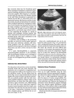
Fig. 7.2 Image of implantable central venous port [44]. Vanek VW, Nutrition in Clinical Practice, 17(3), pp. 142-155,
copyright © 2002 by SAGE Publications. Reprinted by permission of SAGE Publications
As with other long-term access, the most common complication of implantable central venous
ports is infection. Infectious complications in ports are of similar types to other tunneled central
venous catheters, including catheter colonization, tunnel infections, and CLABSIs. In addition, ports
have a subcutaneous pocket that houses the port. This potential space is vulnerable to so-called
“pocket infections” [54]. Repeated daily puncture for access to ports increases the chance that any
particular access will introduce infection. An indwelling needle for access to a port provides a ready
entrance for bacteria through the relatively short needle tract. For these reasons, most clinicians avoid
using ports for home PN [44, 54].
Treatment of infected ports is similar to tunneled central venous catheters with the mainstay being
removal and replacement. However, a trial of intravenous antibiotics may be reasonable should the
patient have poor or difficult vascular access and the patient is not hemodynamically compromised or
have other signs of septic shock. Compared to tunneled central catheters, implantable central venous
ports have a significantly lower rate of CLABSIs overall and a trend towards lower site infections.
However, it is important to remember that implantable central venous ports are used primarily for
intermittent therapy such as chemotherapy, blood draws or infusion, while tunneled central catheters
are used primarily for daily therapies including intravenous antibiotics and PN [2, 44].
Ports are advantageous, as they are entirely beneath the skin when not accessed and no external
tubing is visible to interfere with daily activities. Ports also come in single and double lumens and
require less maintenance; a monthly flush when not in use. However, ports do require repeated skin
puncture for access to the port, and require a physician and an operating room for insertion and removal
[19]. The monthly flush must be done with full sterile precautions and may not be easily done at
home. Ports may also interfere with MRI and CT scans due to scattering of radiation beams [44].
Each vascular access device type has a different useful life-expectancy, and the average duration
of insertion is 23 days for PICC, 125 days for tunneled central venous catheter, and 221 days for
implanted ports [24, 44, 46].
Other Vascular Access
There are other, less common vascular access options for those patients requiring PN whom have
exhausted the traditional access locations. The use of arteriovenous fistulaehas been used as dual
7 Access and Complications of Parenteral Nutrition
113
access for hemodialysis and PN in patients with end-stage renal disease, a lack of alternative venous
access, and intestinal failure with success in three patients [24, 57]. The use of AV fistulas was less
successful, however, in those without chronic renal disease [24]. There is even a case report of accessing the intercostal vein for patients who have exhausted normal vascular access sites [61]. In the setting of SVC occlusion, the azygous vein can be used [19]. Finally, direct placement of a catheter into
the IVC via trans-lumbar approach, trans-hepatic approach or directly into the right atrial appendage
via thoracotomy, an extremely invasive and “last-ditch” maneuver, have been reported [8, 15, 19, 61].
Recommendations
Ideal vascular accessis specific to the patient, the disease-state, the use, and the projected duration.
Should the patient have an adequate basilic vein between the biceps and triceps or in the antecubital
fossa, and the need for access estimated to be weeks to months, a PICC may be a favorable selection.
However, should the patient need a longer duration of PN, months to years, or the patient does not
have adequate superficial veins, a tunneled central catheter should be used. Implantable ports may be
used, but are less favored for PN. If the therapy is intermittent, such as chemotherapy, an implantable
port is likely to be favored because of improved cosmetic appearance and decreased maintenance. If
the therapy is daily and long term, as with PN, a tunneled catheter should be used [2].
Complications of Parenteral Nutrition
Parenteral nutrition is an extremely complex mixture of often more than 70 distinct components,
including dextrose, fat emulsions, water, electrolytes, amino acids, trace elements, and vitamins [2].
Serious harm can occur with an inappropriate mixture [6]. Mirtallo et al. noted the deaths of two individuals from microvascular pulmonary emboli as a result of calcium phosphate precipitation [2]. Care
must be exquisite for the creation of a safe product, as further discussed in Chapter 13. The major
complications can be divided into catheter complications and metabolic complications.
Catheter Complications
Specific catheter complications and infections have been discussed in detail above. Complications
associated with central lines occur at a rate of 1–4% [33, 35, 55]. Generally, complications associated
with line placement are easily treated, but surgical intervention may be required if serious sequelae
develop. It is important to remember that the patient’s disease state, the experience of the physician
placing the catheter, and the specific type of line itself all impact complication rate in catheters [35].
Catheter Occlusion
Patients on PN are specifically vulnerable to catheter occlusion resulting from precipitation of medications or solutions [4, 8, 35]. Mineral solutions, intravenous lipids and medications can precipitate
and lead to catheter occlusion. Complete occlusion from precipitated medications, lipids, or calcium
phosphate can be treated by the instillation of bicarbonate, ethanol, or 0.1 M hydrochloric acid
114
D.R. Neel
solutions, respectively [3, 4, 8, 33, 35, 36, 51]. Ethanol (70%) solution can help dissolve triglyceride
deposits [4, 8]. Line occlusion requires removal and replacement if the precipitate does not dissolve with treatment [4, 35].
Metabolic Complications of Parenteral Nutrition
Glycemic Control
Hyperglycemia is common with patients using PN due to the glucose loads, and the increased blood
sugar levels associated with PN calories relative to enteral nutrition, likely due to the loss of the firstpass effect of the liver [6]. Patient factors such as pre-existing diabetes mellitus, systemic inflammation, postoperative changes, and disease-induced insulin resistance can make glucose control
challenging [35]. Maintaining appropriate glucose control can reduce morbidity and mortality in critically ill patients. Along the same line, providing the appropriate amount of glucose is necessary to
prevent both overfeeding and underfeeding. Overfeeding results in excess carbon dioxide production
and may even lead to respiratory compromise. Underfeeding results in starvation [9, 35]. Hyperglycemia
may lead to increased glycation of certain proteins resulting in their dysfunction. It is also associated
with increased infection rates and decreased wound healing [9]. Hyperglycemia associated with
excessive dextrose administration may also predispose to PN associated hepatic steatosis [8, 9].
Hypoglycemia is less common but can be more devastating. Certain patient populations, including
infants, patients in renal and liver failure, patients with adrenal insufficiency, patients with diabetes at
baseline, septic and severely malnourished patients, and any patient with impaired insulin clearance are more prone to hypoglycemia due to imparied gluconeogenesis. Stopping the infusion of PN
abruptly has historically been reported to result in occasional precipitous hypoglycemia. This is
thought to be due to the continued circulation of insulin due to more rapid clearance of glucose than
insulin, and lack of substrate [6, 35]. Fear of post-cessation hypoglycemia still drives protocols replacing suddenly halted PN with 10 or 20% dextrose solution infusions. However, in the current era, in
which calorie prescriptions are far more conservative than in the earlier days of PN, hypoglycemia
associated with PN cessation is an unusual occurrence, and with frequent point-of-care glucose determinations this practice is unnecessary and may lead to complications such as hypokalemia and hypophosphatemia. Tapered cessation of PN is often practiced, and should help prevent hypoglycemia in
this setting, but is not always feasible in the ICU. For example, in patients with septic shock due to
presumed line-related sepsis, immediate removal of the offending foreign body, the central line, may
be lifesaving. Comparative trials of tapered versus abrupt cessation have indeed shown no difference
in hypoglycemia incidence [2, 35, 62]. Tapered stopping of PN may no longer be necessary, and automatic replacement with dextrose infusions is certainly made obsolete, in the era of conservative calories and frequent point-of-care glucose determinations in the ICU. Close monitoring of glucose levels
during PN administration and cessation remains an important component of PN management.
Lipid Metabolism
Hyperlipidemia can be induced by the lipid and calorie content of the PN. Disease states such as critical illness, diabetes, sepsis, renal and liver failure, and familial hyperlipidemia can lead to decreased
lipid clearance and increased hyperlipidemia. Interestingly, underfeeding leads to ketogenesis, and
may also ultimately result in hypertriglyceridemia [35, 63]. PN-related hyperlipidemia is generally
benign and self-limited when lipid infusion is stopped. However, severe elevations, in the range of
7 Access and Complications of Parenteral Nutrition
115
1000 mg/dl, may be associated with pancreatitis [35]. Elevated lipid infusion rates, greater than 1 g/
kg/day, may lead to cholestasis, resulting in hepatic dysfunction [8].
Essential fatty acid deficiency develops if an insufficient amount of linoleic acid and/or linolenic
acid is provided in the PN [11, 63]. Fatty acid deficiencies can lead to impaired lipoprotein synthesis
resulting in triglyceride accumulation in the liver and causing hepatic steatosis [4]. Clinical signs of
essential fatty acid deficiency include neuropathy, hepatosplenomegaly, dry skin with a flaky rash,
poor wound healing and thrombocytopenia [2, 4, 63]. A minimum of 4–8% of calories should be
provided from lipid emulsion, 50% of which should be linoleic acid, to prevent essential fatty acid
deficiency in patients completely dependent on PN [35]. Essential fatty acid deficiency is rare today
as long as fat supplementation is not withheld for more than 2 weeks [4]. Diagnosis is made by fatty
acid level analysis and specifically the triene–tetraene ratio.
Hepatobiliary Complications
The first description of TPN-associated liver disease was in 1971 [4, 64]. Hepatic dysfunction is quite
common, seen in approximately 47% of home PN patients, and has a broad spectrum of presentation
and severity [35, 65]. In children and neonates, hepatic complications occur in 50% of those on
chronic PN, while 15–30% of adults have hepatic complications [4, 8]. Shortened bowel length, specifically less than 100 cm, is associated with increased liver dysfunction.
Elevations of bilirubin and liver function tests (LFTs) greater than 1.5 times the upper limit of
normal are the mildest form of hepatic dysfunction, and usually develop 1–2 weeks after PN initiation
[4, 8, 65]. A hepatocellular pattern is commonly seen in adult patients demonstrating steatosis, while
a cholestatic pattern is often seen in children [4, 8, 35]. These abnormalities are consistent with periportal steatosis. A prolonged elevation of LFTs for over 6 months is associated with patients on prolonged PN, generally from prolonged intestinal failure [35]. The prevalence in an earlier study was
26% at 2 years and 50% at 6 years. There was a 22% mortality associated with liver disease as a cause
of death of those patients on home PN [66]. Again, in this study, shorter bowel length, less than 50 cm,
played a significant role in the formation of liver disease in home PN. Other factors for hepatobiliary
complications included chronic cholestasis, excess protein administration and elevated lipid intake of
1 g/kg/d or more [4, 8, 35, 65, 66]. Lecithin and choline administered parenterally may help decrease
hepatic steatosis in patients on PN. In previous generations of additives, aluminum was also known to
increase hepatic cholestasis. As mentioned above, hyperglycemia and hyperlipidemia can also lead to
hepatic dysfunction and steatosis [8, 35, 65].
In addition, cholestasis is thought to develop from lack enteral stimulation and cholecystokinin stimulation, resulting in biliary stasis and sludge formation. Data on the incidence of this complication
requires updating, as a large part of the incidence seen in the early days of PN therapy was due to
overfeeding resulting in steatohepatitis. Historically, elevation of bilirubin and alkaline phosphatase,
suggestive of stasis, have been found to occur in as little as 4 weeks for 50% of patients, and in 100%
by 6 weeks [4, 8, 66]. Gallstones or acalculous cholecystitis from biliary stasis and sludge [3, 4, 8, 35]
led to recommendations that considered prophylactic cholecystectomy reasonable in the early era of
PN [3], but this is no longer appropriate. Lack of enteral stimulation can also allow for bacterial overgrowth and the production of lithocholate, which is a hepatotoxic bile acid [35, 65, 66]. Daily oral
intake, even if the patient requires PN to meet caloric needs, may help decrease the risk of biliary
stasis and cholecystitis [4, 65, 66]. Liver injury associated with PN varies from reversible injury,
including cholestasis and steatosis, to more permanent steatohepatitis and cirrhosis [35, 65]. Early
cycling was proposed to help limit or prevent the progression of liver disease and complications, but
this is unproven. PN-dependent patients with intestinal failure and permanent hepatobiliary
116
D.R. Neel
complications should be listed early for combination liver-small bowel transplants. Historically, the
death rate is higher for liver failure associated with PN than for other liver diseases, with essentially
no survival at 5 years [4, 35, 67].
Gastrointestinal Complications
Obviously, when the patient is fully dependent on PN, the “gut” is not used. The lack of intestinal
stimulation has consequences. Mucosal atrophy has been demonstrated in patients that do not receive
enteral feeding, although the significance is not quite understood. The mucosal atrophy of jejunal villi
is quite profound in animal models, but is less pronounced in humans. Cellular permeability is also
altered in profound intestinal isolation. Cellular edema and decreased intraluminal mucosal lining
contribute to increased permeability, but is not associated with bacterial translocation. Marked pancreas atrophy due to lack of trophic substances also develops in patients without enteral stimulation.
Exocrine function decreases in those dependent on PN long-term. The incidence of delayed gastric
emptying also increases over time with chronic PN [4, 8].
Bone Disease
Shike et al. and Klein et al. first described PN-associated bone disease in 1980 [4, 68, 69]. This syndrome was originally characterized by transient hypercalcemia, normal or low serum parathyroid
hormone, high normal plasma 25-OH vitamin D3, hypercalciuria, and a negative calcium balance
with normal phosphorus levels and decreased mineralization and increased osteoid on bone biopsy [4,
69, 70]. Patients on home PN have an increased risk of bone disease manifesting as osteoporosis
(41%), osteopenia (81%), bone pain (35%), and fractures (10%) [3, 35, 68, 71]. Most will be asymptomatic [70]. The cause of bone disease in PN patients is not completely understood, but preexisting
disease including intestinal failure contributes. Obesity, inactivity, hypogonadism, timing of intestinal
failure, smoking, alcohol abuse, and prolonged steroid therapy are all pre-existing disease states that
contribute to bone disease in long-term home PN patients [35, 70, 71]. PN-specific factors predisposing to bone disease include deficiency of phosphorus, calcium, or magnesium, vitamin D excess or
deficiency, and aluminum toxicity. Hypercalciuria in PN denotes an increase in bone loss [8, 68].
Aluminum toxicity can lead to decreased parathyroid hormone secretion due to inhibition [4, 8, 35].
Despite the efforts to remove aluminum from solution additives, patients on PN still receive a significant amount [8, 70, 71]. Vitamin D excess can decrease PTH secretion and stimulate bone resorption.
Bone mineral density loss and PN-associated bone diseases are treated with bisphosphonates, calcium
supplementation, and calcitonin [35, 70].
Kidney Injury
Nephromegaly develops in chronic PN, perhaps due to glomerular hyperfiltration associated with an
elevated creatinine clearance, although the exact mechanism is unknown and may be due to repeated
metabolic insults [4, 8]. It is not associated with amino acid content, but creatinine clearance
decreases by an average of 3.5% per year while on PN [72]. Glomerular necrosis can develop with
long-term PN, resulting in decreased renal function [8, 72]. Increasing age, use of nephrotoxic
7 Access and Complications of Parenteral Nutrition
117
drugs, and episodes of bacteremia/fungemia all contribute to the development of renal dysfunction
and nephromegaly. However, it is unclear to what degree each participates.
Hyperoxaluria results from abnormalities in bile absorption. Oxalate is normally absorbed in the
colon after binding to bile salts and fatty acids. However, in PN patients in whom bacterial overgrowth occurs, increased glycolate formation creates increased oxalate formation and absorption [35].
Hyperoxaluria is especially common in those patients with ileal resection and can result in a nonreversible oxalate nephropathy.
Refeeding Syndrome
Patients who are extremely malnourished, particularly if they have electrolyte losses due to high output enterocutaneous fistulae, recurrent vomiting, etc., are at increased risk for refeeding syndrome if
they are initially fed too aggressively. The syndrome results in severe electrolyte abnormalities and
Wernicke syndrome [1, 73]. Refeeding is not isolated to PN alone but can also occur in those patients
who are malnourished receiving oral or enteral nutrition, or even intravenous hydration containing 5%
dextrose. Refeeding syndrome is characterized by hypokalemia, hypophosphatemia, and hypomagnesemia, and is likely mediated by a sudden rise in insulin as the patient shifts from starvation to a
postprandial state. Early symptoms may be vague and include weakness, myalgia, and shortness of
breath. Patients that experience refeeding syndrome have an increased morbidity and mortality from
cardiac arrhythmias and respiratory failure [35, 73]. Being astute to the correction of electrolyte
abnormalities and supplementation of thiamine before and during nutritional support, including measuring and supplementing electrolytes repeatedly during a single day in high-risk patients, as well as
starting PN with a reduction in dextrose, or all components, to approximately 50% of goal, are of the
utmost importance in preventing complications from this syndrome [73].
Conclusion
PN has come a long way since Dudrick et al first showed positive nitrogen balance and growth in
beagle puppies using solely intravenous alimentation (5,12). However, with great advances come
unintended complications. Fortunately most of these complications are treatable (4). Mean expected
survival rate is 90% at one year and 60% at five years on chronic PN (71). In fact, PN is life-saving
in many instances! Patients with intestinal failure can survive on PN and live a relatively normal life
(5,7,65). In the correct patient population, the benefits of patient survival outweighs the significant
risks of complications. Careful choice of catheter placement, proper monitoring of patients and the
prevention and treatment of complications will result in better outcomes.
References
1. Spencer CT, Compher CW. Total parenteral nutrition, an ally in the management of patients with intestinal failure
and malnutrition: a long-term view. JPEN J Parenter Enteral Nutr. 2003;27(5):374–81. PubMed.
2. Mirtallo J, Canada T, Johnson D, Kumpf V, Petersen C, Sacks G, et al. Safe practices for parenteral nutrition. JPEN
J Parenter Enteral Nutr. 2004;28(6):S39–70. Erratum in: JPEN J Parenter Enteral Nutr. 2006;30(2):177. PubMed.
3. Steiger E. Home parenteral nutrition. Components, application, and complications. Postgrad Med.
1984;75(6):95–102.
118
D.R. Neel
4. Buchman AL. Complications of long-term home total parenteral nutrition their identification, prevention and treatment. Dig Dis Sci. 2001;46:1–18.
5. Dudrick SJ, Palesty JA. Historical highlights of the development of total parenteral nutrition. Surg Clin North Am.
2011;91(3):693–717.
6. Kumpf VJ, Tillman EM. Home parenteral nutrition: safe transition from hospital to home. Nutr Clin Pract.
2012;27(6):749–57. doi:10.1177/0884533612464888. Epub 2012 Oct 22. Review. PubMed.
7. Scolapio JS, Fleming CR, Kelly DG, Wick DM, Zinsmeister AR. Survival of home parenteral nutrition-treated
patients: 20 years of experience at the Mayo Clinic. Mayo Clin Proc. 1999;74:217–22.
8. Montalvo-Jave EE, Zarraga JL, Sarr MG. Specific topics and complications of parenteral nutrition. Arch Surg.
2007;392:119–26.
9. Schloerb PR. TPN or intravenous food poisoning? Nutrition. 2001;17(7–8):680–1.
10. Dudrick SJ, Wilmore DW, Vars HM, Rhoads JE. Long-term total parenteral nutrition with growth, development,
and positive nitrogen balance. Surgery. 1968;64(1):134–42. PubMed.
11. Macht SD. Three hundred years of parenteral nutrition: the history of intravenous nutritional therapy. Conn Med.
1980;44(1):27–30. PubMed.
12. Dudrick SJ, Pimiento JM. Parenteral nutrition and nutritional support of surgical patients: reflections, controversies
and challenges. Surg Clin North Am. 2011;91(3):675–92.
13. Dudrick SJ. A three and one-half decade nutritional and metabolic iliad. J Am Coll Surg. 2007;205:S59–64.
14. Cuthbertson D. Historical background to parenteral nutrition. Acta Chir Scand. 1980;498(Suppl):20–5.
15. Asch MR. Venous access: options, approaches, and issues. Can Assoc Radiol J. 2001;52:153–64.
16. Segarra-Newnham M, Martin-Cooper EM. Antibiotic lock technique: a review of the literature. Ann Pharmacother.
2005;39(2):311–8. Epub 2004 Dec 28. Review. PubMed.
17. Bestul MB, Vandenbussche HL. Antibiotic lock technique: review of the literature. Pharmacotherapy.
2005;25(2):211–27. Review. PubMed.
18. Sansivero GE. Venous anatomy and physiology. Considerations for vascular access device placement and function.
J Intraven Nurs. 1998;21(5 Suppl):S107–14. Review. PubMed.
19. Sansivero GE. Features and selection of vascular access devices. Semin Oncol Nurs. 2010;26(2):88–101.
doi:10.1016/j.soncn.2010.02.006. Review. PubMed.
20. Pettit J. Assessment of infants with peripherally inserted central catheters: part 1. Detecting the most frequently
occurring complications. Adv Neonatal Care. 2002;2(6):304–15. PubMed PMID: 12881943 Review.
21. Vanek VW. The ins and outs of venous access: part I. Nutr Clin Pract. 2002;17(2):85–98. PubMed.
22. Lum PS, Soski M. Management of malpositioned central venous catheters. J Intraven Nurs. 1989;12(6):356–65.
PubMed.
23. Moran BJ. Access methods in nutritional support. Proc Nutr Soc. 1994;53(3):465–71.
24. Meguid MM, Eldar S, Wahba A. The delivery of nutritional support. A potpourri of new devices and methods.
Cancer. 1985;55(1 Suppl):279–89. Review. PubMed.
25. Gil ME, Mateu J. Treatment of extravasation from parenteral nutrition solution. Ann Pharmacother. 1998;32(1):51–5.
PubMed.
26. Maki DG, Ringer M. Risk factors for infusion-related phlebitis with small peripheral venous catheters. A randomized
controlled trial. Ann Intern Med. 1991;114(10):845–54. PubMed.
27. Rosenthal K. Are you up-to-date with the infusion nursing standards? Nursing. 2007;37(7):15. PubMed.
28. Boyd S, Aggarwal I, Davey P, Logan M, Nathwani D. Peripheral intravenous catheters: the road to quality improvement and safer patient care. J Hosp Infect. 2011;77(1):37–41. doi:10.1016/j.jhin.2010.09.011. Epub 2010 Dec 3.
PubMed.
29. Harden JL, Kemp L, Mirtallo J. Femoral catheters increase risk of infection in total parenteral nutrition patients. Nutr
Clin Pract. 1995;10(2):60–6. Review. PubMed.
30. O’Grady NP, Alexander M, Burns LA, Dellinger EP, Garland J, Heard SO, et al. Guidelines for the prevention of
intravascular catheter-related infections. Am J Infect Control. 2011;39(4 Suppl 1):S1–34. doi:10.1016/j.
ajic.2011.01.003. PubMed.
31. Timsit JF. What is the best site for central venous catheter insertion in critically ill patients? Crit Care. 2003;7(6):397–
9. Epub 2003 Mar 28.
32. Ingle RJ. Rare complications of vascular access devices. Semin Oncol Nurs. 1995;11:184–93.
33. Grant J. Recognition, prevention, and treatment of home total parenteral nutrition central venous access complications. JPEN. 2002;26(Suppl):21–8.
34. Timsit JF, L’Hériteau F, Lepape A, Francais A, Ruckly S, Venier AG, et al. A multicentre analysis of catheter-related
infection based on a hierarchical model. Intensive Care Med. 2012;38(10):1662–72. Epub 2012 Jul 14. PubMed.
35. Ghabril MS, Aranda-Michel J, Scolapio JS. Metabolic and catheter complications of parenteral nutrition. Curr
Gastroenterol Rep. 2004;6:327–34.
7 Access and Complications of Parenteral Nutrition
119
36. Nakazawa N. Infectious and thrombotic complications of central venous catheters. Semin Oncol Nurs.
2010;26(2):121–31. doi:10.1016/j.soncn.2010.02.007. Review. PubMed.
37. Hemphill DJ, Sniderman KW, Allard JP. Management of total parenteral nutrition-related superior vena cava obstruction with expandable metal stents. JPEN J Parenter Enteral Nutr. 1996;20(3):222–7. PubMed.
38. Raad II, Luna M, Khalil SA, Costerton JW, Lam C, Bodey GP. The relationship between the thrombotic and infectious complications of central venous catheters. JAMA. 1994;271(13):1014–6. PubMed.
39. Sandstedt S, Heselvik F, Marklund T, Stenport G. Percutaneous, tunneled silicone elastomer central venous catheters
for TPN: low sepsis and thrombosis rate. A prospective study of 325 catheters. Nutrition. 1989;5:23–6.
40. Timsit JF, Farkas JC, Boyer JM, Martin JB, Misset B, Renaud B, et al. Central vein catheter-related thrombosis in
intensive care patients: incidence, risks factors, and relationship with catheter-related sepsis. Chest. 1998;114(1):207–
13. PubMed.
41. Eastridge BJ, Lefor AT. Complications of indwelling venous access devices in cancer patients. J Clin Oncol.
1995;13:233–8.
42. Buchman AL, Misra S, Moukarzel A, Ament ME. Catheter thrombosis and superior/inferior vena cava syndrome are
rare complications of long term parenteral nutrition. Clin Nutr. 1994;13:356–60.
43. Mustafa S, Stein PD, Patel KC, Otten TR, Holmes R, Silbergleit A. Upper extremity deep venous thrombosis. Chest.
2003;123:1953–6.
44. Vanek VW. The ins and outs of venous access: part II. Nutr Clin Pract. 2002;17(3):142–55. PubMed.
45. Hoshal Jr VL. Total intravenous nutrition with peripherally inserted silicone elastomer central venous catheters. Arch
Surg. 1975;110(5):644–6. PubMed.
46. Smith JR, Friedell ML, Cheatham ML, Martin SP, Cohen MJ, Horowitz JD. Peripherally inserted central catheters
revisited. Am J Surg. 1998;176:208–11.
47. Ng PK, Ault MJ, Maldonado LS. Peripherally inserted central catheters in general medicine. Mayo Clin Proc.
1997;72:225–33.
48. Duerksen DR, Papineau N, Siemens J, Yaffe C. Peripherally inserted central catheters for parenteral nutrition: a
comparison with centrally inserted catheters. JPEN. 1999;23:85–9.
49. Broviac JW, Cole JJ, Scribner BH. A silicone rubber atrial catheter for prolonged parenteral alimentation. Surg
Gynecol Obstet. 1973;136:602–6.
50. Hickmann RO, Buckner CD, Clift RA, Sanders JE, Stewart P, Thomas ED. A modified right atrial catheter for access
to the venous system in marrow transplant recipients. Surg Gynecol Obstet. 1979;148:871–5.
51. Rumsey KA, Richardson DK. Management of infection and occlusion associated with vascular access devices.
Semin Oncol Nurs. 1995;11:17483.
52. Prandoni P, Polistena P, Bernandi E, Cogo A, Casara D, Verlato F, et al. Upper-extremity deep vein thrombosis. Risk
factors, diagnosis, and complications. Arch Intern Med. 1997;157:57–62.
53. Atiken DR, Minton JP. The “pinch-off sign”: a warning of impending problems with permanent subclavian catheters.
Am Surg. 1984;148:633–6.
54. Krzywda EA, Andris DA, Edmiston CE. Catheter infections: diagnosis, etiology, treatment and prevention. NCP.
1999;14(4):178–90.
55. Cowl CT, Weinstock JV, Al-Jurf A, Ephgrave K, Murray JA, Dillon K. Complications and cost associated with parenteral nutrition delivered to hospitalized patients through either subclavian or peripherally-inserted central catheters. Clin Nutr. 2000;19:237–43.
56. Pronovost PJ, Goeschel CA, Colantuoni E, Watson S, Lubomski LH, Berenholtz SM, et al. Sustaining reductions in
catheter related bloodstream infections in Michigan intensive care units: observational study. BMJ. 2010;340:c309.
doi:10.1136/bmj.c309. PubMed PMID: 20133365; PubMed Central PMCID: PMC2816728.
57. Ytang VC, Morsey MA, Chemla ES. Using arteriovascular fistulae as a dual access for hemodialysis and total parenteral nutrition administration is feasible with a good outcome: a case series. J Vasc Access. 2007;8:305–8.
58. Madeo M, Lowry L. Infection rates associated with total parenteral nutrition. J Hosp Infect. 2011;79(4):373–4.
doi:10.1016/j.jhin.2011.08.012. Epub 2011 Sep 29. PubMed.
59. Stratov I, Gottlieb T, Bradbury R, O’Kane GM. Candidaemia in an Australian teaching hospital: relationship to central line and TPN use. J Infect. 1998;36(2):203–7. PubMed.
60. Niederhuber JE, Ensminger W, Gyves JW, Liepman M, Doan K, Cozzi E. Totally implanted venous and arterial
access system to replace external catheters in cancer treatment. Surgery. 1982;92:706–12.
61. Lammermeier D, Steiger E, Cosgrove D, Zelch M. Use of an intercostal vein for central venous access in home
parenteral nutrition: a case report. JPEN. 1986;10:659–61.
62. Nirula R, Yamada K, Waxman K. The effect of abrupt cessation of total parenteral nutrition on serum glucose: a
randomized trial. Am Surg. 2000;66:866–9.
63. Crook MA. Lipid clearance and total parenteral nutrition: the importance of monitoring plasma lipids. Nutrition.
2000;16:774–5.
120
D.R. Neel
64. Peden VH, Witzleben CL, Skelton MA. Total parenteral nutrition. J Pediatr. 1971;78(1):180–1.
65. Luman W, Shaffer H. Prevalence, outcome, and associated factors of deranged liver function tests in patients on
home parenteral nutrition. Clin Nutr. 2002;21:337–43.
66. Cavichi M, Beau P, Crenn P, Degott C, Messing B. Prevalence of liver disease and contributing factors in patients
receiving home parenteral nutrition for permanent intestinal failure. Ann Intern Med. 2000;132:525–32.
67. Fryer J, Pellar S, Ormond D, et al. Mortality in candidates waiting for combined liver-intestine transplants exceeds
that for other candidates waiting for liver transplants. Liver Transpl. 2003;9:748–53.
68. Klein GL, Targoff CM, Ament ME, Sherrard DJ, Bluestone R, Young JH, et al. Bone disease associated with total
parenteral nutrition. Lancet. 1980;2:1041–4.
69. Shike M, Harrison JE, Sturtridge WC, Tam CS, Bobechko PE, Jones G, et al. Metabolic bone disease in patients
receiving long-term total parenteral nutrition. Ann Intern Med. 1980;92(3):343–50.
70. Seidner DL. Parenteral nutrition-associated metabolic bone disease. JPEN J Parenter Enteral Nutr. 2002;26(5
Suppl):S37–42. Review. PubMed.
71. Pironi I, Labate AM, Pertkiewicz M, Przedlacki J, Tjellesen L, Staun M, et al. Prevalence of bone disease in patients
on home parenteral nutrition. Clin Nutr. 2002;49:497–500.
72. Buchman AL, Moukarzel A, Ament ME, Gornbein J, Goodson B, Carlson C, et al. Serious renal impairment is associated with long-term parenteral nutrition. JPEN. 1993;17:438–44.
73. Crook MA, Hally V, Panteli JV. The importance of the refeeding syndrome. Nutrition. 2001;17:632–7.
Chapter 8
Surgical Intensive Care Considerations
Charles W. Van Way III
Keywords Stress response • Hypermetabolism • Trauma • Operation • Head injury • Gastrointestinal
tract injury • Liver injury • Burn injury • Intestinal obstruction • Pancreatitis • Short bowel syndrome
• Enterocutaneous fistula
Key Points
• Surgical patients are characterized by response to an acute event, either surgery or injury.
• The stress response is a coordinated neuroendocrine, circulatory, inflammatory, and metabolic response.
• Injured patients should receive enteral or parenteral nutrition support relatively early, usually within
1–2 days post-injury.
• Enteral nutrition is best, if the gastrointestinal tract can be used.
• Patients with gastrointestinal injuries may require parenteral nutrition, but most centers start it after
2–4 days, because of the added risk involved.
• Head injured patients should be fed early, using enteral nutrition.
• Burn injured patients are often dependent on enteral nutrition for up to several weeks.
• Postoperative patients may tolerate several days without adequate nutrition, but should be supported
with enteral nutrition by 5–7 days.
• Use of parenteral nutrition in the postoperative patient should be reserved for patients who cannot
tolerate enteral nutrition.
• Prolonged ileus following gastrointestinal surgery often requires the use of parenteral nutrition
after 5–7 days.
• Glutamine, arginine, and other “immunotherapy” nutrients are often used in surgical patients, but
the evidence in favor of their use is equivocal at best.
• Management of intestinal obstruction may require parenteral nutrition if the obstruction does not
resolve.
• Pancreatitis is best managed using either oral intake or enteral nutrition, contrary to earlier practice
using parenteral nutrition and “bowel rest.”
• Patients with short bowel syndrome, including patients with enterocutaneous fistula, often require
parenteral nutrition, and frequently must be treated with home parenteral nutrition.
C.W. Van Way III, MD, FACS, FCCP, FCCM, FASPEN (*)
Department of Surgery, Truman Medical Center, School of Medicine,
University of Missouri–Kansas City, 2301 Holmes Street, Kansas City, MO 64108, USA
e-mail:
D.S. Seres, C.W. Van Way, III (eds.), Nutrition Support for the Critically Ill, Nutrition and Health,
DOI 10.1007/978-3-319-21831-1_8, © Springer International Publishing Switzerland 2016
121
122
C.W. Van Way III
Introduction
What is surgical intensive care, and why are we giving it special consideration? Is not one critically ill
patient much like another? In fact, no. Some patients are commonly regarded as “surgical,” and for
good reasons. They have a number of characteristics in common, and are different from the so-called
“medical” patients. To generalize, “medical” patients are in the ICU because of exacerbations of
chronic diseases—most commonly respiratory, cardiac, or renal, often in combination, and frequently
with diabetes as well. “Surgical” patients need intensive care because they have had a major acute
event, usually either an injury or an operation. To be sure, there is much overlap. Surgical patients may
also have major chronic diseases. They may develop organ failure syndromes. But that being said,
“surgical” patients have a set of problems that are different from those seen in “medical” patients.
Caring for them requires a somewhat different mindset than dealing with chronic disease.
The prototype of the surgical intensive care patient is the injured patient. A patient is suddenly
injured, perhaps massively, and often requires one or more operations. Oral nutrition is cut off abruptly.
Now, healthy people can tolerate a day or two without food easily, and several days without apparent
ill effects. At some point, starvation begins to interfere with healing, and to impair the patient’s
recovery. There may be delayed wound healing, wound infection, or dehiscence. Prolonged lack of
nutrition may also predispose to other infections such as urinary tract infections and pneumonia, and
to decubitus ulcers.
Postoperative patients are not unlike injured patients. The metabolic response to operation is
similar to that of injury. Here again, healthy people can go without food for several days. Most surgical procedures interrupt eating for only a day or two. Even in gastrointestinal surgery, keeping patients
“nil per os” for as long as 5–7 days while the GI tract recovers has long been surgical practice. But in
the postoperative patient, like the injured patient, there comes a time after which the patient simply
must be fed.
In most hospital settings, care of the “surgical” critical care patient is carried out by surgeons.
Traditionally, attending surgeons care for their own patients in the ICU. More recently, this is commonly
done by Surgical Critical Care (SCC) specialists. SCC requires an extra year or two of training. SCC
specialists usually practice surgery as well, often trauma or acute care surgery. While any surgical specialist can become trained and qualified in SCC, most who do so are general or cardiothoracic surgeons.
Anesthesiologists and emergency medicine specialists can also be trained in and practice SCC.
It is the purpose of this chapter to outline the particular requirements of nutrition support in the
surgical patient, emphasizing largely the injured and/or postoperative patient. To begin this discussion, it is best to start with the metabolic characteristics that are seen in the surgical patient, and can
be said to define this group of patients.
Metabolic Characteristics of the Surgical Patient
The acute response to any type of stress is characterized by endocrine events, inflammatory response,
and metabolic response. In brief, the body undergoes a number of changes that collectively prepares it
for the challenge of surviving injury. The basics of this have been known since the pioneering work of
Cuthbertson during the 1930s [1]. Many others have elaborated on it since [2–4]. Indeed, the response
to injury and to operation are similar, as both are modifications of the stress response. This response
may be somewhat arbitrarily divided into neuroendocrine, inflammatory, and metabolic.
Neuroendocrine response begins with activation of the hypothalamus. This stimulates the pituitary to
release ACTH, which in turn stimulates the adrenal cortex to produce cortisol. Cortisol in turn raises the
blood sugar and mobilizes fatty acids by lipolysis of fat stores. It also depresses the immune system.
8
Surgical Intensive Care Considerations
123
At the same time, the sympathetic nervous system is activated. This stimulates the adrenal medulla to
produce catecholamines, largely epinephrine, whose secretion may increase 20-fold. These changes
produce increases in blood pressure, heart rate, and cardiac output. The metabolic rate increases.
These changes produce increases in blood pressure, heart rate, and cardiac output. Temperature
increases modestly. There is increased blood flow to muscles, skin, heart, and viscera. There is
increased blood flow to muscles, skin, and heart, with constriction elsewhere in the body, such as the
viscera. Gastrointestinal activity is depressed by sympathetic nervous stimulation. All of this—mobilization of energy stores, redistribution of blood flow, tachycardia, and increased cardiac output—
prepares the organism to respond to stress [1–4].
There are increases in secretion of both glucagon and insulin from pancreatic islet cells. At the
same time, there is decreased responsiveness to insulin [5]. This insulin resistance characterizes not
only injured patients, but postoperative patients as well [6]. As further discussed, it is actually deleterious to recovery.
Hemorrhagic shock exaggerates the “normal” stress response. Such patients show all of the above
changes. Resuscitation with crystalloid and blood products may restore the blood pressure and appear
to restore perfusion to normal. It may take up to 3 days to completely resuscitate patients from hemorrhagic shock. Current practice is to limit the time of initial operation to an hour, the so-called “damage
control” procedure. The intent is to control hemorrhage and prevent further blood loss, removing
devitalized tissue (including bowel), but making little effort to reconstruct the injuries. Then the
patient is taken to the ICU for continued resuscitation. Second and third operations follow at intervals
of 12–24 h, until the wounds are completely debrided, reconstruction of bowel and/or vascular continuity carried out, and the patient is completely resuscitated. Current thinking on resuscitation emphasizes the simultaneous administration of packed red cells, platelets, and clotting factors (fresh frozen
plasma) in a 1:1:1 ratio [7, 8].
A number of metabolic changes are produced, many in response to the neuroendocrine changes
already outlined [9]. Hypermetabolism is a part of the stress reaction. Lipolysis releases ketone bodies
and fatty acids into the circulation, to be metabolized for energy by skeletal muscles, heart, and most
viscera. Stored glycogen is broken down for glucose, to provide energy to those systems that require
glucose—the central nervous system, the hematopoietic system, and healing tissues. With many parts
of the body being inadequately perfused, anaerobic glucose metabolism occurs, releasing lactate into
the circulation. Indeed, lactate levels are often used as an index of the degree to which a patient has
been adequately resuscitated.
After the stress response begins, the body runs out of stored glycogen in 4–6 h. Proteolysis begins,
producing amino acids, notably alanine and glutamine. Alanine is the main substrate for gluconeogenesis by the liver, which provides glucose after glycogen runs out in about 24 h. Glutamine can be
used directly for energy by the gut. The protein breakdown of stress can be modulated by providing
at least modest amounts of glucose; in an adult, 400–500 cal, or 100–125 g, is sufficient. For this
reason, 5 % dextrose in water is commonly used, usually with added sodium and potassium to meet
maintenance requirements [4]. Unfortunately, many physicians use normal saline or lactated Ringer’s
solution for routine maintenance. These overload patients with sodium and chloride, and provide
little or no potassium. Worse, these fluids contain no calories, and their use fails to attenuate protein
breakdown.
The systemic inflammatory response is the final element of the response to injury [3, 4, 10]. It is
mediated in part by the hormonal changes discussed above. But the greatest part of systemic inflammation is mediated through a number of mediators, most of which are still being actively investigated.
Pro-inflammatory cytokines are prominent. These include TNF-α, IL-1β, IL-6, and IL-8. Besides
TNF and the interleukins, cytokines include chemokines, lymphokines, and interferons. All of these
are small proteins, 5–10 kDa in size, produced by cells, and generally act on other cells through
cell-surface receptors. Cytokines may be pro- or anti-inflammatory cytokines [10].