Ebook Human anatomy (7th edition): Part 2
Bạn đang xem bản rút gọn của tài liệu. Xem và tải ngay bản đầy đủ của tài liệu tại đây (32.13 MB, 504 trang )
The Nervous System
The Spinal Cord and
Spinal Nerves
Student Learning Outcomes
After completing this chapter, you should
be able to do the following:
1
Discuss the structure and functions of
the spinal cord.
2
Locate the spinal meninges, describe
their structure, and compare and
contrast their functions.
3
Discuss the structure and location of
gray matter and white matter, and
compare and contrast the roles of
both in processing and relaying
sensory and motor information.
4
Identify the regional groups of spinal
nerves.
5
Discuss the connective tissue layers
associated with a spinal nerve.
6
Describe the various branches of a
representative spinal nerve.
7
Define dermatomes and explain their
significance.
8
Define nerve plexus and compare and
contrast the anatomical organization
of the four main spinal nerve plexuses.
9
Identify the spinal nerves originating
at the four major nerve plexuses, list
their major branches, and analyze
their primary functions.
10
Describe the structures and steps
involved in a neural reflex, classify
reflexes, and differentiate among their
structural components.
11
Explain the types of motor responses
produced by spinal reflexes.
368 Introduction
368 Gross Anatomy of the Spinal Cord
368 Spinal Meninges
373 Sectional Anatomy of the Spinal Cord
375 Spinal Nerves
386 Reflexes
368
The Nervous System
THE CENTRAL NERVOUS SYSTEM (CNS) CONSISTS of the spinal cord and
brain. Despite the fact that the two are anatomically connected, the spinal cord and
brain show significant degrees of functional independence. The spinal cord is far
more than just a highway for information traveling to or from the brain. Although
most sensory data is relayed to the brain, the spinal cord also integrates and
processes information on its own. This chapter describes the anatomy of the spinal
cord and examines the integrative activities that occur in this portion of the CNS.
Gross Anatomy of the Spinal
Cord [Figures 14.1 to 14.3]
The adult spinal cord (Figure 14.1a) measures approximately 45 cm (18 in.) in
length and extends from the foramen magnum of the skull to the inferior border
of the first lumbar vertebra (L1). The dorsal surface of the spinal cord bears a
shallow longitudinal groove, the posterior median sulcus. The deep crease
along the ventral surface is the anterior median fissure (Figure 14.1d). Each region of the spinal cord (cervical, thoracic, lumbar, and sacral) contains tracts involved with that particular segment and those associated with it. Figure 14.1d
provides a series of sectional views that demonstrate the variations in the relative
mass of gray matter versus white matter along the length of the spinal cord.
The amount of gray matter is increased substantially in segments of the
spinal cord concerned with the sensory and motor innervation of the limbs.
These areas contain interneurons responsible for relaying arriving sensory information and coordinating the activities of the somatic motor neurons that control the complex muscles of the limbs. These areas of the spinal cord are
expanded to form the enlargements of the spinal cord seen in Figure 14.1a. The
cervical enlargement supplies nerves to the pectoral girdle and upper limbs; the
lumbosacral enlargement provides innervation to structures of the pelvis and
lower limbs. Inferior to the lumbosacral enlargement, the spinal cord tapers to a
conical tip called the conus medullaris, at or inferior to the level of the first lumbar vertebra. A slender strand of fibrous tissue, the filum terminale (“terminal
thread”), extends from the inferior tip of the conus medullaris along the length
of the vertebral canal as far as the dorsum of the coccyx (Figure 14.1a,c). There
it provides longitudinal support to the spinal cord as a component of the
coccygeal ligament.
The entire spinal cord can be divided into 31 segments. Each segment is
identified by a letter and number designation. For example, C3 is the third cervical segment (Figures 14.1a and 14.3).
Every spinal segment is associated with a pair of dorsal root ganglia that
contain the cell bodies of sensory neurons. These sensory ganglia lie between the
pedicles of adjacent vertebrae. ∞ pp. 167–168 On either side of the spinal cord, a
typical dorsal root contains the axons of the sensory neurons in the dorsal root
ganglion (Figure 14.1b,c). Anterior to the dorsal root, a ventral root leaves the
spinal cord. The ventral root contains the axons of somatic motor neurons and,
at some levels, visceral motor neurons that control peripheral effectors. The dorsal and ventral roots of each segment enter and leave the vertebral canal between
adjacent vertebrae at the intervertebral foramina. ∞ p. 168 The dorsal roots are
usually thicker than the ventral roots.
Distal to each dorsal root ganglion, the sensory and motor fibers form a single spinal nerve (Figures 14.1d, 14.2c, and 14.3). Spinal nerves are classified as
mixed nerves because they contain both afferent (sensory) and efferent (motor)
fibers. Figure 14.3 shows the spinal nerves as they emerge from intervertebral
foramina.
The spinal cord continues to enlarge and elongate until an individual is approximately 4 years old. Up to that time, enlargement of the spinal cord keeps
pace with the growth of the vertebral column, and the segments of the spinal
cord are aligned with the corresponding vertebrae. The ventral and dorsal roots
are short, and leave the vertebral canal through the adjacent intervertebral
foramina. After age 4 the vertebral column continues to grow, but the spinal cord
does not. This vertebral growth carries the dorsal root ganglia and spinal nerves
farther and farther away from their original position relative to the spinal cord.
As a result, the dorsal and ventral roots gradually elongate. The adult spinal cord
extends only to the level of the first or second lumbar vertebra; thus spinal cord
segment S2 lies at the level of vertebra L1 (Figure 14.1a).
When seen in gross dissection, the filum terminale and the long ventral and
dorsal roots that extend caudal to the conus medullaris reminded early
anatomists of a horse’s tail. With this in mind the complex was called the cauda
equina (KAW-da ek-WI-na; cauda, tail ϩ equus, horse) (Figure 14.1a,c).
Spinal Meninges [Figures 14.1b,c • 14.2 • 14.3]
The vertebral column and its surrounding ligaments, tendons, and muscles isolate the spinal cord from the external environment. ∞ p. 221 The delicate neural
tissues also must be protected against damaging contacts with the surrounding
bony walls of the vertebral canal. Specialized membranes, collectively known as
the spinal meninges (men-IN-jez), provide protection, physical stability, and
shock absorption (Figure 14.1b,c). The spinal meninges cover the spinal cord
and surround the spinal nerve roots (Figure 14.2). Blood vessels branching
within these layers also deliver oxygen and nutrients to the spinal cord. There are
three meningeal layers: the dura mater, the arachnoid mater, and the pia mater.
At the foramen magnum of the skull, the spinal meninges are continuous with
the cranial meninges that surround the brain. (The cranial meninges, which
have the same three layers, will be described in Chapter 16.)
The Dura Mater [Figures 14.1b,c • 14.2]
The tough, fibrous dura mater (DOO-ra MA-ter; dura, hard ϩ mater, mother)
forms the outermost covering of the spinal cord and brain (Figure 14.1b,c). The
dura mater of the spinal cord consists of a layer of dense irregular connective tissue whose outer and inner surfaces are covered by a simple squamous epithelium. The outer epithelium is not bound to the bony walls of the vertebral canal,
and the intervening epidural space contains areolar tissue, blood vessels, and
adipose tissue (Figure 14.2b,d).
Localized attachments of the dura mater to the edge of the foramen magnum of the skull, the second and third cervical vertebrae, the sacrum, and to the
posterior longitudinal ligament serve to stabilize the spinal cord within the vertebral canal. Caudally, the spinal dura mater tapers from a sheath to a dense cord
of collagen fibers that ultimately blend with components of the filum terminale
to form the coccygeal ligament. The coccygeal ligament extends along the
sacral canal and is interwoven into the periosteum of the sacrum and coccyx.
The cranial and sacral attachments provide longitudinal stability. Lateral support
is provided by the connective tissues within the epidural space and by the extensions of the dura mater that accompany the spinal nerve roots as they pass
through the intervertebral foramina. Distally, the connective tissue of the spinal
dura mater is continuous with the connective tissue sheath that surrounds each
spinal nerve (Figure 14.2a,c,d).
Chapter 14 • The Nervous System: The Spinal Cord and Spinal Nerves
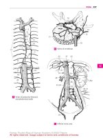
Figure 14.1 Gross Anatomy of the Spinal Cord The spinal cord extends inferiorly from the base of the
brain along the vertebral canal.
Posterior median sulcus
Dorsal root
Dorsal root
ganglion
Cervical
spinal cord
Rootlets
of C8
Cervical spinal
nerves
Dorsal root
ganglion of C8
Dura mater
Dorsal root
ganglia of T4
and T5
C1
C2
C3
C4
C5
C6
C7
C8
T1
T2
T3
T4
T5
T6
White matter
Gray
matter
Central
canal
Cervical
enlargement
Spinal
nerve
Ventral
root
Anterior median fissure
C3
T7
Thoracic
spinal
nerves
T8
T9
Posterior
median sulcus
T10
b Posterior view of a dissection
T11
of the cervical spinal cord
Lumbosacral
enlargement
T3
T12
L1
Conus medullaris
of spinal cord
Cauda equina
Dura mater
Conus
medullaris
L2
Lumbar
spinal
nerves
L3
L4
Inferior
tip of
spinal cord
Cauda equina
L5
Dorsal root
ganglia of L2
and L3
Sacral spinal
nerves
1st sacral
nerve root
Sacrum
(cut)
Filum
terminale
c
Posterior view of a dissection
of the conus medullaris,
cauda equina, filum
terminale, and associated
spinal nerve root
L1
S1
S2
S3
S4
S5
Coccygeal
nerve (Co1)
Filum terminale
(in coccygeal ligament)
S2
d Inferior views of cross sections
a Superficial anatomy and orientation of the adult spinal cord. The
numbers to the left identify the spinal nerves and indicate where
the nerve roots leave the vertebral canal. The spinal cord, however,
extends from the brain only to the level of vertebrae L1–L2.
through representative
segments of the spinal cord
showing the arrangement of
gray and white matter
369
370
The Nervous System
Figure 14.2 The Spinal Cord and Spinal Meninges
Spinal cord
Anterior median
fissure
Gray matter
White matter
Ventral root
Pia mater
Spinal nerve
Dorsal root
Dorsal root
ganglion
Pia mater
Denticulate
ligaments
Arachnoid mater
Dura mater
Arachnoid mater
(reflected)
Dura mater
(reflected)
Spinal blood
vessel
Dorsal root of
sixth cervical
nerve
c
Ventral root of
sixth cervical
nerve
Posterior view of the spinal cord showing the meningeal
layers, superficial landmarks, and distribution of gray and
white matter
a Anterior view of spinal cord showing meninges and spinal nerves. For this
Dura mater
view, the dura and arachnoid membranes have been cut longitudinally
and retracted (pulled aside); notice the blood vessels that run in the
subarachnoid space, bound to the outer surface of the delicate pia mater.
Arachnoid
mater
ANTERIOR
Subarachnoid
space
Vertebral
body
Autonomic
(sympathetic)
ganglion
Spinal cord
Pia mater
Ventral
root of
spinal
nerve
Rami
communicantes
Ventra
ramus
Filum terminale
L5 vertebra
Subarachnoid space
containing cerebrospinal
fluid and spinal nerve roots
Terminal portion
of filum terminale
S2 vertebra
b An MRI scan of the inferior portion of the spinal cord
showing its relationship to the vertebral column
Dorsal
ramus
Spinal cord
Adipose tissue
in epidural
space
Denticulate
ligament
Dorsal root
ganglion
POSTERIOR
d Sectional view through the spinal cord and meninges
showing the peripheral distribution of the spinal nerves
Chapter 14 • The Nervous System: The Spinal Cord and Spinal Nerves
Figure 14.3 Posterior View of Vertebral Column and Spinal Nerves
Occipital bone
Spinal cord
emerging from
foramen magnum
Cervical
plexus
(C1–C5)
Cervical
spinal
nerves
(C1–C8)
Brachial
plexus
(C5–T1)
Sacral
plexus
(L4–S4)
Coccygeal nerves (Co1)
In most anatomical and histological preparations, a narrow subdural space
separates the dura mater from deeper meningeal layers. It is likely, however,
that in life no such space exists, and the inner surface of the dura is in contact with the outer surface of the arachnoid (a-RAK-noyd; arachne, spider)
mater (Figure 14.2a,c,d). The arachnoid mater, the middle meningeal layer,
consists of a simple squamous epithelium. It is separated from the innermost
layer, the pia mater, by the subarachnoid space. This space contains
cerebrospinal fluid (CSF) that acts as a shock absorber as well as a diffusion
medium for dissolved gases, nutrients, chemical messengers, and waste
products. The cerebrospinal fluid flows through a meshwork of collagen and
elastin fibers produced by modified fibroblasts. Bundles of fibers, known as
arachnoid trabeculae, extend from the inner surface of the arachnoid mater
to the outer surface of the pia mater. The subarachnoid space and the role of
cerebrospinal fluid will be discussed in Chapter 16. The subarachnoid space
of the spinal meninges can be accessed easily between L3 and L4 (Figure 14.2
and Clinical Note on p. 372) for the clinical examination of cerebrospinal
fluid or for the administration of anesthetics.
The Pia Mater [Figure 14.2]
Thoracic
spinal
nerves
(T1–T12)
Lumbar
spinal
nerves
(L1–L5)
The Arachnoid Mater [Figures 14.2a,c,d • 14.3]
Lumbar
plexus
(T12–L4)
The subarachnoid space bridges the gap between the arachnoid epithelium
and the innermost meningeal layer, the pia mater (pia, delicate ϩ mater,
mother) as seen in Figure 14.2a,c,d. The elastic and collagen fibers of the pia
mater are interwoven with those of the arachnoid trabeculae. The blood vessels supplying the spinal cord are found here. The pia mater is firmly bound
to the underlying neural tissue, conforming to its bulges and fissures. The
surface of the spinal cord consists of a thin layer of astrocytes, and cytoplasmic extensions of these glial cells lock the collagen fibers of the spinal pia
mater in place.
Along the length of the spinal cord, paired denticulate ligaments are extensions of the spinal pia mater that connect the pia mater and spinal arachnoid
mater to the dura mater (Figure 14.2a,d). These ligaments originate along either
side of the spinal cord, between the ventral and dorsal roots. They begin at the
foramen magnum of the skull, and collectively they help prevent side-to-side
movement and inferior movement of the spinal cord. The connective tissue
fibers of the spinal pia mater continue from the inferior tip of the conus
medullaris as the filum terminale. As noted earlier, the filum terminale blends
into the coccygeal ligament; this arrangement prevents superior movement of
the spinal cord.
The spinal meninges surround the dorsal and ventral roots within the intervertebral foramina. As seen in Figure 14.2c,d, the meningeal membranes are
continuous with the connective tissues surrounding the spinal nerves and their
peripheral branches.
Concept Check
Sciatic
nerve
Sacral spinal
nerves (S1–S5)
emerging from
sacral foramina
See the blue ANSWERS tab at the back of the book.
1
Damage to which root of a spinal nerve would interfere with motor function?
2
Identify the location of the cerebrospinal fluid that surrounds the
spinal cord.
3
What are the two spinal enlargements? Why are these regions of
the spinal cord increased in diameter?
4
What is found within a dorsal root ganglion?
371
372
The Nervous System
C L I N I C A L N OT E
Spinal Taps and Spinal Anesthesia
TISSUE SAMPLES, OR BIOPSIES, are taken from many organs to assist in
diagnosis. Samples are seldom removed from nervous tissue because any
extracted or damaged neurons will not be replaced. Instead, small volumes of cerebrospinal fluid (CSF) are collected and analyzed. CSF is intimately associated with the neural tissue of the CNS, and pathogens, cell
debris, and metabolic wastes in the CNS are detectable in the CSF.
The withdrawal of cerebrospinal fluid, known as a spinal tap, must
be done with care to avoid injuring the spinal cord. The adult spinal
cord extends only as far as vertebra L1 or L2. Between vertebra L2 and
the sacrum, the meningeal layers remain intact, but they enclose only
the relatively sturdy components of the cauda equina and a significant
quantity of CSF. With the vertebral column flexed, a needle can be inserted between the lower lumbar vertebrae and into the subarachnoid
space with minimal risk to the cauda equina. In this procedure, known
as a lumbar puncture (LP), 3–9 ml of fluid are taken from the subarachnoid space between vertebrae L3 and L4. Spinal taps are performed
when CNS infection is suspected or when diagnosing severe headaches,
disc problems, some types of strokes, and other altered mental states.
Spinal Taps
Dura mater
Epidural space
Body of third
lumbar vertebra
Interspinous
ligament
Lumbar puncture
needle
Cauda equina in
subarachnoid
space
Filum terminale
The position of the lumbar puncture needle is in the subarachnoid
space, near the nerves of the cauda equina. The needle has been
inserted in the midline between the third and fourth lumbar vertebral
spines, pointing at a superior angle toward the umbilicus. Once the
needle correctly punctures the dura and enters the subarachnoid
space, a sample of CSF may be obtained.
Anesthetics can be used to control the functioning of spinal nerves
in specific locations. Injecting a local anesthetic around a spinal nerve
produces a temporary blockage of sensory and motor nerve function.
This procedure can be done peripherally, as when skin lacerations are
sewn up, or at sites around the spinal cord to obtain more widespread
anesthetic effects. An epidural block—the injection of an anesthetic into
the epidural space of the spinal cord—has the advantage of (1) affecting
only the spinal nerves in the immediate area of the injection, and (2)
providing mainly sensory anesthesia. If a catheter is left in place, continued injection allows sustained anesthesia. Epidural anesthesia can be
difficult to achieve in the upper cervical and midthoracic regions,
where the epidural space is extremely narrow. It is more effective in the
lower lumbar region, inferior to the conus medullaris, because the
epidural space is somewhat broader.
Chapter 14 • The Nervous System: The Spinal Cord and Spinal Nerves
Sectional Anatomy of the Spinal Cord [Figure 14.4]
The anterior median fissure and the posterior median sulcus are longitudinal landmarks that follow the division between the left and right sides of the spinal cord
(Figure 14.4). There is a central, H-shaped mass of gray matter, dominated by the
cell bodies of neurons and glial cells. The gray matter surrounds the narrow central
canal, which is located in the horizontal bar of the H. The projections of gray matter toward the outer surface of the spinal cord are called horns (Figure 14.4a,b).
The peripherally situated white matter contains large numbers of myelinated and
unmyelinated axons organized in tracts and columns. ∞ pp. 348, 351
direction. Small commissural tracts carry sensory or motor signals between segments of the spinal cord; other, larger tracts connect the spinal cord with the
brain. Ascending tracts carry sensory information toward the brain, and
descending tracts convey motor commands into the spinal cord. Within each
column, the tracts are segregated according to the destination of the motor information or the source of the sensory information being carried. As a result, the
tracts show a regional organization comparable to that found in the nuclei of the
gray matter (Figure 14.4b,c). The identities of the major CNS tracts will be discussed when we consider sensory and motor pathways in Chapter 15.
C L I N I C A L N OT E
Organization of Gray Matter [Figure 14.4b,c]
The cell bodies of neurons in the gray matter of the spinal cord are organized into
groups, called nuclei, with specific functions. Sensory nuclei receive and relay
sensory information from peripheral receptors, such as touch receptors located
in the skin. Motor nuclei issue motor commands to peripheral effectors, such as
skeletal muscles (Figure 14.4b). Sensory and motor nuclei may extend for a considerable distance along the length of the spinal cord. A frontal section along the
axis of the central canal separates the sensory (dorsal) nuclei from the motor
(ventral) nuclei. The posterior (dorsal) gray horns contain somatic and visceral
sensory nuclei, whereas the anterior (ventral) gray horns contain neurons concerned with somatic motor control. Lateral gray horns (intermediate horns),
found between segments T1 and L2, contain visceral motor neurons. The gray
commissures (commissura, a joining together) contain axons crossing from one
side of the cord to the other before reaching a destination within the gray matter
(Figure 14.4b). There are two gray commissures, one posterior to and one anterior to the central canal.
Figure 14.4b shows the relationship between the function of a particular nucleus (sensory or motor) and its relative position within the gray matter of the
spinal cord. Sensory nuclei are arranged within the white matter such that fibers
entering the spinal cord more inferiorly (such as from the leg or hip) are located
more medially than fibers entering at a higher level (trunk or arm). The nuclei
within each gray horn are also highly organized. Motor nuclei are organized such
that nerves innervating skeletal muscles of more proximal structures (such as the
trunk and shoulder) would be located more medially within the gray matter than
nuclei innervating the skeletal muscles of more distal structures (forearm and
hand). Figure 14.4b,c illustrates the distribution of somatic motor nuclei in the
anterior gray horns of the cervical enlargement. The size of the anterior horns
varies with the number of skeletal muscles innervated by that segment. Thus, the
anterior horns are largest in cervical and lumbar regions, which control the muscles associated with the limbs.
Organization of White Matter [Figure 14.4]
The white matter can be divided into regions, or columns (also termed funiculi,
singular, funiculus) (Figure 14.4c). The posterior white columns are sandwiched between the posterior gray horns and the posterior median sulcus. The
anterior white columns lie between the anterior gray horns and the anterior
median fissure; they are interconnected by the anterior white commissure. The
white matter on either side between the anterior and posterior columns represents the lateral white columns.
Each column contains tracts, or fasciculi, whose axons share functional and
structural characteristics (specific tracts are detailed in Chapter 15). A specific
tract conveys either sensory information or motor commands, and the axons
within a tract are relatively uniform with respect to diameter, myelination, and
conduction speed. All of the axons within a tract relay information in the same
Spinal Cord Injuries
INJURIES AFFECTING THE SPINAL CORD produce
symptoms of sensory loss or motor paralysis that reflect
the specific nuclei and tracts involved. At the outset, any severe injury to the spinal cord produces a period of sensory and motor
paralysis termed spinal shock. The skeletal muscles become flaccid; neither somatic nor visceral reflexes function; and the brain no
longer receives sensations of touch, pain, heat, or cold. The location and severity of the injury determine the extent and duration of
these symptoms and how much recovery takes place.
Violent jolts, such as those associated with blows or gunshot
wounds, may cause spinal concussion without visibly damaging
the spinal cord. Spinal concussion produces a period of spinal
shock, but the symptoms are only temporary and recovery may
be complete in a matter of hours. More serious injuries, such as
whiplash or falls, usually involve physical damage to the spinal
cord. In a spinal contusion, hemorrhages occur in the meninges
and within the spinal cord, pressure rises in the cerebrospinal
fluid, and the white matter of the spinal cord may degenerate at
the site of injury. Gradual recovery over a period of weeks may
leave some functional losses. Recovery from a spinal laceration
by vertebral fragments or other foreign bodies will usually be far
slower and less complete. Spinal compression occurs when the
spinal cord becomes physically squeezed or distorted within the
vertebral canal. In a spinal transection the spinal cord is completely severed. Current surgical procedures cannot repair a severed spinal cord, but experimental techniques have restored
partial function in laboratory rats.
Spinal injuries often involve some combination of compression, laceration, contusion, and partial transection. Relieving
pressure and stabilizing the affected area through surgery may
prevent further damage and allow the injured spinal cord to recover as much as possible. Extensive damage at or above the
fourth or fifth cervical vertebra will eliminate sensation and motor control of the upper and lower limbs. The extensive paralysis
produced is called quadriplegia. If the damage extends from C3
to C5, the motor paralysis will include all of the major respiratory muscles, and the patient will usually need mechanical assistance in breathing. Paraplegia, the loss of motor control of the
lower limbs, may follow damage to the thoracic vertebrae and
spinal cord. Injuries to the inferior lumbar vertebrae may compress or distort the elements of the cauda equina, causing problems with peripheral nerve function.
373
374
The Nervous System
Figure 14.4 Sectional Organization of the Spinal Cord
POSTERIOR
Posterior
median sulcus
Posterior gray
commissure
Dura mater
Posterior
gray horn
Arachnoid mater
(broken)
Lateral
gray horn
Dorsal root
Central canal
Anterior gray
horn
Anterior gray
commissure
Anterior median
fissure
Pia mater
Dorsal root
ganglion
ANTERIOR
a Histology of the spinal cord,
Ventral root
transverse section
Posterior median sulcus
From dorsal root
Posterior
gray horn
Posterior gray
commissure
Somatic
Visceral
Lateral
gray horn
Visceral
Anterior
gray horn
Somatic
b The left half of this sectional view
shows important anatomical
landmarks; the right half indicates
the functional organization of the
gray matter in the anterior, lateral,
and posterior gray horns.
To ventral
root
Anterior gray
commissure
Anterior median
fissure
Leg
Posterior white
column (funiculus)
Hip
Trunk
Arm
c
The left half of this sectional view
shows the major columns of white
matter. The right half indicates the
anatomical organization of sensory
tracts in the posterior white column
for comparison with the organization
of motor nuclei in the anterior gray
horn. Note that both sensory and
motor components of the spinal cord
have a definite regional organization.
Lateral
white
column
(funiculus)
Flexors
Extensors
Hand
Forearm
Arm
Shoulder
Trunk
Anterior white
column (funiculus)
Anterior white
commissure
Sensory
nuclei
Motor
nuclei
375
Chapter 14 • The Nervous System: The Spinal Cord and Spinal Nerves
Concept Check
See the blue ANSWERS tab at the back of the book.
1
A patient with polio has lost the use of his leg muscles. In what
area of the spinal cord would you expect to locate the virally infected motor neurons in this individual?
2
How is white matter organized within the spinal cord?
3
What is the term used to describe the projections of gray matter
toward the outer surface of the spinal cord?
4
What is the difference between ascending tracts and descending
tracts in the white matter?
Figure 14.5 Anatomy of a Peripheral Nerve A peripheral nerve
consists of an outer epineurium enclosing a variable number of fascicles
(bundles of nerve fibers). The fascicles are wrapped by the perineurium, and
within each fascicle the individual axons, which are ensheathed by Schwann
cells, are surrounded by the endoneurium.
Blood vessels
Connective Tissue
Layers
Spinal Nerves [Figures 14.1 • 14.5]
There are 31 pairs of spinal nerves: 8 cervical spinal nerves, 12 thoracic, 5 lumbar, 5 sacral, and 1 coccygeal spinal nerve. Each can be identified by its association with adjacent vertebrae. Every spinal nerve has a regional number, as
indicated in Figure 14.1, p. 369.
In the cervical region the first pair of spinal nerves, C1, exits between the
skull and the first cervical vertebra. For this reason, cervical nerves take their
names from the vertebra immediately following them. In other words, cervical
nerve C2 precedes vertebra C2, and the same system is used for the rest of the cervical spinal nerves. The transition from this identification method occurs between the last cervical and first thoracic vertebrae. The spinal nerve lying
between these two vertebrae has been designated C8 and is shown in
Figure 14.1b. Thus, there are seven cervical vertebrae but eight cervical nerves.
Spinal nerves caudal to the first thoracic vertebra take their names from the vertebra immediately preceding them. Thus, the spinal nerve T1 emerges immediately caudal to vertebra T1, spinal nerve T2 follows vertebra T2, and so forth.
Each peripheral nerve has three layers of connective tissue: an outer
epineurium, a central perineurium, and an inner endoneurium (Figure 14.5).
These are comparable to the connective tissue layers associated with skeletal
muscles. ∞ p. 244 The epineurium is a tough fibrous sheath that forms the outermost layer of a peripheral nerve. It consists of dense irregular connective tissue primarily composed of collagen fibers and fibrocytes. At each intervertebral
foramen, the epineurium of a spinal nerve becomes continuous with the dura
mater of the spinal cord.
The perineurium is composed of collagenous fibers, elastic fibers, and fibrocytes. The perineurium divides the nerve into a series of compartments that contain
bundles of axons. A single bundle of axons is known as a fascicle, or fasciculus.
Peripheral nerves must be isolated and protected from the chemical components of the interstitial fluid and the general circulation. The blood–nerve barrier,
formed by the connective tissue fibers and fibrocyte cells of the epineurium,
serves as this diffusion barrier.
The endoneurium consists of loose, irregularly arranged connective tissue
composed of delicate collagenous and elastic connective tissue fibers and a few
isolated fibrocytes that surround individual axons. Capillaries leaving the perineurium branch in the endoneurium and provide oxygen and nutrients to the
axons and Schwann cells of the nerve.
Peripheral Distribution of Spinal Nerves
[Figures 14.2a,c,d • 14.6 • 14.7]
Each spinal nerve forms through the fusion of dorsal and ventral nerve roots as
those roots pass through an intervertebral foramen; the only exceptions are at C1
and Co1, where some people lack dorsal roots (Figure 14.2a,c,d, p. 370). Distally,
Epineurium covering
peripheral nerve
Perineurium (around
one fascicle)
Endoneurium
Schwann cell
Myelinated
axon
a A typical peripheral nerve
Fascicle
and its connective tissue
wrappings
Blood vessels
Perineurium (around one fascicle)
Endoneurium
b A scanning electron micrograph showing the various layers in great detail
(SEM ϫ 340) [Dr. Richard Kessel & Dr. Randy Kardon/Tissues &
Organs/Visuals Unlimited/Corbis]
376
The Nervous System
the spinal nerve divides into several branches. All spinal
nerves form two branches, a dorsal ramus and a ventral ramus. For spinal nerves T1 to L2 there are four branches: a
white ramus and a gray ramus, collectively known as the rami
communicantes (“communicating branches”), a dorsal ramus, and a ventral ramus (Figure 14.6). The rami communicantes carry visceral motor fibers to and from a nearby
autonomic ganglion associated with the sympathetic division of the ANS. (We will examine this division in Chapter
17.) Because preganglionic axons are myelinated, the branch
carrying those fibers to the ganglion has a light color, and it
is known as the white ramus (ramus, branch). Two groups of
unmyelinated postganglionic fibers leave the ganglion.
Those innervating glands and smooth muscles in the body
wall or limbs form a second branch, the gray ramus, that rejoins the spinal nerve. The gray ramus is typically proximal
to the white ramus. Preganglionic or postganglionic fibers
that innervate internal organs do not rejoin the spinal nerves.
Instead, they form a series of separate autonomic nerves,
such as the splanchnic nerves, involved with regulating the activities of organs in the abdominopelvic cavity.
The dorsal ramus of each spinal nerve provides sensory
innervation from, and motor innervation to, a specific segment of the skin and muscles of the neck and back. The region innervated resembles a horizontal band that begins at
the origin of the spinal nerve. The relatively large ventral ramus supplies the ventrolateral body surface, structures in the
body wall, and the limbs.
The distribution of the sensory fibers within the dorsal
and ventral rami illustrates the segmental division of labor
along the length of the spinal cord (Figure 14.6b). Each pair
of spinal nerves monitors a specific region of the body surface, an area known as a dermatome (Figure 14.7). Dermatomes are clinically important because damage to either a
spinal nerve or dorsal root ganglion will produce a characteristic loss of sensation in specific areas of the skin.
Figure 14.6 Peripheral Distribution of Spinal Nerves Diagrammatic view illustrating the
distribution of fibers in the major branches of a representative thoracic spinal nerve.
Motor Commands
Postganglionic fibers
to smooth muscles,
glands, etc., of back
Dorsal root ganglion
Dorsal
root
Visceral Somatic
motor
motor
Dorsal ramus
Ventral ramus
To skeletal
muscles of body
wall, limbs
Ventral
root
Postganglionic fibers to
smooth muscles, glands,
etc., of body wall, limbs
Spinal nerve
Sympathetic ganglion
Gray ramus
(postganglionic)
Rami
communicantes
Postganglionic fibers to
smooth muscles, glands,
visceral organs in
thoracic cavity
White ramus
(preganglionic)
Sympathetic nerve
KEY
Preganglionic fibers to
sympathetic ganglia
innervating abdominopelvic viscera
Somatic motor
commands
Visceral motor
commands
a The distribution of motor neurons in the spinal cord and motor fibers within the spinal nerve and its
branches. Although the gray ramus is typically proximal to the white ramus, this simplified diagrammatic
view makes it easier to follow the relationships between preganglionic and postganglionic fibers.
Sensory Information
From interoceptors
of back
Nerve Plexuses [Figures 14.3 • 14.6 • 14.8]
The distribution pattern illustrated in Figure 14.6 applies to
spinal nerves T1–L2. White and gray rami communicantes
are found only in these segments; however, gray rami, dorsal
rami, and ventral rami are characteristic of all spinal nerves.
The dorsal rami provide roughly segmental sensory innervation, as evidenced by the pattern of dermatomes. The segmental alignment isn’t exact, because the boundaries are
imprecise, and there is some overlap between adjacent dermatomes. But in segments controlling the skeletal musculature of the neck and the upper and lower limbs, the
peripheral distribution of the ventral rami does not proceed
directly to their peripheral targets. Instead, the ventral rami
of adjacent spinal nerves blend their fibers to produce a series of compound nerve trunks. Such a complex interwoven
network of nerves is called a nerve plexus (PLEK-sus,
“braid”). Nerve plexuses form during development as small
skeletal muscles fuse with their neighbors to form larger
To skeletal
muscles of back
From exteroceptors,
proprioceptors of back
Dorsal
root
Somatic
sensory
Visceral
sensory
Dorsal ramus
Ventral ramus
From exteroceptors,
proprioceptors of
body wall, limbs
Dorsal
root
ganglion
From interoceptors
of body wall, limbs
Rami
communicantes
KEY
Ventral
root
Somatic
sensations
Visceral
sensations
From interoceptors
of visceral organs
b A comparable view detailing the distribution of sensory neurons and sensory fibers
Chapter 14 • The Nervous System: The Spinal Cord and Spinal Nerves
Figure 14.7 Dermatomes Anterior and posterior
Figure 14.8 Peripheral Nerves and Nerve Plexuses
distribution of dermatomes; the related spinal nerves are
indicated for each dermatome.
C2–C3
NV
C2–C3
C2
C3
T2
C6
L1
L2
C8
C7
T1
L3
L4
C3
C4
C5
T1
T2
T3
T4
T5
T6
T7
T8
T9
T10
T11
T12
T2
T3
T4
T5
T6
T7
T8
T9
T10
T11
T12
L1
L2
L4 L3
L5
C4
Cervical
plexus
C5
Brachial
plexus
T2
C6
T1
C7
SS
S2
43
Lesser occipital nerve
Great auricular nerve
Transverse cervical nerve
Supraclavicular nerve
Phrenic nerve
Axillary nerve
T8
Musculocutaneous
nerve
T9
Thoracic nerves
T10
L1
S5
T11
C8
T12
S1 L 5
Radial nerve
L1
L 2 S2
Lumbar
plexus
L5
C1
C2
C3
C4
C5
C6
C7
C8
T1
T2
T3
T4
T5
T6
T7
L2
Ulnar nerve
L3
L3
Median nerve
L4
L5
Sacral
plexus
S1
S2
S3
S4
S5
Co1
L4
ANTERIOR
S1
POSTERIOR
Iliohypogastric
nerve
Ilioinguinal
nerve
Genitofemoral
nerve
Femoral nerve
Obturator nerve
Superior
Inferior
Gluteal
nerves
Pudendal nerve
muscles with compound origins. Although the anatomical
boundaries between the embryonic muscles disappear, the
original pattern of innervation remains intact. Thus the
“nerves” that innervate these compound muscles in the adult
contain sensory and motor fibers from the ventral rami that
innervated the embryonic muscles. Nerve plexuses exist
where ventral rami are converging and branching to form
these compound nerves. The four major nerve plexuses are
the cervical plexus, brachial plexus, lumbar plexus, and sacral
plexus (Figures 14.3, p. 371, and 14.8).
Sciatic nerve
Lateral femoral cutaneous nerve
Saphenous nerve
Common fibular nerve
Tibial nerve
Medial sural cutaneous nerve
377
378
The Nervous System
Table 14.1
Spinal
Segments
The Cervical Plexus [Figures 14.8 • 14.9 • Table 14.1]
The Cervical Plexus
Nerves
Distribution
C1–C4
Ansa cervicalis (superior and
inferior branches)
Five of the extrinsic laryngeal
muscles (sternothyroid,
sternohyoid, omohyoid,
geniohyoid, and thyroyhyoid) by
way of N XII
C2–C3
Lesser occipital, transverse cervical,
supraclavicular, and great auricular
nerves
Skin of upper chest, shoulder, neck,
and ear
C3–C5
Phrenic nerve
Diaphragm
C1–C5
Cervical nerves
Levator scapulae, scalenes,
sternocleidomastoid, and trapezius
muscles (with N XI)
The cervical plexus (Figures 14.8 and 14.9) consists of cutaneous and muscular branches in the ventral rami of spinal nerves C1–C4 and some nerve
fibers from C5. The cervical plexus lies deep to the sternocleidomastoid muscle (∞ pp. 270, 271), and anterior to the middle scalene and levator scapulae
muscles. ∞ pp. 280, 281, 292, 293 The cutaneous branches of this plexus innervate areas on the head, neck, and chest. The muscular branches innervate
the omohyoid, sternohyoid, geniohyoid, thyrohyoid, and sternothyroid muscles of the neck (∞ pp. 271, 277–278), the sternocleidomastoid, scalene, levator scapulae, and trapezius muscles of the neck and shoulder (∞ pp. 270, 271,
292–295, 297), and the diaphragm. ∞ p. 283 The phrenic nerve, the major
nerve of this plexus, provides the entire nerve supply to the diaphragm.
Figures 14.8 and 14.9 identify the nerves responsible for the control of axial
and appendicular skeletal muscles considered in Chapters 10 and 11.
Figure 14.9 The Cervical Plexus
Accessory
nerve (N XI)
Cranial
nerves
Hypoglossal
nerve (N XII)
Great auricular nerve
Lesser occipital
nerve
C1
C2
Nerve roots of
cervical plexus
C3
Geniohyoid muscle
C4
Transverse
cervical nerve
C5
Thyrohyoid muscle
Ansa cervicalis
Omohyoid muscle
Supraclavicular
nerves
Clavicle
Phrenic nerve
Sternohyoid muscle
Sternothyroid muscle
379
Chapter 14 • The Nervous System: The Spinal Cord and Spinal Nerves
The Brachial Plexus [Figures 14.8 • 14.10 • 14.11 • Table 14.2]
The brachial plexus is larger and more complex than the cervical plexus. It innervates the pectoral girdle and upper limb. The brachial plexus is formed by the
ventral rami of spinal nerves C5–T1 (Figures 14.8, 14.10a,b, and 14.11). The ventral rami converge to form the superior, middle, and inferior trunks. Each of
these trunks then divides into an anterior division and a posterior division. All
three posterior divisions will unite to form the posterior cord, while the anterior divisions of the superior and middle trunks unite to form the lateral cord.
The medial cord is formed by a continuation of the anterior division of the in-
Figure 14.10 The Brachial Plexus
ferior trunk. The nerves of the brachial plexus arise from one or more trunks or
cords whose names indicate their positions relative to the axillary artery, a large
artery supplying the upper limb. The lateral cord forms the musculocutaneous
nerve exclusively and, together with the medial cord, contributes to the median
nerve. The ulnar nerve is the other major nerve of the medial cord. The posterior cord gives rise to the axillary nerve and the radial nerve. Figures 14.8 and
14.10 identify these nerves as well as the smaller nerves responsible for the control of axial and appendicular skeletal muscles considered in Chapters 10 and 11.
∞ pp. 279, 284, 296, 299, 305 Table 14.2 provides further information about
these and other major nerves of the brachial plexus.
Dorsal scapular
nerve
C5
Nerve to
subclavius muscle
KEY
Roots (ventral rami)
SUPERIOR TRUNK
Trunks
C6
Divisions
Suprascapular nerve
Cords
Peripheral nerves
MIDDLE
TRUNK
Lateral cord
C7
Posterior cord
C8
Lateral pectoral nerve
Medial pectoral nerve
Subscapular nerves
T1
Axillary nerve
INFERIOR
TRUNK
Medial cord
First
rib
Musculocutaneous
nerve
Medial antebrachial
cutaneous nerve
Median nerve
Posterior brachial
cutaneous nerve
Long thoracic
nerve
Thoracodorsal
nerve
Ulnar nerve
Radial nerve
a The trunks and cords of the brachial plexus
BRACHIAL
PLEXUS
380
The Nervous System
Figure 14.10 (continued)
Dorsal scapular nerve
C4
C5
Suprascapular nerve
BRACHIAL
PLEXUS
C6
Superior trunk
Middle trunk
C7
C8
T1
Inferior trunk
Musculocutaneous
nerve
Median nerve
Musculocutaneous
nerve
Axillary nerve
Ulnar nerve
Radial nerve
Branches of
axillary nerve
Lateral antebrachial
cutaneous nerve
Radial nerve
Ulnar nerve
Deep radial nerve
Superficial branch
of radial nerve
Median nerve
Posterior antebrachial
cutaneous nerve
Ulnar nerve
Median nerve
Anterior interosseous
nerve
Deep branch of
radial nerve
Deep branch of ulnar nerve
Superficial branch of ulnar nerve
Palmar digital
nerves
Superficial branch
of radial nerve
Dorsal digital nerves
Radial
nerve
Ulnar
nerve
b Anterior view of the brachial
plexus and upper limb showing
the peripheral distribution of
major nerves
Median
nerve
Anterior
Posterior
Distribution of cutaneous nerves
c
Posterior view of the brachial plexus
and the innervation of the upper limb
Chapter 14 • The Nervous System: The Spinal Cord and Spinal Nerves
Figure 14.11 The Cervical and Brachial Plexuses This dissection
shows the major nerves arising from the cervical and brachial plexuses.
Cervical plexus
Right common
carotid artery
Clavicle, cut
and removed
Deltoid muscle
Musculocutaneous
nerve
Brachial plexus
(C5–T1)
Sternocleidomastoid muscle,
sternal head
Sternocleidomastoid muscle,
clavicular head
Right axillary artery
over axillary nerve
Median nerve
Radial nerve
Right subclavian
artery
Biceps brachii,
long and short heads
Ulnar nerve
Coracobrachialis
muscle
Skin
Right brachial
artery
Median
nerve
Retractor holding
pectoralis major
muscle (cut and
reflected)
381
382
The Nervous System
Table 14.2
The Brachial Plexus
Spinal Segments
Nerve(s)
Distribution
C4–C6
Nerve to subclavius
Subclavius muscle
C5
Dorsal scapular nerve
Rhomboid and levator scapulae muscles
C5–C7
Long thoracic nerve
Serratus anterior muscle
C5, C6
Suprascapular nerve
Supraspinatus and infraspinatus muscles; sensory from shoulder joint and scapula
C5–T1
Pectoral nerves (medial and lateral)
Pectoralis muscles
C5, C6
Subscapular nerves
Subscapularis and teres major muscles
C6–C8
Thoracodorsal nerve
Latissimus dorsi muscle
C5, C6
Axillary nerve
Deltoid and teres minor muscles; sensory from skin of shoulder
C8, T1
Medial antebrachial cutaneous nerve
Sensory from skin over anterior, medial surface of arm and forearm
C5–T1
Radial nerve
Many extensor muscles on the arm and forearm (triceps brachii, anconeus, extensor carpi radialis, extensor carpi
ulnaris, and brachioradialis muscles); supinator muscle, digital extensor muscles, and abductor pollicis muscle via the
deep branch; sensory from skin over the posterolateral surface of the limb through the posterior brachial cutaneous
nerve (arm), posterior antebrachial cutaneous nerve (forearm), and the superficial branch (radial portion of hand)
C5–C7
Musculocutaneous nerve
Flexor muscles on the arm (biceps brachii, brachialis, and coracobrachialis muscles); sensory from skin over lateral
surface of the forearm through the lateral antebrachial cutaneous nerve
C6–T1
Median nerve
Flexor muscles on the forearm (flexor carpi radialis and palmaris longus muscles); pronator quadratus and pronator
teres muscles; radial half of flexor digitorum profundus muscle, digital flexors (through the anterior interosseous
nerve); sensory from skin over anterolateral surface of the hand
C8, T1
Ulnar nerve
Flexor carpi ulnaris muscle, ulnar half of flexor digitorum profundus muscle, adductor pollicis muscle, and small
digital muscles through the deep branch; sensory from skin over medial surface of the hand through the superficial
branch
The Lumbar and Sacral Plexuses [Figures 14.8 • 14.12 •
14.13 • Table 14.3]
The lumbar plexus and the sacral plexus arise from the lumbar and sacral segments of the spinal cord. The ventral rami of these nerves supply the pelvis and
lower limb (Figures 14.8, p. 377, and 14.12). Because the ventral rami of both
plexuses are distributed to the lower limb, they are often collectively referred to
as the lumbosacral plexus. The nerves that form the lumbar and sacral plexuses
are detailed in Table 14.3.
The lumbar plexus is formed by the ventral rami of T12–L4. The major
nerves of the lumbar plexus are the genitofemoral nerve, lateral femoral cutaneous nerve, and femoral nerve. The sacral plexus contains the ventral rami
from spinal nerves L4–S4. The ventral rami of L4 and L5 form the lumbosacral
trunk, which contributes to the sacral plexus along with the ventral rami of
S1–S4 (Figure 14.12a,b). The major nerves of the sacral plexus are the sciatic
nerve and the pudendal nerve. The sciatic nerve passes posterior to the femur
and deep to the long head of the biceps femoris muscle. As it approaches the
popliteal fossa, the sciatic nerve divides into two branches: the common fibular
nerve and the tibial nerve (Figures 14.8 and 14.13). Figures 14.8, 14.12, and
14.13 show these nerves as well as the smaller nerves responsible for controlling
the axial and appendicular muscles detailed in Chapters 10 and 11.
Although dermatomes can provide clues to the location of injuries along the
spinal cord, the loss of sensation at the skin does not provide precise information
concerning the site of injury, because the boundaries of dermatomes are not precise, clearly defined lines. More exact conclusions can be drawn from the loss of
motor control on the basis of the origin and distribution of the peripheral nerves
originating at nerve plexuses. In the assessment of motor performance, a distinction is made between the conscious ability to control motor activities and the
performance of automatic, involuntary motor responses. These latter, programmed motor patterns, called reflexes, will be described now.
Concept Check
See the blue ANSWERS tab at the back of the book.
1
Injury to which of the nerve plexuses would interfere with the ability to breathe?
2
Describe in order, from outermost to innermost, the three connective tissue layers surrounding each peripheral nerve.
3
Distinguish between a white ramus and a gray ramus.
4
Which nerve plexus may have been damaged if motor activity in
the arm and forearm are affected by injury?
Chapter 14 • The Nervous System: The Spinal Cord and Spinal Nerves
Table 14.3
The Lumbar and Sacral Plexuses
Spinal Segment(s)
Nerve(s)
Distribution
T12–L1
Iliohypogastric nerve
Abdominal muscles (external and internal oblique muscles, transverse abdominis muscles); skin over inferior abdomen
and buttocks
L1
Ilioinguinal nerve
Abdominal muscles (with iliohypogastric nerve); skin over superior, medial thigh and portions of external genitalia
L1, L2
Genitofemoral nerve
Skin over anteromedial surface of thigh and portions of external genitalia
L2, L3
Lateral femoral cutaneous nerve
Skin over anterior, lateral, and posterior surfaces of thigh
L2–L4
Femoral nerve
Anterior muscles of thigh (sartorius muscle and quadriceps group); adductors of hip (pectineus and iliopsoas muscles);
skin over anteromedial surface of thigh, medial surface of leg and foot
L2–L4
Obturator nerve
Adductors of hip (adductors magnus, brevis, and longus); gracilis muscle; skin over medial surface of thigh
L2–L4
Saphenous nerve
Skin over medial surface of leg
LUMBAR PLEXUS
SACRAL PLEXUS
L4–S2
Gluteal nerves:
Superior
Abductors of hip (gluteus minimus, gluteus medius, and tensor fasciae latae)
Inferior
Extensor of hip (gluteus maximus)
S1–S3
Posterior femoral cutaneous nerve
Skin of perineum and posterior surface of thigh and leg
L4–S3
Sciatic nerve:
Two of the hamstrings (semimembranosus and semitendinosus); adductor magnus (with obturator nerve)
S2–S4
Tibial nerve
Flexors of knee and extensors (plantar flexors) of ankle (popliteus, gastrocnemius, soleus, and tibialis posterior muscles
and long head of the biceps femoris muscle); flexors of toes; skin over posterior surface of leg; plantar surface of foot
Fibular nerve
Short head of biceps femoris muscle; fibularis (brevis and longus) and tibialis anterior muscles; extensors of toes; skin
over anterior surface of leg and dorsal surface of foot; skin over lateral portion of foot (through the sural nerve)
Pudendal nerve
Muscles of perineum, including urogenital diaphragm and external anal and urethral sphincter muscles; skin of
external genitalia and related skeletal muscles (bulbospongiosus and ischiocavernosus muscles)
C L I N I C A L N OT E
Peripheral Neuropathies
PERIPHERAL NEUROPATHIES, or peripheral nerve palsies, are characterized by regional losses of sensory and motor function as a result of
nerve trauma or compression. Brachial palsies result from injuries to
the brachial plexus or its branches.
The pressure palsies are especially interesting; a familiar, but mild,
example is the experience of having an arm or leg “fall asleep.” The limb
becomes numb, and afterward an uncomfortable “pins-and-needles”
sensation, or paresthesia, accompanies the return to normal function.
These incidents are seldom clinically significant, but they provide
graphic examples of the effects of more serious palsies that can last for
days to months. In radial nerve palsy, pressure on the back of the arm
interrupts the function of the radial nerve, so the extensors of the wrist
and fingers are paralyzed. This condition is also known as “Saturday
night palsy,” because falling asleep on a couch with your arm over the
seat back (or beneath someone’s head) can produce the right combination of pressures. Students may also be familiar with ulnar palsy, which
can result from prolonged contact between an elbow and a desk. The
ring finger and little finger lose sensation, and the fingers cannot be ad-
ducted. Carpal tunnel syndrome is a neuropathy resulting
from compression of the median nerve at the wrist, where it
passes deep to the flexor retinaculum with the flexor tendons. Repetitive flexion/extension at the wrist can irritate these tendon sheaths; the
swelling that results is what compresses the median nerve.
Crural palsies involve the nerves of the lumbosacral plexus. Persons who carry large wallets in their hip pockets may develop symptoms of sciatic compression after they drive or sit in one position for
extended periods. As nerve function declines, the individuals notice
lumbar or gluteal pain, numbness along the back of the leg, and weakness in the leg muscles. Similar symptoms result from the compression
of nerve roots that form the sciatic nerve by a distorted lumbar intervertebral disc. This condition is termed sciatica, and one or both lower
limbs may be affected, depending on the site of compression. Finally,
sitting with your legs crossed can produce symptoms of a fibular palsy
(peroneal palsy). Sensory losses from the top of the foot and side of the
leg are accompanied by a decreased ability to dorsiflex (“foot drop”) or
evert the foot.
383
384
The Nervous System
Figure 14.12 The Lumbar and Sacral Plexuses, Part I
T12
T12 subcostal nerve
L5
L1
Lumbosacral trunk
Iliohypogastric nerve
L2
LUMBAR
PLEXUS
S1
Superior gluteal nerve
Ilioinguinal nerve
L3
Inferior gluteal nerve
Genitofemoral nerve
Lateral femoral
cutaneous nerve
Branches of
genitofemoral
nerve
S3
S4
L4
Sciatic nerve
Femoral branch
Genital branch
Femoral nerve
Obturator nerve
S2
SACRAL
PLEXUS
Co1
Posterior femoral
cutaneous nerve
Pudendal nerve
L5
a The lumbar plexus, anterior view
S5
Lumbosacral
trunk
b The sacral plexus, anterior view
Subcostal nerve
Iliohypogastric nerve
Superior gluteal nerve
Ilioinguinal nerve
Inferior gluteal nerve
Genitofemoral nerve
Pudendal
nerve
Lateral femoral
cutaneous nerve
Femoral nerve
Posterior femoral
cutaneous nerve
Sciatic nerve
Superior gluteal nerve
Inferior gluteal nerve
Pudendal nerve
Posterior femoral
cutaneous nerve (cut)
Obturator nerve
Sciatic nerve
Saphenous
nerve
Saphenous nerve
Sural
nerve
Fibular
nerve
Tibial nerve
Common fibular
nerve
Medial sural
cutaneous nerve
Common fibular
nerve
Deep fibular
nerve
Lateral sural
cutaneous nerve
Tibial
nerve
Superficial fibular
nerve
Sural
nerve
Saphenous
nerve
Sural nerve
Saphenous
nerve
c
The lumbar and sacral
plexuses, anterior view
Tibial
nerve
Sural
nerve
Fibular
nerve
Medial plantar
nerve
Lateral plantar
nerve
d The sacral plexus,
posterior view
385
Chapter 14 • The Nervous System: The Spinal Cord and Spinal Nerves
Figure 14.13 The Lumbar and Sacral Plexuses, Part II Posterior
views of lumbar and sacral plexuses and distribution of peripheral nerves.
Major nerves are seen in three views.
Gluteus maximus
(cut)
Gluteus maximus
Superior gluteal
nerve
Inferior gluteal
nerve
Inferior gluteal
nerve
Gluteus
minimus
Pudendal nerve
Superior
gluteal
nerve
Perineal branch
Gluteus medius
Gluteus minimus
Hemorrhoidal
branch
Tibial branch
Internal
pudendal
artery
Common fibular
branch
Perineal branches
Sciatic
nerve
Posterior femoral
cutaneous nerve
Nerve to
gemellus
and
obturator
internus
Piriformis
Posterior
femoral
cutaneous
nerve
Components of
sciatic nerve
Greater trochanter
of femur
Pudendal
nerve
Gluteus
medius
(cut)
Descending
cutaneous
branch
Gluteus
maximus
Semitendinosus
a A dissection of the right gluteal region
Biceps femoris
Tibial nerve
Tibial nerve
Popliteal artery
and vein
Sartorius
Gracilis
Lateral sural
cutaneous nerve
Medial sural
cutaneous
nerve
Semimembranosus
Popliteal artery
Semitendinosus
Nerve to
medial head
of gastrocnemius
Gastrocnemius,
medial head
Common fibular
nerve
Biceps
femoris
(cut)
Common
fibular
nerve
Lateral sural
cutaneous
nerve
Gastrocnemius
Plantaris
Nerve to lateral
head of
gastrocnemius
Small saphenous
vein
Gastrocnemius,
lateral head
Sural nerve
Medial sural
cutaneous nerve
Calcaneal tendon
Tibial nerve
(medial calcaneal
branch)
b A dissection of the popliteal fossa
c
A diagrammatic posterior view of the
right hip and lower limb detailing the
distribution of peripheral nerves
386
The Nervous System
STEP 5. Response of a Peripheral Effector. Activation of the motor neuron
causes a response by a peripheral effector, such as a skeletal muscle or gland. In
general, this response is aimed at removing or counteracting the original stimulus. Reflexes play an important role in opposing potentially harmful changes in
the internal or external environment.
Reflexes [Figures 14.14 to 14.17]
Conditions inside or outside the body can change rapidly and unexpectedly. A
reflex is an immediate involuntary motor response to a specific stimulus
(Figures 14.14 to 14.17). Reflexes help preserve homeostasis by making rapid
adjustments in the function of organs or organ systems. The response shows little variability—activation of a particular reflex always produces the same motor
response. The neural “wiring” of a single reflex is called a reflex arc. A reflex arc
begins at a receptor and ends at a peripheral effector, such as a muscle or gland
cell. Figure 14.14 illustrates the five steps involved in a neural reflex:
Classification of Reflexes [Figures 14.15 • 14.16]
Reflexes can be classified according to (1) their development (innate and
acquired reflexes), (2) the site where information processing occurs (spinal
and cranial reflexes), (3) the nature of the resulting motor response (somatic
and visceral, or autonomic reflexes), or (4) the complexity of the neural circuit involved (monosynaptic and polysynaptic reflexes). These categories, presented in Figure 14.15, are not mutually exclusive; they represent different
ways of describing a single reflex.
In the simplest reflex arc, a sensory neuron synapses directly on a motor
neuron. Such a reflex is termed a monosynaptic reflex (Figure 14.16a). Transmission across a vesicular synapse always involves a synaptic delay, but with only
one synapse, the delay between stimulus and response is minimized.
Polysynaptic reflexes (Figure 14.16b) have a longer delay between stimulus and response, the length of the delay being proportional to the number of
synapses involved. Polysynaptic reflexes can produce far more complicated responses because the interneurons can control several different muscle groups.
Many of the motor responses are extremely complicated; for example, stepping
on a sharp object not only causes withdrawal of the foot, but triggers all of the
muscular adjustments needed to prevent a fall. Such complicated responses result from the interactions between multiple interneuron pools.
STEP 1. Arrival of a Stimulus and Activation of a Receptor. There are many
types of sensory receptors, and general categories were introduced in Chapter 13.
∞ p. 357 Each receptor has a characteristic range of sensitivity; some receptors,
such as pain receptors, respond to almost any stimulus. These receptors, the dendrites of sensory neurons, are stimulated by pressure, temperature extremes,
physical damage, or exposure to abnormal chemicals. Other receptors, such as
those providing visual, auditory, or taste sensations, are specialized cells that respond to only a limited range of stimuli.
STEP 2. Relay of Information to the CNS. Information is carried in the form
of action potentials along an afferent fiber. In this case, the axon conducts the action potentials into the spinal cord via one of the dorsal roots (Figure 14.16).
STEP 3. Information Processing. Information processing begins when a neurotransmitter released by synaptic terminals of the sensory neuron reaches the
postsynaptic membrane of either a motor neuron or an interneuron. ∞ p. 360 In
the simplest reflexes, such as the one diagrammed in Figure 14.14, this processing is performed by the motor neuron that controls peripheral effectors. In more
complex reflexes, several pools of interneurons are interposed between the sensory and motor neurons, and both serial and parallel processing occur.
∞ pp. 361–362 The goal of this information processing is the selection of an appropriate motor response through the activation of specific motor neurons.
Spinal Reflexes [Figures 14.16 • 14.17]
The neurons in the gray matter of the spinal cord participate in a variety of reflex arcs. These spinal reflexes range in complexity from simple monosynaptic reflexes involving a single segment of the spinal cord to polysynaptic reflexes that
integrate motor output from many different spinal cord segments to produce a
coordinated motor response.
STEP 4. Activation of a Motor Neuron. A motor neuron stimulated to threshold conducts action potentials along its axon into the periphery, in this example,
through the ventral root of a spinal nerve.
Figure 14.14 A Reflex Arc This diagram illustrates the five steps involved in a neural reflex.
1
2
Arrival of stimulus and
activation of receptor
Dorsal
root
Activation of a
sensory neuron
Sensation
relayed to
the brain by
collateral
REFLEX
ARC
Receptor
Stimulus
Ventral
root
Effector
5
Response by effector
4
Activation of a
motor neuron
3
Information processing
in CNS
KEY
Sensory neuron
(stimulated)
Excitatory
interneuron
Motor neuron
(stimulated)
387
Chapter 14 • The Nervous System: The Spinal Cord and Spinal Nerves
Figure 14.15 The Classification of Reflexes Four different methods are used to classify reflexes.
Reflexes
can be classified by
development
response
complexity of circuit
processing site
Innate Reflexes
Somatic Reflexes
Monosynaptic
Spinal Reflexes
• Genetically
determined
• Control skeletal muscle contractions
• Include superficial and stretch reflexes
• One synapse
• Processing in
the spinal cord
Acquired Reflexes
Visceral (Autonomic) Reflexes
Polysynaptic
Cranial Reflexes
• Learned
• Control actions of smooth and
cardiac muscles, glands
• Multiple synapses
(two to several hundred)
• Processing in
the brain
The best-known spinal reflex is the stretch reflex. It is a simple monosynaptic reflex that provides automatic regulation of skeletal muscle length
(Figure 14.17a). The stimulus stretches a relaxed muscle, thus activating a sensory neuron and triggering the contraction of that muscle. The stretch reflex
also provides for the automatic adjustment of muscle tone, increasing or decreasing it in response to information provided by the stretch receptors of
muscle spindles (Figure 14.16a). Muscle spindles, which will be considered in
Chapter 18, consist of specialized muscle fibers whose lengths are monitored
by sensory neurons.
The most familiar stretch reflex is probably the knee jerk, or patellar reflex.
In this reflex, a sharp rap on the patellar ligament stretches muscle spindles in
the quadriceps muscles (Figure 14.17b). With so brief a stimulus, the reflexive
contraction occurs unopposed and produces a noticeable kick. Physicians often
test this reflex to check the status of the lower segments of the spinal cord. A
normal patellar reflex indicates that spinal nerves and spinal segments L1–L4 are
undamaged.
The stretch reflex is an example of a postural reflex, a reflex that maintains normal upright posture. Postural muscles usually have a firm muscle tone
Figure 14.16 Neural Organization and Simple Reflexes A comparison of monosynaptic and
polysynaptic reflexes.
Sensory
receptor
Ganglion
CENTRAL NERVOUS
SYSTEM
Sensory
neuron
Ganglion
Sensory
neuron
CENTRAL NERVOUS
SYSTEM
Interneurons
Circuit 2
Motor
neuron
Motor
neurons
Circuit 1
Sensory
receptor
(muscle
spindle)
Skeletal muscle 1
Skeletal muscle
a A monosynaptic reflex circuit involves a peripheral sensory neuron
and a central motor neuron. In this example, stimulation of the
receptor will lead to a reflexive contraction in a skeletal muscle.
Skeletal muscle 2
b A polysynaptic reflex circuit involves a sensory neuron, interneurons,
and motor neurons. In this example, the stimulation of the receptor
leads to the coordinated contractions of two different skeletal muscles.
388
The Nervous System
Figure 14.17 Stretch Reflexes
1
2
Stimulus. Stretching of muscle
stimulates muscle spindles
Activation of a
sensory neuron
3
5
4
Response. Contraction
of muscle
Information processing
at motor neuron
Activation of
motor neuron
a Steps 1–5 are common to all stretch reflexes.
Receptor
(muscle
spindle)
Spinal cord
Stretch
REFLEX
ARC
Stimulus
Effector
KEY
Sensory neuron
(stimulated)
Contraction
Motor neuron
(stimulated)
b The patellar reflex is controlled by muscle spindles in the quadriceps group.
Response
The stimulus is a reflex hammer striking the muscle tendon, stretching the
spindle fibers. This results in a sudden increase in the activity of the sensory
neurons, which synapse on spinal motor neurons. The response occurs upon
the activation of motor units in the quadriceps group, which produces an
immediate increase in muscle tone and a reflexive kick.
and extremely sensitive stretch receptors. As a result, very fine adjustments are
continually being made, and you are not aware of the cycles of contraction and
relaxation that occur.
Embryology Summary
For a summary of the development of the spinal cord and spinal nerves,
see Chapter 28 (Embryology and Human Development).
Higher Centers and Integration of Reflexes
Reflexive motor activities occur automatically, without instructions from higher
centers in the brain. However, higher centers can have a profound effect on reflex performance. For example, processing centers in the brain can enhance or
suppress spinal reflexes via descending tracts that synapse on interneurons and
motor neurons throughout the spinal cord. Motor control therefore involves a
series of interacting levels. At the lowest level are monosynaptic reflexes that are
rapid but stereotyped and relatively inflexible. At the highest level are centers in
the brain that can modulate or build on reflexive motor patterns.
Concept Check
See the blue ANSWERS tab at the back of the book.
1
What is a reflex?
2
In order, list the five steps in a reflex arc.
3
Distinguish between a monosynaptic and polysynaptic reflex.
4
What are the four methods of classifying reflexes?
Chapter 14 • The Nervous System: The Spinal Cord and Spinal Nerves
Clinical Terms
epidural block: Regional anesthesia produced
by the injection of an anesthetic into the epidural
space near targeted spinal nerve roots.
lumbar puncture: A spinal tap performed between adjacent lumbar vertebrae.
paraplegia: Paralysis involving loss of motor
patellar reflex: The “knee jerk” reflex; often
used to provide information about the related
spinal segments.
spinal shock: A period of sensory and motor
paralysis following any severe injury to the spinal
cord.
quadriplegia: Paralysis involving loss of sensa-
spinal tap: A procedure in which fluid is ex-
tion and motor control of the upper and lower
limbs.
tracted from the subarachnoid space through a
needle inserted between the vertebrae.
control of the lower limbs.
Study Outline
Introduction
1
The central nervous system (CNS) consists of the spinal cord and brain.
Although they are connected, they have some functional independence. The
spinal cord integrates and processes information on its own, in addition to
relaying information to and from the brain.
Gross Anatomy of the Spinal Cord
1
2
3
4
5
space also contains cerebrospinal fluid, which acts as a shock absorber and a
diffusion medium for dissolved gases, nutrients, chemical messengers, and
waste products. (see Figure 14.2)
368
The Pia Mater 371
4
368
The adult spinal cord has a posterior median sulcus (shallow) and an anterior
median fissure (wide). It includes localized enlargements (cervical and lumbar),
which are expanded regions where there is increased gray matter to provide
innervation of the limbs. (see Figures 14.1 to 14.3)
The adult spinal cord extends from the foramen magnum to L1. The spinal cord
tapers to a conical tip, the conus medullaris. The filum terminale (a strand of
fibrous tissue) originates at this tip and extends through the vertebral canal to
the second sacral vertebra, ultimately becoming part of the coccygeal ligament.
(see Figures 14.1 to 14.3)
The spinal cord has 31 segments, each associated with a pair of dorsal root
ganglia (containing sensory neuron cell bodies), and pairs of dorsal roots and
ventral roots. The first cervical and first coccygeal nerves represent exceptions,
in that the dorsal roots are absent in many individuals. (see Figures 14.1 to 14.3)
Sensory and motor fibers unite as a single spinal nerve distal to each dorsal
root ganglion. Spinal nerves emerge from intervertebral foramina and are
mixed nerves since they contain both sensory and motor fibers. (see Figures 14.1
to 14.3)
The cauda equina is the inferior extension of the ventral and dorsal roots and
the filum terminale in the vertebral canal. (see Figures 14.1/14.3)
Sectional Anatomy of the Spinal Cord
1
1
2
The Dura Mater 368
2
1
2
The spinal dura mater is the tough, fibrous outermost layer that covers the
spinal cord; caudally it forms the coccygeal ligament with the filum terminale.
The epidural space separates the dura mater from the inner walls of the
vertebral canal. (see Figures 14.1b,c/14.2)
Internal to the inner surface of the dura mater is the subdural space. When
present it separates the dura mater from the middle meningeal layer, the
arachnoid mater. Internal to the arachnoid mater is the subarachnoid space,
which has a network of collagen and elastic fibers, the arachnoid trabeculae. This
Neuron cell bodies in the spinal cord gray matter are organized into groups,
termed nuclei. The posterior gray horns contain somatic and visceral sensory
nuclei, while nuclei in the anterior gray horns are involved with somatic motor
control. The lateral gray horns contain visceral motor neurons. The gray
commissures are posterior and anterior to the central canal. They contain the
axons of interneurons that cross from one side of the cord to the other. (see
Figure 14.4)
The white matter can be divided into six columns (funiculi), each of which
contains tracts (fasciculi). Ascending tracts relay information from the spinal
cord to the brain, and descending tracts carry information from the brain to the
spinal cord. (see Figure 14.4)
Spinal Nerves
The Arachnoid Mater 371
3
The central gray matter surrounds the central canal and contains cell bodies of
neurons and glial cells. The gray matter projections toward the outer surface of
the spinal cord are called horns. The peripheral white matter contains
myelinated and unmyelinated axons in tracts and columns. (see Figure 14.4)
Organization of White Matter 373
368
The spinal meninges are a series of specialized membranes that provide
physical stability and shock absorption for neural tissues of the spinal cord; the
cranial meninges are membranes that surround the brain (Chapter 16). There
are three meningeal layers: the dura mater, the arachnoid mater, and the pia
mater. (see Figure 14.2)
373
Organization of Gray Matter 373
3
Spinal Meninges
The pia mater is the innermost meningeal layer. It is bound firmly to the
underlying neural tissue. Paired denticulate ligaments are supporting fibers
extending laterally from the spinal cord surface, binding the spinal pia mater
and arachnoid mater to the dura mater to prevent either side-to-side or inferior
movement of the spinal cord. (see Figure 14.2)
375
There are 31 pairs of spinal nerves; each is identified through its association with
an adjacent vertebra (cervical, thoracic, lumbar, and sacral). (see Figures 14.1/14.3)
Each spinal nerve is ensheathed by a series of connective tissue layers. The
outermost layer, the epineurium, is a dense network of collagen fibers; the
middle layer, the perineurium, partitions the nerve into a series of bundles
(fascicles) and forms the blood–nerve barrier; and the inner layer, the
endoneurium, is composed of delicate connective tissue fibers that surround
individual axons. (see Figure 14.5)
Peripheral Distribution of Spinal Nerves 375
3
The first branch of each spinal nerve in the thoracic and upper lumbar regions is
the white ramus, which contains myelinated axons going to an autonomic
ganglion. Two groups of unmyelinated fibers exit this ganglion: a gray ramus,
389
390
The Nervous System
carrying axons that innervate glands and smooth muscles in the body wall or
limbs back to the spinal nerve, and an autonomic nerve carrying fibers to
internal organs. Collectively, the white and gray rami are termed the rami
communicantes. (see Figures 14.2/14.6)
Each spinal nerve has both a dorsal ramus (provides sensory/motor innervation
to the skin and muscles of the back) and a ventral ramus (supplies ventrolateral
body surface, body wall structures, and limbs). Each pair of spinal nerves
monitors a region of the body surface, an area called a dermatome. (see Figures
14.2/14.6/14.7)
4
Nerve Plexuses 376
5
7
8
Classification of Reflexes 386
5
6
7
A complex, interwoven network of nerves is called a nerve plexus. The four
major plexuses are the cervical plexus, the brachial plexus, the lumbar plexus, and
the sacral plexus. (see Figures 14.3/14.8 to 14.13 and Tables 14.1 to 14.3)
The cervical plexus consists of the ventral rami of C1–C4 and some fibers from
C5. Muscles of the neck are innervated; some branches extend into the thoracic
cavity to the diaphragm. The phrenic nerve is the major nerve in this plexus.
(see Figures 14.3/14.8/14.9/14.11 and Table 14.1)
The brachial plexus innervates the pectoral girdle and upper limbs by the
ventral rami of C5–T1. The nerves in this plexus originate from cords or trunks:
superior, middle, and inferior trunks give rise to the lateral cord, medial
cord, and posterior cord. (see Figures 14.3/14.8/14.10/14.11 and Table 14.2)
Collectively the lumbar plexus and sacral plexus originate from the posterior
abdominal wall and ventral rami of nerves supplying the pelvic girdle and lower
limb. The lumbar plexus contains fibers from spinal segments T12–L4 , and the
sacral plexus contains fibers from spinal segments L4–S4 . (see Figures
14.3/14.8/14.12/14.13 and Table 14.3)
6
processing; (4) activation of a motor neuron; and (5) response by a peripheral
effector. (see Figure 14.14)
8
9
Spinal Reflexes 386
10
11
Reflexes
1
386
A neural reflex is a rapid, automatic, involuntary motor response to stimuli.
Reflexes help preserve homeostasis by rapidly adjusting the functions of organs
or organ systems. (see Figure 14.14)
A reflex arc is the neural “wiring” of a single reflex. (see Figure 14.14)
A receptor is a specialized cell that monitors conditions in the body or external
environment. Each receptor has a characteristic range of sensitivity.
There are five steps involved in a neural reflex: (1) arrival of a stimulus and
activation of a receptor; (2) relay of information to the CNS; (3) information
2
3
4
Reflexes are classified by (1) their development (innate, acquired); (2) where
information is processed (spinal, cranial); (3) motor response (somatic, visceral
[autonomic]); and (4) complexity of the neural circuit (monosynaptic,
polysynaptic). (see Figure 14.15)
Innate reflexes are genetically determined. Acquired reflexes are learned
following repeated exposure to a stimulus. (see Figure 14.15)
Reflexes processed in the brain are cranial reflexes. In a spinal reflex the
important interconnections and processing occur inside the spinal cord. (see
Figure 14.15)
Somatic reflexes control skeletal muscle contractions, and visceral (autonomic)
reflexes control the activities of smooth and cardiac muscles and glands. (see
Figure 14.15)
A monosynaptic reflex is the simplest reflex arc. A sensory neuron synapses
directly on a motor neuron that acts as the processing center. Polysynaptic
reflexes have at least one interneuron placed between the sensory afferent and
the motor efferent. Thus, they have a longer delay between stimulus and
response. (see Figures 14.15/14.16)
12
13
Spinal reflexes range from simple monosynaptic reflexes (involving only one
segment of the cord) to more complex polysynaptic reflexes (in which many
segments of the cord interact to produce a coordinated motor response). (see
Figure 14.16)
The stretch reflex is a monosynaptic reflex that automatically regulates skeletal
muscle length and muscle tone. The sensory receptors involved are stretch
receptors of muscle spindles. (see Figure 14.17a)
A patellar reflex is the familiar knee jerk, wherein a tap on the patellar ligament
stretches the muscle spindles in the quadriceps muscles. (see Figure 14.17b)
A postural reflex is a stretch reflex that maintains normal upright posture.
Higher Centers and Integration of Reflexes 388
14
Higher centers in the brain can enhance or inhibit reflex motor patterns based in
the spinal cord.
Chapter Review
Level 1 Reviewing Facts and Terms
Match each numbered item with the most closely
related lettered item. Use letters for answers in the
spaces provided.
1.
2.
3.
4.
5.
6.
7.
8.
9.
10.
ventral root..............................................................
epidural space........................................................
white matter...........................................................
fascicle.......................................................................
dermatome .............................................................
phrenic nerve.........................................................
brachial plexus.......................................................
obturator nerve.....................................................
reflex ..........................................................................
pudendal nerve.....................................................
For answers, see the blue ANSWERS tab at the back of the book.
a.
b.
c.
d.
e.
f.
g.
h.
i.
j.
tracts and columns
specific region of body surface
cervical plexus
motor neuron axons
sacral plexus
lumbar plexus
single bundle of axons
involuntary motor response
loose connective tissue, adipose tissue
pectoral girdle/upper extremity
11. The _______________ is a strand of fibrous tissue that provides longitudinal support as a component of the coccygeal ligament.
(a) conus medullaris
(b) filum terminale
(c) cauda equina
(d) dorsal root
12. Axons crossing from one side of the spinal
cord to the other within the gray matter are
found in the
(a) anterior gray horns
(b) white commissures
(c) gray commissures
(d) lateral gray horns
13. The paired structures that contain cell bodies of
sensory neurons and are associated with each
segment of the spinal cord are the
(a) dorsal rami
(b) ventral rami
(c) dorsal root ganglia
(d) ventral root ganglia
14. The deep crease on the ventral surface of the
spinal cord is the
(a) posterior median sulcus
(b) posterior median fissure
(c) anterior median sulcus
(d) anterior median fissure
Chapter 14 • The Nervous System: The Spinal Cord and Spinal Nerves
15. Sensory and motor innervations of the skin of
the lateral and ventral surfaces of the body are
provided by the
(a) white rami communicantes
(b) gray rami communicantes
(c) dorsal ramus
(d) ventral ramus
16. The brachial plexus
(a) innervates the shoulder girdle and the upper
extremity
(b) is formed from the ventral rami of spinal
nerves C5–T1
(c) is the source of the musculocutaneous, radial,
median, and ulnar nerves
(d) all of the above
17. The middle layer of connective tissue that surrounds each peripheral nerve is the
(a) epineurium
(b) perineurium
(c) endoneurium
(d) endomysium
18. The expanded area of the spinal cord that supplies nerves to the pectoral girdle and upper
limbs is the
(a) conus medullaris
(b) filum terminale
(c) lumbosacral enlargement
(d) cervical enlargement
19. Spinal nerves are called mixed nerves because
(a) they contain sensory and motor fibers
(b) they exit at intervertebral foramina
(c) they are associated with a pair of dorsal root
ganglia
(d) they are associated with dorsal and ventral
roots
20. The gray matter of the spinal cord is dominated by
(a) myelinated axons only
(b) cell bodies of neurons and glial cells
(c) unmyelinated axons only
(d) Schwann cells and satellite cells
Level 2 Reviewing Concepts
1. What nerve is likely to transmit pain when a person receives an intramuscular injection into the
deltoid region of the arm?
(a) ulnar nerve
(b) radial nerve
(c) intercostobrachial nerve
(d) upper lateral cutaneous nerve of the arm
2. Which of the following actions would be compromised if a person suffered an injury to lumbar spinal segments L3 and L4?
(a) a plié (shallow knee bend) in ballet
(b) sitting cross-legged (lateral side of the foot on
the medial side of opposite thigh) to form the
lotus position
(c) riding a horse
(d) all of the above
3. Tingling and numbness in the palmar region of
the hand could be caused by
(a) compression of the median nerve in the
carpal tunnel
(b) compression of the ulnar nerve
(c) compression of the radial artery
(d) irritation of the structures that form the superficial arterial loop
4. What is the role of the meninges in protecting the
spinal cord?
5. How does a reflex differ from a voluntary muscle
movement?
6. If the dorsal root of the spinal cord were damaged,
what would be affected?
7. Why is response time in a monosynaptic reflex
much faster than the response time in a polysynaptic reflex?
8. Why are there eight cervical spinal nerves but only
seven cervical vertebrae?
9. What prevents side-to-side movements of the
spinal cord?
10. Why is it important that a spinal tap be done between the third and fourth lumbar vertebrae?
Level 3 Critical Thinking
1. The incision that allows access to the abdominal
cavity involves cutting the sheath of the rectus abdominis muscle. This muscle is always retracted
laterally, never medially. Why?
2. Cindy is in an automobile accident and injures her
spinal cord. She has lost feeling in her right hand,
and her doctor tells her that it is the result of
swelling compressing a portion of her spinal cord.
Which part of her cord is likely to be compressed?
3. Karen falls down a flight of stairs and suffers spinal
cord damage due to hyperextension of the cord
during the fall. The injury results in edema of the
spinal cord with resulting compression of the anterior horn cells of the spinal region. What symptoms would you expect to observe as a result of
this injury?
Online Resources
Access more review
material online in
the Study Area at
www.masteringaandp.com. There, you’ll find:
Chapter guides
Chapter quizzes
Chapter practice tests
Labeling activities
Animations
Flashcards
A glossary with
pronunciations
Practice Anatomy Lab™ (PAL)
is an indispensable virtual
anatomy practice tool. Follow these navigation
paths in PAL for concepts in this chapter:
PAL Ͼ Human Cadaver Ͼ Nervous System Ͼ
Central Nervous System
PAL Ͼ Human Cadaver Ͼ Nervous System Ͼ
Peripheral Nervous System
PAL Ͼ Anatomical Models Ͼ Nervous System Ͼ
Central Nervous System
PAL Ͼ Anatomical Models Ͼ Nervous System Ͼ
Peripheral Nervous System
391